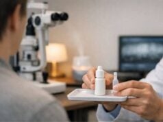
Bullous keratopathy is a visually debilitating eye disorder characterized by the formation of fluid-filled blisters (bullae) on the corneal surface, often resulting in pain, blurred vision, and sensitivity to light. The condition most frequently arises after endothelial cell loss, following cataract surgery, trauma, or as a result of inherited dystrophies. Effective management aims to relieve symptoms, restore corneal clarity, and prevent complications. In this comprehensive guide, we will delve into bullous keratopathy’s underlying mechanisms, established and advanced treatments, innovative technologies, and the latest clinical research to help patients, families, and professionals make confident, informed decisions.
Table of Contents
- Understanding Bullous Keratopathy and Its Epidemiology
- Current Non-Surgical and Drug-Based Management
- Operative and Interventional Approaches
- Recent Innovations and Future Technologies
- Clinical Trials and What Lies Ahead
- Frequently Asked Questions
Understanding Bullous Keratopathy and Its Epidemiology
Bullous keratopathy occurs when the corneal endothelium—the single cell layer responsible for maintaining corneal clarity—loses its ability to pump fluid out of the corneal stroma. The resulting fluid accumulation leads to stromal edema and the formation of painful epithelial blisters.
Key Points:
- Causes:
- Most often follows cataract surgery (pseudophakic bullous keratopathy).
- Can result from Fuchs’ endothelial dystrophy, trauma, glaucoma, or intraocular inflammation.
- Epidemiology:
- More common in older adults, especially after complicated ocular surgery.
- Incidence varies globally, but remains a leading cause for corneal transplantation in developed countries.
- Symptoms:
- Persistent blurry vision
- Eye pain, especially in the morning or after waking
- Glare and photophobia (light sensitivity)
- Recurrent erosions (if bullae rupture)
Practical Advice:
Routine ophthalmic evaluations are crucial for anyone with a history of eye surgery or endothelial dystrophy. Early detection of corneal swelling allows for more effective intervention and may delay or prevent the need for surgery.
Current Non-Surgical and Drug-Based Management
Non-surgical management primarily focuses on alleviating symptoms, promoting healing of the corneal surface, and minimizing further damage until definitive treatment is possible.
Conventional Therapies:
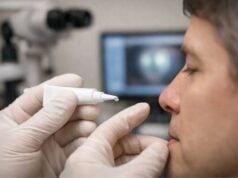
- Hypertonic Saline Drops and Ointments:
- Draw fluid out of the cornea, reducing swelling and discomfort.
- Typically used four times daily; ointment may be applied at bedtime for overnight relief.
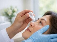
- Lubricating Artificial Tears:
- Soothe irritated corneas and reduce the sensation of a foreign body.
- Preservative-free options are preferred to minimize surface toxicity.
Additional Medical Interventions:
- Topical Steroids:
- May decrease inflammation but should be used with caution, as they can raise eye pressure.
- Bandage Contact Lenses:
- Protect the cornea from mechanical trauma caused by eyelid movement and ruptured bullae.
- Require careful monitoring for infection.
- Cycloplegic Drops:
- Relieve pain from ciliary muscle spasm, particularly in severe cases.
Adjunctive Strategies:
- Reducing Intraocular Pressure:
- Medications to lower eye pressure may slow corneal swelling.
- Avoiding Irritants:
- Shield the eye from wind, dust, and smoke.
- Use sunglasses for light sensitivity.
- Home Care:
- Cool compresses can ease discomfort.
- Avoid rubbing or pressing on the affected eye.
When Medical Treatment Isn’t Enough:
While these therapies can temporarily manage symptoms, they cannot restore lost endothelial function. Patients with persistent pain or declining vision may ultimately require surgical intervention.
Operative and Interventional Approaches
Surgical treatment is often the definitive solution for bullous keratopathy, aiming to restore corneal clarity and alleviate pain.
Surgical Procedures:
- Corneal Transplantation:
- Penetrating Keratoplasty (PK):
- Full-thickness transplant for severe or extensive disease.
- Traditional approach, but associated with higher rejection rates and longer recovery.
- Endothelial Keratoplasty (EK):
- Partial-thickness techniques (DSAEK, DMEK) replace only the diseased endothelium.
- Faster visual recovery, fewer complications, and less induced astigmatism.
- Anterior Stromal Puncture:
- Small needle punctures create adhesions, reducing bullae formation in patients unfit for transplant.
- Provides symptom relief, but does not improve vision.
- Amniotic Membrane Transplantation:
- Promotes healing and reduces pain for non-healing epithelial defects.
- Often used as a temporary measure before keratoplasty.
- Conjunctival Flap:
- Covers the affected cornea with conjunctiva, providing comfort in eyes with poor visual potential.
- Phototherapeutic Keratectomy (PTK):
- Laser ablation of the superficial cornea to remove diseased tissue and relieve pain.
Decision-Making and Patient Selection:
- Choice of surgery depends on age, general health, vision potential, and the presence of other eye diseases.
- Visual prognosis is best when intervention occurs before permanent corneal scarring.
Practical Advice:
Discuss with your corneal surgeon the pros and cons of each surgical option. If possible, prepare for surgery by optimizing your overall health and arranging post-operative support.
Recent Innovations and Future Technologies
Bullous keratopathy management is rapidly evolving, with several promising new technologies and experimental therapies on the horizon.
Gene and Cell Therapies:
- Cultured Endothelial Cell Injection:
- Early studies show that injection of lab-grown endothelial cells, sometimes combined with ROCK inhibitors, may restore corneal clarity without transplantation.
- Gene Editing:
- Research is ongoing to correct genetic defects in inherited endothelial dystrophies, offering hope for future generations.
Advanced Biomaterials:
- Artificial Corneas (Keratoprostheses):
- Modern designs are being developed for patients who are poor candidates for traditional transplantation.
- Novel Contact Lenses:
- Lenses embedded with drugs or bioactive molecules to accelerate healing and relieve pain.
Enhanced Diagnostic Tools:
- In Vivo Confocal Microscopy and Anterior Segment OCT:
- Provide high-resolution imaging of corneal cells, allowing for earlier diagnosis and better treatment planning.
- AI-Powered Algorithms:
- Assist clinicians in predicting disease progression and customizing treatment strategies.
Telemedicine and Home Monitoring:
Smartphone applications and home vision monitoring can facilitate early identification of changes, prompt specialist input, and improve patient engagement in their care.
Practical Advice:
Stay informed about clinical trials or innovative procedures that might be available to you. Ask your provider about new diagnostic and treatment options if your current management plan is not meeting your needs.
Clinical Trials and What Lies Ahead
The landscape of bullous keratopathy treatment is expanding, with ongoing research focused on long-term solutions and preventing recurrence.
Current Research Initiatives:
- Optimizing Transplantation:
- Studies are exploring ways to improve graft survival, reduce rejection, and minimize the need for lifelong immunosuppression.
- New Drug Delivery Systems:
- Extended-release formulations and sustained drug implants for pain control and anti-edema therapies.
- Tissue Engineering:
- Development of bioengineered corneal tissues and synthetic matrices to address donor shortages.
- Predictive Biomarkers:
- Genetic and protein markers may soon allow earlier detection of endothelial dysfunction.
Future Directions:
- Personalized Medicine:
- Tailoring therapies based on individual risk profiles and genetic backgrounds.
- Regenerative Approaches:
- Harnessing stem cells or molecular therapies to regenerate damaged endothelial cells.
- Global Health Solutions:
- Expanding access to corneal transplantation and eye banking in underserved regions.
Participating in Clinical Trials:
Participation offers access to cutting-edge care and contributes to the future of bullous keratopathy management. Ask your doctor about active trials or upcoming studies that may be suitable for you.
Practical Advice:
Regular follow-up is essential after any surgical procedure. Adhering to post-operative regimens and reporting new symptoms promptly will help ensure the best outcome.
Frequently Asked Questions
What causes bullous keratopathy?
Bullous keratopathy is mainly caused by endothelial cell loss from prior eye surgery, trauma, glaucoma, or inherited conditions like Fuchs’ dystrophy, leading to corneal swelling and blister formation.
What is the best treatment for bullous keratopathy?
The most effective long-term treatment is corneal transplantation, especially endothelial keratoplasty (DSAEK/DMEK). Temporary symptom relief may be achieved with medications, bandage contact lenses, or laser therapy.
How can I relieve pain from bullous keratopathy?
Pain is often relieved by using lubricating drops, hypertonic saline, bandage contact lenses, or protective eyewear. In persistent cases, surgical measures may be required.
Is bullous keratopathy reversible?
Permanent reversal is usually only possible with a corneal transplant or cell therapy. Symptom control can be achieved, but restoring normal corneal function typically requires surgery.
Can bullous keratopathy come back after treatment?
Recurrence is rare after successful endothelial transplantation, but underlying diseases (like Fuchs’ dystrophy) may affect the new graft over time. Regular follow-up is crucial.
Are there new treatments for bullous keratopathy?
Yes, innovations such as lab-grown cell injections, gene therapy, and advanced keratoprostheses are being studied and may soon be widely available.
Disclaimer:
This article is for educational purposes only and should not be considered as a substitute for professional medical advice. Always consult an eye care professional for an accurate diagnosis and personalized treatment plan.
If this guide was helpful, please share it on Facebook, X (formerly Twitter), or your favorite social platform—and follow us for more eye health insights. Your support helps us continue providing valuable content to the community. Thank you!


