
MyoD1 IHC Test: Rhabdomyosarcoma Marker, Sarcoma Diagnosis, and Positive Staining
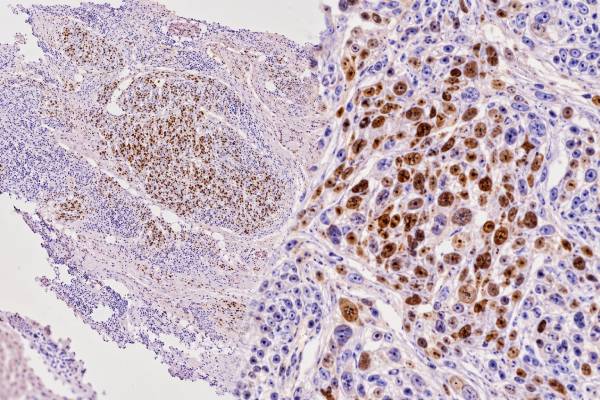
MyoD1 immunohistochemistry (IHC) is a tissue stain used to identify skeletal-muscle differentiation in tumors. MyoD1 is a myogenic regulatory transcription factor, so the diagnostically...
Myogenin IHC Test: Rhabdomyosarcoma Marker, Skeletal Muscle Differentiation, and Positive Staining
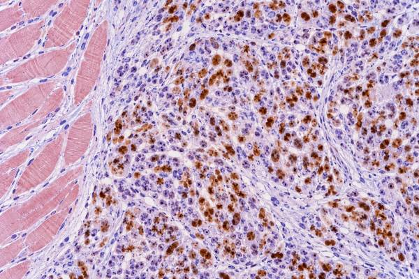
Myogenin immunohistochemistry (IHC) is a nuclear tissue stain used to demonstrate skeletal-muscle differentiation in tumors. The myogenin protein, encoded by the MYOG gene, is...
Napsin A IHC Test: Lung Adenocarcinoma Marker, Tumor Origin, and Positive Staining
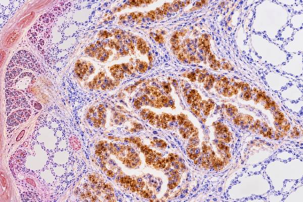
Napsin A immunohistochemistry (IHC) is a tissue stain commonly used to support a diagnosis of lung adenocarcinoma and to help evaluate the likely origin...
OCT4 IHC Test: Germ Cell Tumor Marker, Seminoma, Embryonal Carcinoma, and Positive Staining
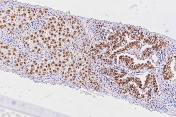
The OCT4 IHC test is a tissue stain that helps pathologists identify certain germ cell tumors, especially seminoma and embryonal carcinoma. OCT4, also called...
p40 IHC Test: Squamous Cell Carcinoma Marker, Lung Tumor Typing, and Positive Staining
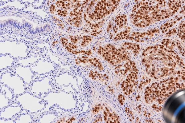
The p40 immunohistochemistry (IHC) test is a tissue stain that helps pathologists identify squamous differentiation, especially when classifying poorly differentiated lung cancers. p40 is...
p53 IHC Test: Tumor Suppressor Marker, Abnormal Staining, and Cancer Subtype
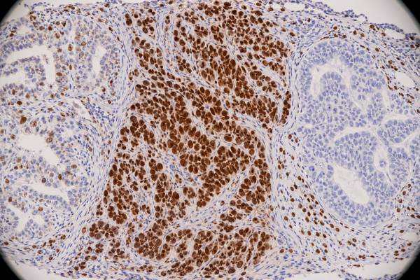
The p53 immunohistochemistry (IHC) test is a tissue stain that shows the pattern of p53 protein expression in tumor cells. Pathologists often use that...
p63 IHC Test: Squamous Cell Cancer, Urothelial Cancer, Tumor Origin, and Staining Meaning
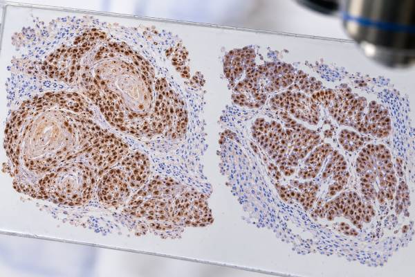
The p63 immunohistochemistry (IHC) test is a tissue stain used to identify a nuclear protein associated with basal cells, squamous differentiation, urothelial differentiation, and...
Pancytokeratin IHC Test: Epithelial Tumors, Carcinoma Marker, and Positive Staining
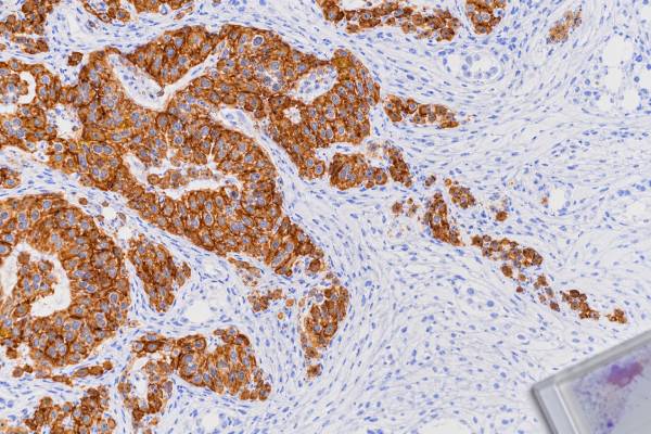
Pancytokeratin immunohistochemistry (IHC) is a broad tissue stain used to look for keratin proteins that are typical of epithelial cells. In a poorly differentiated...
PAX5 IHC Test: B-Cell Lymphoma Marker, B-Cell Lineage, and Tumor Classification
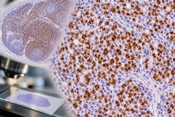
The PAX5 IHC test is a tissue stain used mainly to identify B-cell lineage in lymphomas and other hematolymphoid neoplasms. PAX5 is a nuclear...
PAX8 IHC Test: Ovarian, Kidney, Thyroid Cancer Marker, and Tumor Origin
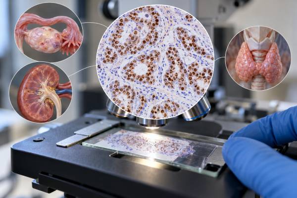
PAX8 immunohistochemistry (IHC) is a tissue stain that helps pathologists recognize tumors with Müllerian, renal, or thyroid follicular-cell differentiation. The PAX8 protein is a...
PLAP IHC Test: Germ Cell Tumor Marker, Seminoma Pattern, and Positive Staining
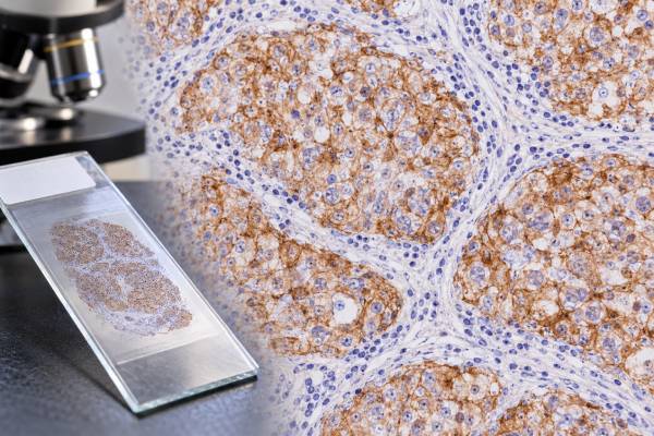
The PLAP IHC test detects placental alkaline phosphatase-related protein in tissue and is used mainly as a supporting marker for germ cell tumors. PLAP...
Progesterone Receptor (PR) IHC Test: Breast Cancer Hormone Receptor Status and Positive Score
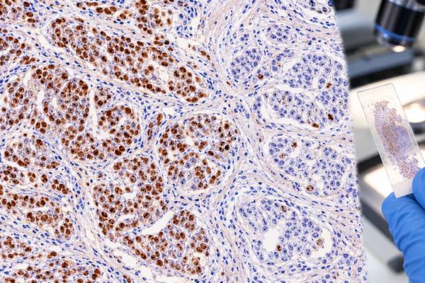
The progesterone receptor (PR) immunohistochemistry (IHC) test measures PR protein in the nuclei of breast cancer cells. It is usually performed with estrogen receptor...
S100 IHC Test: Melanoma, Nerve Sheath Tumor Marker, and Positive Staining
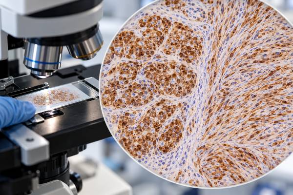
The S100 immunohistochemistry (IHC) test is a sensitive tissue stain used in the diagnosis of melanoma, peripheral nerve sheath tumors, and several other neoplasms....
SALL4 IHC Test: Germ Cell Tumor Marker, Embryonal Pattern, and Positive Staining
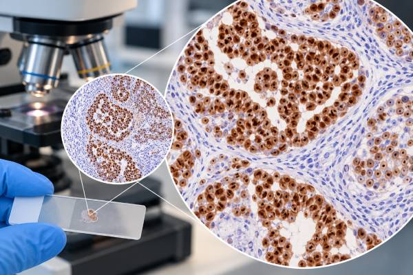
SALL4 immunohistochemistry (IHC) is a tissue stain used mainly to support germ cell differentiation in a tumor. SALL4 is a nuclear transcription factor associated...
SATB2 IHC Test: Colorectal Cancer Marker, Lower GI Tumor Origin, and Positive Staining
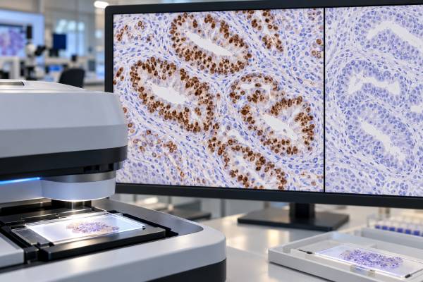
SATB2 immunohistochemistry (IHC) is a tissue stain that helps pathologists recognize colorectal and other lower gastrointestinal differentiation in a tumor. SATB2 is a nuclear...
SMAD4 IHC Test: Pancreatic Cancer Marker, Tumor Suppressor Loss, and Meaning
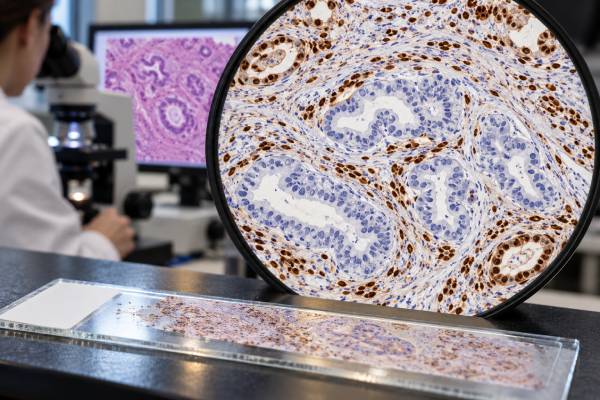
SMAD4 immunohistochemistry (IHC), also called DPC4 IHC, is a tissue stain that shows whether tumor cells retain or lose detectable SMAD4 protein. SMAD4 is...