
Fractional Excretion of Uric Acid (FEUA) Test: Gout, Kidney Handling, and Uric Acid Disorders

Fractional excretion of uric acid (FEUA) estimates what percentage of urate filtered by the kidneys is excreted in urine. It combines paired blood and...
Transtubular Potassium Gradient (TTKG) Test: Potassium Disorders and Aldosterone Effect

The transtubular potassium gradient (TTKG) is a calculated urine index historically used to estimate potassium secretion in the cortical collecting duct and to judge...
Urine Ammonium Test: Kidney Acid Excretion and Metabolic Acidosis Evaluation
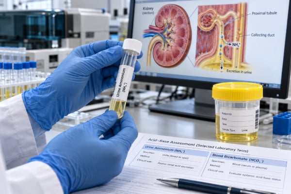
A urine ammonium test measures ammonium (NH₄⁺), the major form in which the kidneys excrete daily acid. It is most useful when blood tests...
Urine Anion Gap Test: Metabolic Acidosis, Kidney Acid Excretion, and Meaning
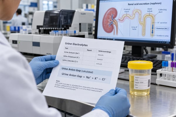
The urine anion gap (UAG) is a calculated value used mainly during normal-anion-gap metabolic acidosis to estimate whether the kidneys are increasing ammonium excretion....
Urine Calcium Test: High, Low, Kidney Stones, Parathyroid Causes, and Meaning

A urine calcium test measures how much calcium is being excreted by the kidneys. It is most often used to evaluate recurrent kidney stones,...
Urine Chloride Test: High, Low, Acid-Base Balance, and Volume Status

A urine chloride test measures how much chloride, a major negatively charged electrolyte, is being excreted in urine. The result is most useful when...
Urine Citrate Test: Kidney Stone Risk, Low Citrate, and Urine Chemistry

A urine citrate test measures citrate excreted by the kidneys, usually as part of a 24-hour urine evaluation for kidney stones. Citrate is protective...
Urine Creatinine Test: Kidney Function, Collection Check, Ratio Tests, and Meaning
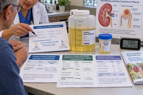
A urine creatinine test measures creatinine, a waste product generated from normal muscle metabolism, in a urine sample. It can be reported as a...
Urine Electrolyte Panel: Sodium, Potassium, Chloride, Osmolality, and Kidney Handling
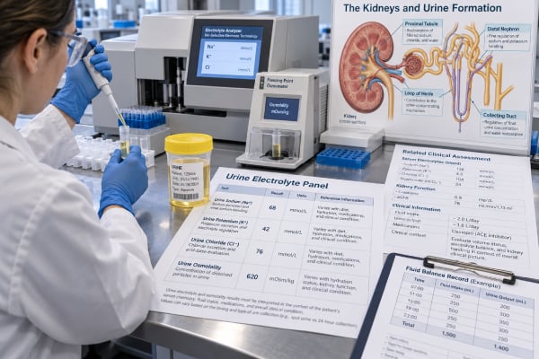
A urine electrolyte panel measures selected substances in urine—most often sodium, potassium, and chloride, with urine osmolality frequently added—to show how the kidneys are...
Urine Magnesium Test: High, Low, Kidney Loss, Mineral Balance, and Meaning

A urine magnesium test measures how much magnesium the kidneys are excreting. It is most useful when blood magnesium is low and the clinician...
Urine Osmolal Gap Test: Ammonium Excretion, Metabolic Acidosis, and Kidney Acid Handling
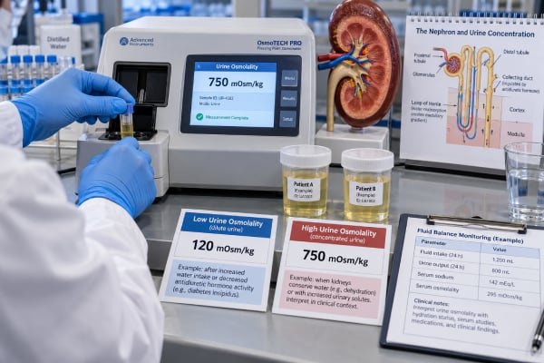
The urine osmolal gap is a calculated estimate of unmeasured particles in urine. In clinical nephrology, its main use is to roughly estimate urinary...
Urine Osmolality Test: High, Low, Hydration, Kidney Concentration, and Meaning
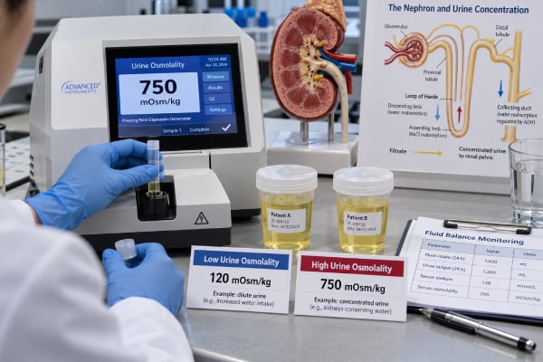
A urine osmolality test measures how concentrated or dilute urine is by counting the number of dissolved particles per kilogram of water. The kidneys...
Urine Oxalate Test: Kidney Stone Risk, High Oxalate, and Calcium Oxalate Stones
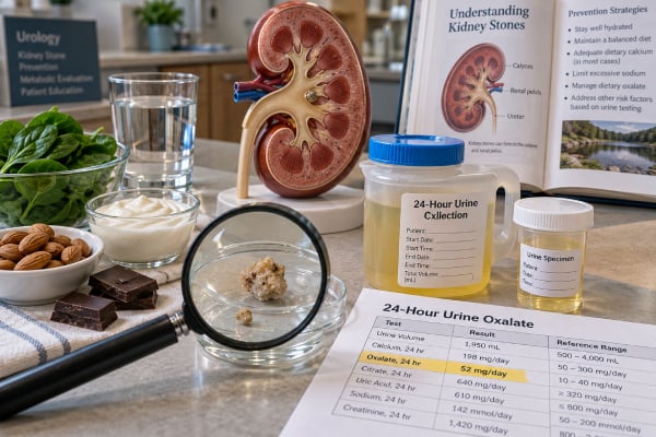
A urine oxalate test measures how much oxalate is excreted in urine, usually over 24 hours. Oxalate is a small organic compound that comes...
Urine pH Test: Acid-Base Balance, Kidney Stones, UTI, and Meaning
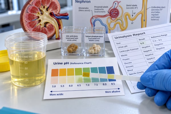
A urine pH test shows how acidic or alkaline urine is. It is a simple measurement, but its meaning depends heavily on context because...
Urine Phosphorus Test: High, Low, Kidney Function, Bone Mineral Balance, and Meaning
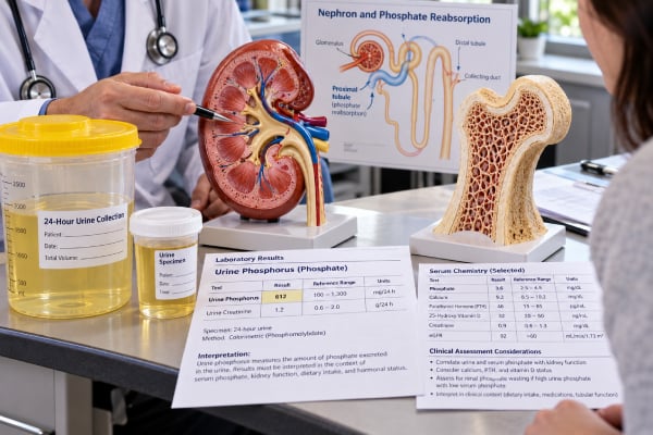
A urine phosphorus test measures how much phosphorus, mainly in the form of phosphate, your kidneys remove into urine. The result can help show...
Urine Potassium Test: High, Low, Kidney Potassium Loss, and Balance Meaning
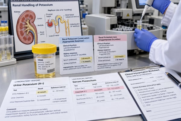
A urine potassium test measures how much potassium your kidneys are excreting. It is most useful when a blood potassium level is abnormal and...