
Urine Protein-Creatinine Ratio (UPCR) Test: Proteinuria, Kidney Disease, and Result Meaning

A urine protein-creatinine ratio (UPCR) estimates how much total protein is being lost in the urine while correcting for how concentrated the urine sample...
Urine Total Protein Test: High Levels, Kidney Disease, Proteinuria, and Meaning
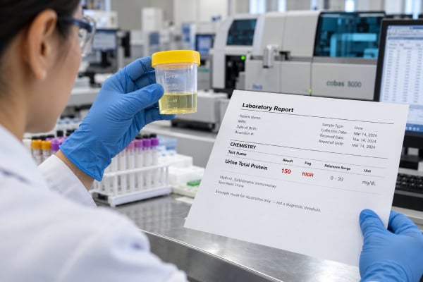
A urine total protein test measures the combined amount of protein present in urine rather than albumin alone. It can help detect and quantify...
Abnormal Urinalysis Results: Protein, Blood, Glucose, Ketones, Leukocytes, and Meaning

An abnormal urinalysis result is a clue, not a diagnosis. A dipstick can detect protein, blood, glucose, ketones, leukocyte esterase, nitrites, bilirubin, and changes...
Complete Urinalysis Test: Chemical, Microscopic, and Physical Urine Findings
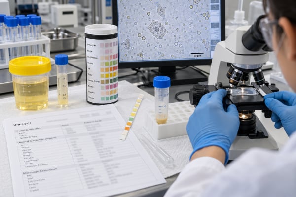
A complete urinalysis examines urine in three connected ways: physical appearance, chemical dipstick testing, and microscopic analysis of the sediment. Together, these measurements can...
Urinalysis for Diabetes: Glucose, Ketones, Protein, Specific Gravity, and Meaning
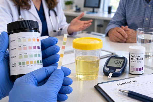
Urinalysis can provide useful clues about diabetes, but it does not diagnose or monitor diabetes as accurately as blood glucose, A1C, or continuous glucose...
Urinalysis for Kidney Disease: Protein, Blood, Casts, Albumin, and Microscopy Findings
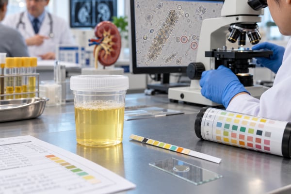
Urinalysis can reveal kidney damage before symptoms appear and can help identify which part of the kidney may be affected. The most useful findings...
Urinalysis for UTI: Leukocytes, Nitrites, Bacteria, WBCs, and Culture Follow-Up
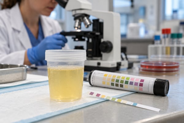
Urinalysis can support a urinary tract infection diagnosis by detecting leukocyte esterase, nitrites, white blood cells, bacteria, blood, and other changes, but it cannot...
Urinalysis Test: Dipstick, Microscopy, Normal Results, and What It Means
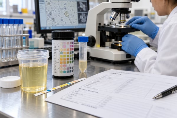
A urinalysis is a common test that checks urine for physical, chemical, and microscopic clues about the urinary tract, kidneys, metabolism, and several other...
Urine Albumin Dipstick Test: Positive Result, Albuminuria, Kidney Damage, and Meaning
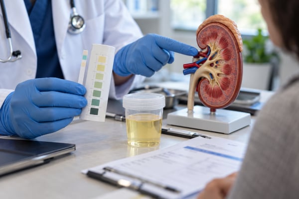
A positive urine albumin dipstick means albumin was detected in the sample, but it does not by itself prove chronic kidney disease or show...
Urine Ascorbic Acid Test: Vitamin C Interference and False-Negative Dipstick Results

A urine ascorbic acid test detects vitamin C in urine mainly so laboratories can recognize possible interference with other dipstick results. Vitamin C is...
Urine Bilirubin Test: Positive Result, Liver Disease, Bile Duct Problems, and Meaning

A urine bilirubin test checks for conjugated bilirubin, a water-soluble form of the yellow pigment produced when red blood cells are broken down. Normal...
Urine Blood Dipstick Test: Positive Blood, Hematuria, Causes, and Follow-Up
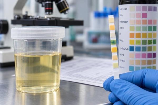
Urine Blood Dipstick Test is most useful when it is read as part of a clinical pattern, not as a stand-alone verdict. In practical...
Urine Clarity Test: Cloudy Urine, Sediment, Infection, and Abnormal Findings
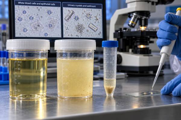
Urine Clarity Test is most useful when it is read as part of a clinical pattern, not as a stand-alone verdict. In practical terms,...
Urine Color Test: Dark, Cloudy, Red, Orange Urine, and What It Means
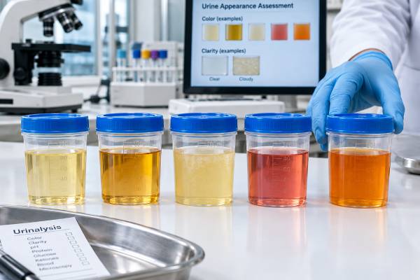
Urine Color Test is most useful when it is read as part of a clinical pattern, not as a stand-alone verdict. In practical terms,...
Urine Creatinine Spot Test: Urine Concentration, Ratio Tests, and Kidney Evaluation
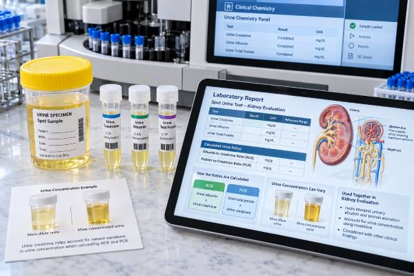
Urine Creatinine Spot Test is most useful when it is read as part of a clinical pattern, not as a stand-alone verdict. In practical...
Urine Dipstick Test: Protein, Blood, Glucose, Ketones, Nitrites, and Leukocytes
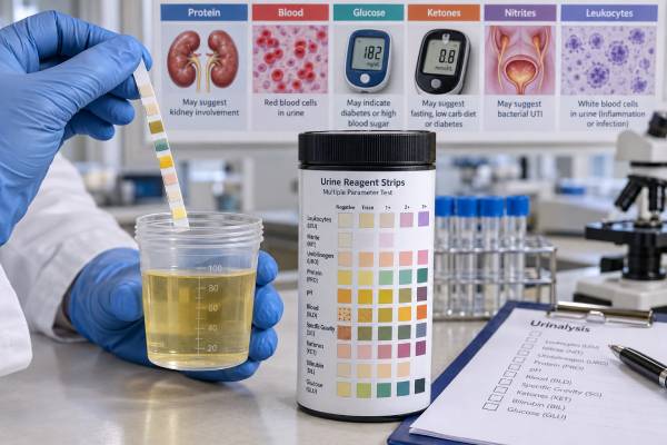
Urine Dipstick Test is most useful when it is read as part of a clinical pattern, not as a stand-alone verdict. In practical terms,...