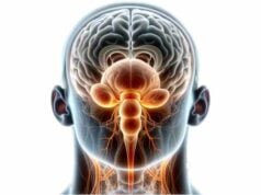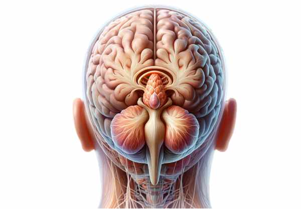
The pineal gland is a tiny, pine-cone shaped endocrine organ nestled deep within the brain that plays a pivotal role in regulating circadian rhythms and overall health. Acting as the master clock, it secretes melatonin—a hormone that governs sleep patterns, mood, and seasonal biological cycles—in response to light-dark cues. Despite its small size, its influence extends to neuroprotection, immune modulation, and even aging processes. This guide provides an in-depth exploration of the pineal gland’s complex anatomy, multifaceted physiological roles, common disorders, advanced diagnostic techniques, and modern treatment strategies, along with practical tips for maintaining optimal gland function and overall well-being.
Table of Contents
- Anatomical Structure and Histology
- Physiological Functions and Mechanisms
- Common Disorders and Conditions
- Diagnostic Techniques and Assessments
- Treatment Options and Therapies
- Nutritional Strategies and Supplementation
- Preventative Care and Healthy Practices
- Trusted Resources and Further Reading
- Frequently Asked Questions
Anatomical Structure and Histology
The pineal gland is a small, solitary structure whose name is derived from its resemblance to a pine cone. Despite its diminutive size—approximately 5–8 millimeters in length and weighing around 100–150 milligrams in adults—it holds significant neuroendocrine influence.
Macroscopic Anatomy
The gland is located in the epithalamus, near the center of the brain, adjacent to critical structures such as the thalamus, hypothalamus, and midbrain. It sits atop the roof of the third ventricle in a small cavity known as the pineal recess, which allows cerebrospinal fluid (CSF) to interact closely with its secretory cells. Encased by a delicate capsule derived from the pia mater, the pineal gland is protected yet remains intimately connected with the brain’s vascular and neural networks.
Key macroscopic features include:
- Size and Shape: A flattened, ovoid structure, typically 5–8 mm long and 3–5 mm wide.
- Coloration: Generally reddish-gray, which distinguishes it from surrounding brain tissue.
- Capsule: A thin, protective sheath formed from pia mater that provides structural support.
Microscopic Architecture
Under the microscope, the pineal gland reveals a rich tapestry of cellular components and specialized structures essential for its neuroendocrine function.
- Pinealocytes:
These are the primary secretory cells responsible for melatonin production. They are large, polygonal cells characterized by a prominent, round nucleus, abundant mitochondria, a well-developed Golgi apparatus, and dense-core secretory vesicles. Pinealocytes are organized into clusters or cords separated by delicate connective tissue septa, which help maintain their spatial arrangement and communication. - Interstitial (Glial) Cells:
Interspersed among the pinealocytes, these supportive cells share similarities with astrocytes in other regions of the brain. They provide metabolic support, help regulate the extracellular environment, and assist in the clearance of metabolic byproducts. - Perivascular Phagocytes:
Resembling macrophages, these cells are strategically located around blood vessels to remove cellular debris and play a role in immune surveillance. Their presence underscores the gland’s vulnerability to inflammatory processes. - Calcified Deposits (Corpora Arenacea):
Over time, calcium deposits, known as brain sand, accumulate within the pineal gland. These calcifications are common with advancing age and can be visualized in radiographic imaging. While their exact function remains under investigation, they are generally considered a benign aspect of aging.
Vascularization and Innervation
The pineal gland’s rich blood supply is provided primarily by the posterior choroidal arteries—branches of the posterior cerebral artery. These vessels form an intricate capillary network within the gland, ensuring a steady influx of oxygen and nutrients vital for melatonin synthesis. Venous drainage occurs through the internal cerebral veins, which eventually converge into the great cerebral vein (of Galen).
Innervation is equally complex, with both sympathetic and parasympathetic fibers playing key roles:
- Sympathetic Innervation:
Originating from the superior cervical ganglion, sympathetic fibers release norepinephrine, a critical regulator of melatonin production. These fibers travel along established neural pathways, linking the retina’s light information to the gland’s secretory machinery. - Parasympathetic Innervation:
Though less clearly defined, parasympathetic fibers from the pterygopalatine and otic ganglia are thought to modulate the gland’s activity, possibly influencing melatonin’s release in a complementary manner to sympathetic signals.
Developmental Origins
During embryonic development, the pineal gland arises from the roof of the diencephalon. By the seventh week of gestation, the pineal primordium is evident, and its maturation continues through childhood and adolescence. This developmental process is marked by differentiation into pinealocytes and supporting glial cells, setting the stage for the gland’s lifelong role in regulating circadian rhythms.
Physiological Functions and Mechanisms
The pineal gland’s primary function is the synthesis and secretion of melatonin—a hormone central to the regulation of circadian rhythms. However, its role extends into numerous physiological domains, influencing sleep, mood, immune function, and even aging.
Melatonin Synthesis
Melatonin production is a multistep enzymatic process that begins with the neurotransmitter serotonin. The key steps include:
- Conversion by AANAT:
Serotonin is first acetylated by the enzyme serotonin N-acetyltransferase (AANAT). This step is the rate-limiting phase in melatonin synthesis and is highly sensitive to the light-dark cycle. - Final Conversion by HIOMT:
The enzyme hydroxyindole O-methyltransferase (HIOMT) then converts N-acetylserotonin into melatonin. Although HIOMT activity remains relatively constant, the overall production of melatonin is largely dictated by AANAT activity.
Regulation of Melatonin Secretion
Melatonin secretion is intricately linked to the environmental light-dark cycle:
- Light Exposure:
During daylight, retinal photoreceptors detect light and transmit signals via the retinohypothalamic tract to the suprachiasmatic nucleus (SCN). The SCN, acting as the body’s master clock, inhibits melatonin production. - Darkness:
In the absence of light, the inhibition is lifted, allowing AANAT activity to increase and melatonin to be secreted in a rhythmic pattern. This nocturnal surge in melatonin is crucial for inducing sleep and regulating circadian rhythms.
Functions of Melatonin
Melatonin’s effects are wide-ranging, impacting various bodily functions:
- Circadian Rhythm Regulation:
By conveying information about environmental light conditions, melatonin helps synchronize the body’s internal clock with the external day-night cycle. This synchronization is critical for sleep-wake patterns, hormone release, and metabolic processes. - Antioxidant Defense:
Melatonin acts as a potent antioxidant, scavenging free radicals and protecting cells—particularly neurons—from oxidative stress. This neuroprotective function is being explored for its potential in mitigating neurodegenerative diseases. - Immune Modulation:
Melatonin influences immune cell activity, enhancing the body’s defense against pathogens. It modulates both innate and adaptive immune responses, contributing to an anti-inflammatory state. - Mood and Reproductive Regulation:
Emerging evidence suggests that melatonin may play a role in mood regulation and even influence reproductive hormones, affecting the timing of puberty and seasonal breeding patterns in some species.
Neuroendocrine Integration
The pineal gland acts as a bridge between the nervous and endocrine systems, translating neural signals into hormonal messages. This neuroendocrine integration is fundamental for the gland’s ability to influence circadian biology, linking environmental light cues with physiological processes throughout the body.
Additional Functions
Beyond melatonin synthesis, the pineal gland may have roles in:
- Aging:
Alterations in melatonin production have been associated with the aging process, and reduced melatonin levels are observed in older individuals. - Neuroprotection:
Its antioxidant and anti-inflammatory properties contribute to neuronal health, potentially guarding against diseases like Alzheimer’s and Parkinson’s. - Seasonal Adaptations:
In many animals, melatonin regulates seasonal rhythms such as reproductive cycles and hibernation, and similar mechanisms may subtly influence human physiology.
Common Disorders and Conditions
Though small, the pineal gland is susceptible to several disorders that can have significant systemic effects. Disruptions in its structure or function can alter melatonin secretion and, consequently, affect circadian rhythms and overall health.
Pineal Gland Tumors
Tumors of the pineal gland are rare but represent a significant portion of brain tumors, particularly in children and young adults. They include:
- Pineocytomas:
Benign tumors that arise from pinealocytes, usually growing slowly and associated with a favorable prognosis following surgical removal. - Pineoblastomas:
Malignant, aggressive tumors that often affect pediatric populations. Pineoblastomas require intensive treatment, including surgery, radiation, and chemotherapy. - Germ Cell Tumors:
These tumors, such as germinomas, can arise in the pineal region and are generally sensitive to radiation therapy. - Other Neoplasms:
Occasionally, metastases, meningiomas, or gliomas may involve the pineal gland, either primarily or as secondary lesions.
Pineal Cysts
Pineal cysts are fluid-filled cavities within the gland. They are usually asymptomatic and discovered incidentally on imaging studies. However, if a cyst enlarges, it may cause headaches, visual disturbances, or signs of increased intracranial pressure.
Calcification
Calcification of the pineal gland, known as corpora arenacea or “brain sand,” increases with age. While often a normal finding, excessive calcification may correlate with decreased melatonin production and has been linked in some studies to neurodegenerative processes.
Melatonin Secretion Disorders
Disorders in melatonin synthesis and secretion can lead to significant sleep and circadian rhythm disturbances:
- Insomnia:
Low melatonin levels are associated with difficulty in falling and staying asleep. - Delayed Sleep Phase Disorder (DSPD):
Abnormal timing of melatonin release can delay sleep onset, particularly in adolescents and young adults. - Seasonal Affective Disorder (SAD):
Reduced daylight in winter months may disrupt melatonin patterns, contributing to depressive symptoms. - Jet Lag:
Rapid transmeridian travel can disturb circadian rhythms, leading to temporary sleep disturbances that may be alleviated with melatonin supplementation.
Pineal Gland Dysfunction and Neurodegenerative Diseases
Emerging research suggests that pineal gland dysfunction may be linked to neurodegenerative diseases such as Alzheimer’s and Parkinson’s. Altered melatonin secretion in these conditions may contribute to sleep disturbances, cognitive impairment, and mood disorders.
Inflammatory and Infectious Processes
Though rare, the pineal gland can be affected by:
- Pinealitis:
Inflammation of the gland, which may result from autoimmune reactions or infections, potentially leading to headaches and circadian rhythm disturbances. - Infectious Conditions:
Certain viral or bacterial infections can involve the pineal region, though these are uncommon and typically require targeted antimicrobial therapy.
Diagnostic Techniques and Assessments
Diagnosing pineal gland disorders requires a combination of clinical evaluations, advanced imaging, laboratory tests, and sometimes invasive procedures. Each diagnostic tool contributes to a comprehensive understanding of the gland’s structure and function.
Clinical Evaluation
A detailed clinical evaluation is the first step:
- Medical History:
Physicians review the patient’s sleep patterns, neurological symptoms (such as headaches or visual changes), and any endocrine abnormalities. - Physical Examination:
A neurological exam is conducted to assess cranial nerve function, reflexes, and signs of increased intracranial pressure (e.g., papilledema).
Imaging Modalities
Imaging studies are crucial for visualizing pineal gland abnormalities:
- Magnetic Resonance Imaging (MRI):
MRI is the gold standard for evaluating the pineal region, providing high-resolution images to detect tumors, cysts, and calcifications. Contrast-enhanced studies further delineate lesion characteristics. - Computed Tomography (CT):
CT scans are particularly effective in identifying calcifications within the pineal gland and can help in the assessment of mass effects or hydrocephalus. - Ultrasound:
Transcranial ultrasound is occasionally used in neonates and infants, taking advantage of open fontanelles to view deep brain structures.
Laboratory Tests
Laboratory evaluations can help assess endocrine function and identify biochemical markers:
- Melatonin Assays:
Measuring melatonin levels in blood or saliva over a 24-hour period provides insights into circadian rhythm disturbances and pineal function. - Hormonal Panels:
Tests for other hormones (such as cortisol, thyroid hormones, and gonadotropins) may be performed if pineal tumors are suspected to affect broader endocrine functions. - Tumor Markers:
For suspected germ cell tumors, markers such as alpha-fetoprotein (AFP) and beta-human chorionic gonadotropin (β-hCG) can be measured.
Biopsy and Histological Examination
In cases where imaging indicates a neoplastic process:
- Stereotactic Biopsy:
A minimally invasive technique that uses a stereotactic frame to guide a biopsy needle to the pineal lesion, allowing for precise tissue sampling. - Open Surgical Biopsy:
In certain cases, especially with large lesions causing hydrocephalus, an open biopsy may be combined with therapeutic intervention.
Electrophysiological Studies
- Polysomnography:
Sleep studies may be conducted in patients with suspected melatonin secretion disorders, evaluating sleep architecture and disturbances. - Electroencephalography (EEG):
EEG can help assess any cortical disturbances that may be secondary to pineal lesions, especially if seizures are a concern.
Advanced Imaging Techniques
Emerging diagnostic modalities offer additional insights:
- Positron Emission Tomography (PET):
PET scans, particularly when combined with CT or MRI, evaluate the metabolic activity of pineal lesions to differentiate benign from malignant processes. - Magnetic Resonance Spectroscopy (MRS):
MRS provides a metabolic profile of the pineal gland, identifying biochemical markers that can aid in the diagnosis and characterization of tumors or cysts.
Treatment Options and Therapeutic Approaches
Treatment for pineal gland disorders depends on the specific condition, its severity, and the overall health of the patient. Options range from conservative management to surgical intervention and targeted therapies.
Management of Pineal Tumors
Surgical Approaches
- Craniotomy:
The traditional method involves creating an opening in the skull to access and remove pineal tumors. This approach is often necessary for large or invasive lesions. - Endoscopic Surgery:
Minimally invasive endoscopic techniques are used for smaller tumors or cysts, offering reduced recovery times and fewer complications.
Radiation Therapy
- External Beam Radiation:
High-energy beams target tumor cells, often used as adjunct therapy following surgery. - Stereotactic Radiosurgery (SRS):
This highly precise method delivers focused radiation to small lesions while minimizing damage to surrounding tissue.
Chemotherapy and Targeted Therapies
- Chemotherapy:
Utilized particularly for malignant tumors such as pineoblastomas and germ cell tumors. Treatment regimens may combine multiple agents to inhibit tumor growth. - Targeted Molecular Therapy:
Emerging treatments that focus on specific molecular pathways involved in tumor proliferation, offering a less toxic alternative to conventional chemotherapy.
Management of Pineal Cysts
For asymptomatic pineal cysts:
- Observation:
Regular monitoring with MRI to track changes in size or behavior. - Surgical Intervention:
In symptomatic cases, cyst drainage or excision may be indicated to relieve pressure on adjacent structures.
Addressing Melatonin Secretion Disorders
Pharmaceutical Interventions
- Melatonin Supplementation:
Over-the-counter melatonin supplements are widely used to treat insomnia, jet lag, and circadian rhythm disturbances. Dosage and timing are tailored to individual needs. - Sedative-Hypnotics:
In cases of severe insomnia, short-term use of benzodiazepines or non-benzodiazepine sleep aids may be prescribed, though these require cautious use due to dependency risks.
Light Therapy
- Bright Light Therapy:
Exposure to bright light during the day, especially in the morning, helps reset the circadian clock and suppress daytime melatonin production, thereby improving sleep patterns.
Neuroprotective and Anti-inflammatory Strategies
Research continues into therapies aimed at protecting pinealocytes from oxidative stress and inflammation:
- Antioxidant Therapy:
Supplements such as vitamins E and C, as well as coenzyme Q10, may protect the gland’s cells from free radical damage. - Anti-inflammatory Medications:
NSAIDs or corticosteroids can be used in cases of pinealitis or inflammatory damage to reduce tissue inflammation.
Multimodal Approaches
For complex cases where multiple dysfunctions occur:
- Combined Therapy:
Integration of surgery, radiation, and chemotherapy may be required to manage aggressive tumors. - Supportive Care:
Addressing symptoms such as sleep disturbances with a combination of behavioral therapy, dietary adjustments, and stress management techniques is crucial.
Nutritional Strategies and Supplementation
Proper nutrition and targeted supplementation play an essential role in supporting the pineal gland’s function and overall neuroendocrine health. A balanced diet rich in antioxidants, vitamins, and healthy fats can enhance melatonin production and protect against oxidative stress.
Key Nutrients for Pineal Health
- Melatonin:
Supplementing with melatonin can help regulate sleep-wake cycles, particularly for individuals suffering from insomnia or jet lag. It is available in various formulations and dosages. - Vitamin D:
Vital for overall brain health, vitamin D supports proper endocrine function and may influence melatonin synthesis. - Magnesium:
This mineral promotes relaxation and is essential for the enzymatic processes involved in melatonin production. - Omega-3 Fatty Acids:
Found in fish oil, these fatty acids help reduce inflammation and oxidative stress, supporting neuroprotection and healthy pineal function. - Zinc:
Zinc plays a role in hormone synthesis and immune function, and adequate levels are essential for optimal pineal activity.
Herbal and Natural Supplements
- Valerian Root:
Known for its sedative effects, valerian root can promote relaxation and improve sleep quality. - Passionflower and Chamomile:
These herbs have mild anxiolytic and sedative properties that support a healthy circadian rhythm. - Antioxidant-Rich Supplements:
Supplements such as glutathione, vitamin E, and vitamin C help neutralize free radicals, protecting the pineal gland from oxidative damage.
Probiotics
Maintaining a healthy gut flora can indirectly support pineal function by enhancing overall immune health and reducing systemic inflammation.
Integrating these nutritional strategies with a balanced diet—rich in fruits, vegetables, lean proteins, and whole grains—provides a solid foundation for optimal pineal gland health.
Preventative Care and Healthy Practices
Taking proactive steps to maintain the health of the pineal gland is essential for ensuring proper sleep patterns, hormone regulation, and neuroprotection. Adopting healthy habits can help prevent dysfunction and promote longevity.
Sleep Hygiene
- Consistent Sleep Schedule:
Going to bed and waking up at the same time every day helps maintain stable melatonin rhythms. - Minimize Light Exposure at Night:
Reducing blue light exposure from screens and using dim lighting in the evening can prevent melatonin suppression. - Create a Sleep-Friendly Environment:
A cool, dark, and quiet bedroom, along with a relaxing bedtime routine, enhances sleep quality.
Lifestyle Modifications
- Regular Physical Activity:
Exercise not only improves overall health but also promotes better sleep and hormonal balance. - Stress Management:
Techniques such as meditation, yoga, and deep breathing reduce stress, which can otherwise impair melatonin production. - Balanced Diet:
Eating nutrient-rich foods supports brain health and helps regulate circadian rhythms. - Hydration:
Staying well-hydrated ensures proper cellular function, including that of the pineal gland. - Moderate Caffeine and Alcohol Intake:
Limiting stimulants, especially in the evening, prevents interference with sleep patterns.
Environmental Considerations
- Exposure to Natural Light:
Spending time outdoors in natural sunlight during the day reinforces the body’s internal clock. - Use of Blue Light Filters:
Installing apps or using blue light blocking glasses in the evening can help maintain melatonin levels.
By incorporating these healthy practices into daily routines, you can support the pineal gland’s function, promote restorative sleep, and maintain overall neuroendocrine health.
Trusted Resources and Further Reading
Access to reliable and current information is crucial for understanding and managing pineal gland health. Below are some trusted resources and tools for further education and support.
Recommended Books
- “The Melatonin Miracle: Nature’s Age-Reversing, Disease-Fighting, Sex-Enhancing Hormone” by Walter Pierpaoli and William Regelson:
This book delves into the multifaceted benefits of melatonin, discussing its role in aging, disease prevention, and overall wellness. - “The Circadian Code: Lose Weight, Supercharge Your Energy, and Transform Your Health from Morning to Midnight” by Satchin Panda:
Satchin Panda provides insights into the importance of circadian rhythms and how aligning daily habits with natural cycles can improve overall health. - “Why We Sleep: Unlocking the Power of Sleep and Dreams” by Matthew Walker:
An authoritative exploration of sleep science, this book explains how melatonin and circadian rhythms influence health, performance, and longevity.
Academic Journals
- Journal of Pineal Research:
A peer-reviewed journal dedicated to all aspects of pineal gland physiology, pathology, and therapeutic research. - Chronobiology International:
This journal covers research on biological rhythms, offering extensive insights into circadian biology and the role of melatonin in health and disease.
Mobile Applications
- Sleep Cycle:
An app that monitors sleep patterns and offers insights to improve sleep quality through gentle wake-up techniques. - Headspace:
This mindfulness and meditation app provides guided exercises that help reduce stress and promote relaxation, indirectly supporting circadian health. - f.lux:
An application that adjusts your screen’s color temperature in the evening, reducing blue light exposure and supporting natural melatonin production.
Frequently Asked Questions on the Pineal Gland
What is the primary function of the pineal gland?
The pineal gland primarily produces and regulates melatonin, a hormone that governs sleep-wake cycles and circadian rhythms. It helps synchronize the body’s internal clock with the external light-dark cycle, influencing sleep, mood, and overall health.
How does light affect melatonin production?
Light exposure, especially blue light from screens, suppresses melatonin production by signaling the brain’s suprachiasmatic nucleus. In darkness, melatonin production increases, promoting sleep and regulating circadian rhythms.
What conditions are associated with pineal gland dysfunction?
Pineal gland dysfunction can lead to sleep disorders such as insomnia, delayed sleep phase disorder, and seasonal affective disorder. Additionally, tumors, cysts, and abnormal calcifications can impact melatonin secretion and overall neuroendocrine function.
How are pineal gland disorders diagnosed?
Diagnosis typically involves a combination of clinical evaluation, advanced imaging techniques (MRI, CT), laboratory tests for melatonin and hormone levels, and, in some cases, biopsy for histological analysis of suspected tumors or cysts.
Can lifestyle changes improve pineal gland health?
Yes, maintaining a consistent sleep schedule, reducing nighttime light exposure, managing stress, and consuming a balanced diet rich in antioxidants and healthy fats can support optimal pineal gland function and melatonin production.
Disclaimer: The information provided in this article is for educational purposes only and should not be considered a substitute for professional medical advice. Always consult a healthcare provider for any concerns regarding your health.
Please share this article on Facebook, X (formerly Twitter), or your preferred social media platform to help spread awareness about pineal gland health and the latest treatment strategies.

