
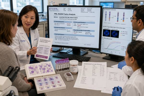
A PML-RARA test detects the fusion gene that defines acute promyelocytic leukemia (APL), a highly treatable but immediately dangerous subtype of acute myeloid leukemia. The fusion usually results from the chromosome translocation t(15;17), joining PML on chromosome 15 to RARA on chromosome 17. It blocks normal maturation of promyelocytes and creates sensitivity to all-trans retinoic acid (ATRA) and arsenic trioxide. When APL is suspected from blood counts, bleeding, coagulation abnormalities, or cell appearance, clinicians often start ATRA before molecular confirmation because life-threatening bleeding can occur early. Rapid fluorescence in situ hybridization or reverse-transcription PCR can confirm the fusion; quantitative RT-PCR later measures molecular response and residual disease. A positive result in the right clinical setting confirms PML::RARA-positive APL. A negative result requires urgent review of sample quality and alternative RARA fusions, especially when morphology remains strongly suggestive. The test’s purpose changes over time—from emergency diagnosis to transcript typing, remission confirmation, and relapse surveillance.
- PML::RARA positivity confirms the defining molecular abnormality of classic APL.
- Suspected APL is a medical emergency; ATRA is commonly started before the final test returns.
- RT-PCR identifies the transcript type and provides a baseline for later quantitative monitoring.
- A negative FISH result does not fully exclude a cryptic PML::RARA fusion or another RARA rearrangement.
- Molecular positivity after consolidation or a confirmed rising transcript can indicate persistent disease or molecular relapse.
Table of Contents
- Why PML-RARA Causes APL
- When Rapid Testing Is Ordered
- FISH, PCR, and Other Methods
- Interpreting the Diagnostic Result
- Molecular Monitoring and MRD
- Treatment Implications
- Negative, Atypical, and Variant RARA Cases
- Urgent Safety and Follow-Up
Why PML-RARA Causes APL
RARA encodes retinoic acid receptor alpha, a transcription factor that helps immature myeloid cells mature in response to retinoic acid. PML participates in nuclear structures involved in tumor suppression, apoptosis, and genome regulation. The PML::RARA fusion combines parts of both proteins and recruits corepressor complexes too strongly, locking promyelocytes in an immature state.
Pharmacologic doses of ATRA release the differentiation block, while arsenic trioxide promotes degradation of the fusion protein through a complementary mechanism. This direct molecular vulnerability transformed APL from one of the most lethal acute leukemias into one of the most curable. The early hazard remains severe coagulopathy, including disseminated intravascular coagulation, intracranial bleeding, pulmonary bleeding, and thrombosis.
The fusion is usually created by t(15;17)(q24;q21). Three main PML breakpoint regions generate transcript isoforms commonly called bcr1, bcr2, and bcr3, corresponding to long, variable, and short forms. Identifying the transcript at diagnosis allows the laboratory to use the correct quantitative assay during follow-up. Transcript type alone generally does not determine treatment.
PML::RARA is a somatic leukemia abnormality, not an inherited mutation. It does not imply that relatives carry an APL gene. Rare therapy-related APL can occur after exposure to certain topoisomerase II inhibitors, but the fusion remains acquired in the leukemia cells.
APL morphology often shows abnormal promyelocytes with heavy granules and bundles of Auer rods, sometimes called faggot cells. A microgranular variant may have bilobed nuclei, few visible granules, and a high white count, mimicking other AML. Flow cytometry commonly shows strong myeloperoxidase, CD33, and CD117, with absent or weak HLA-DR and CD34, but no immunophenotype replaces molecular confirmation.
When Rapid Testing Is Ordered
PML-RARA testing is ordered immediately when APL is suspected. Triggers include abnormal promyelocytes on a blood or marrow smear, severe thrombocytopenia, prolonged coagulation tests, low fibrinogen, high D-dimer, unexplained bleeding, or an AML immunophenotype compatible with APL. The diagnosis can arise in a person with a low, normal, or high white blood cell count.
Because fatal hemorrhage can occur before confirmation, suspected APL is treated as an emergency. ATRA is generally started as soon as the diagnosis is considered and stopped later if testing excludes APL. Coagulation studies and blood counts are repeated frequently, and platelets, cryoprecipitate or fibrinogen concentrate, and plasma are given according to protocol. Invasive procedures are minimized until coagulopathy improves.
The laboratory may run several rapid tests in parallel:
- PML::RARA RT-PCR on blood or marrow;
- PML/RARA dual-fusion FISH;
- RARA break-apart FISH when a variant fusion is possible;
- immunofluorescence for the characteristic PML nuclear pattern in specialized centers;
- chromosome analysis;
- a rapid RNA fusion panel.
Blood can provide a fast result when circulating abnormal cells are present. Bone marrow is often collected once the patient is stabilized enough for the procedure. Treatment should not be delayed solely to obtain marrow if blood is diagnostic.
Rapid communication among the emergency team, hematologist, pathologist, transfusion service, and molecular laboratory is essential. A routine send-out test with a long turnaround is not adequate when local rapid testing or transfer to a center with APL capability is available.
FISH, PCR, and Other Methods
RT-PCR detects the expressed fusion transcript. It is highly sensitive, identifies the breakpoint form, and establishes the target for quantitative MRD testing. Diagnostic qualitative RT-PCR may return detected/not detected, while RT-qPCR reports a normalized amount.
FISH uses fluorescent DNA probes to show fusion of PML and RARA or separation of RARA. It works on interphase nuclei and often returns quickly. Dual-fusion FISH can miss rare cryptic insertions or unusual breakpoint configurations. RARA break-apart FISH may reveal a non-PML partner but cannot identify the partner by itself.
| Method | Best use | Main strength | Main limitation |
|---|---|---|---|
| RT-PCR | Rapid diagnosis and transcript typing | Sensitive and directly detects fusion RNA | Requires intact RNA and primers matching the fusion |
| RT-qPCR | Quantitative MRD monitoring | Can detect very low transcript levels | Results depend on control-gene quality and standardization |
| Dual-fusion FISH | Rapid t(15;17) confirmation | Visual result in nondividing cells | Can miss cryptic or variant rearrangements |
| Karyotype | Chromosome-level diagnosis and additional changes | Shows the classic translocation and broader genome | Requires viable dividing cells and takes longer |
| RNA fusion sequencing | Cryptic or variant cases | Can identify uncommon partners | Turnaround and RNA quality may limit emergency use |
An FISH test and a quantitative PCR test answer related but different questions. FISH is excellent for rapid structural confirmation, while RT-qPCR provides the sensitivity and quantitative continuity needed for follow-up.
The report should state specimen type, fusion transcript, control-gene performance, sensitivity, and whether the assay can detect all common isoforms. At diagnosis, a baseline transcript level may be recorded. A percentage from one laboratory should not be compared directly with a different platform unless harmonization is established.
Interpreting the Diagnostic Result
A PML::RARA detected result in a compatible myeloid neoplasm confirms classic APL. The integrated report may also state the transcript type and cytogenetic result. Additional chromosome changes do not usually negate the diagnosis. FLT3 mutations are common, particularly in high-white-count or microgranular APL, but they do not replace the defining fusion.
A positive result is qualitatively different from a result that says RARA rearranged. RARA break-apart positivity means RARA has another partner unless PML involvement is separately proven. Some variant RARA fusions resemble APL morphologically but respond differently to ATRA and arsenic. Partner identification is therefore urgent.
A not-detected result must be interpreted by method. If both sensitive RT-PCR and appropriate FISH are negative in an adequate sample, classic PML::RARA-positive APL becomes unlikely. If morphology is compelling, the team should review specimen quality, repeat testing, use RARA break-apart probes, and perform RNA fusion analysis. Rare cryptic PML::RARA fusions can be PCR-positive despite normal karyotype and initially negative FISH.
An inadequate result is not negative. RNA can degrade, a marrow aspirate can be hemodiluted, or too few abnormal cells may remain after urgent therapy. The report should state the problem and recommend another specimen or method. Treatment decisions in the first hours may rely on the clinical suspicion while confirmatory testing continues.
A low diagnostic transcript number should not be interpreted as mild disease. Sample dilution and assay normalization influence the value. Clinical risk at presentation is commonly stratified by white blood cell and platelet counts, while coagulopathy severity and organ status drive immediate supportive care.
Molecular Monitoring and MRD
After treatment starts, RT-qPCR tracks the fall in PML::RARA transcripts. Morphologic remission means blasts and abnormal promyelocytes are no longer visible at the usual microscopic threshold. Molecular remission means the fusion is not detected by a much more sensitive assay in an adequate sample. These milestones occur on different timelines.
A positive PCR immediately after induction does not necessarily mean treatment failure. Differentiating cells and residual transcripts can persist while therapy continues. The end-of-consolidation result is more decisive: patients should achieve molecular remission after completing consolidation. Persistent positivity at that point requires confirmation and specialist management.
Bone marrow is generally more sensitive than blood for molecular detection. Blood is easier to sample and may be used in surveillance, sometimes at shorter intervals. Monitoring schedules vary by risk group, regimen, and guideline. Some low-risk patients treated successfully with modern ATRA/arsenic regimens may need less intensive routine surveillance after confirmed molecular remission than historically used; higher-risk patients often receive scheduled PCR monitoring for a defined period.
A molecular relapse generally means confirmed reappearance or a significant rise of PML::RARA after prior negativity, ideally demonstrated in serial samples and according to laboratory criteria. Detecting relapse before overt blood-count or morphologic relapse can permit earlier salvage therapy. A single weak positive near the detection limit is usually repeated promptly before a major treatment decision.
The MRD result should include specimen, transcript, normalized value, sensitivity, prior comparison, and trend. Switching laboratories can complicate interpretation. Keeping a copy of the baseline molecular report helps the follow-up laboratory verify the correct transcript target.
Treatment Implications
The defining fusion predicts sensitivity to differentiation therapy. Non-high-risk APL is commonly treated with ATRA plus arsenic trioxide, often without conventional chemotherapy. High-risk APL, commonly defined by a presenting white blood cell count above 10 × 10⁹/L, may receive ATRA and arsenic with an additional cytoreductive agent or chemotherapy according to protocol. Pediatric, pregnant, relapsed, and medically complex patients need individualized plans.
ATRA and arsenic can cause differentiation syndrome, marked by fever, weight gain, edema, low blood pressure, shortness of breath, lung infiltrates, or kidney dysfunction. It can be fatal without prompt corticosteroids and supportive care. Patients should report symptoms immediately rather than waiting for the next clinic visit.
Arsenic can prolong the QT interval and disturb electrolytes. Electrocardiograms, potassium, magnesium, liver tests, and other monitoring are part of care. ATRA can cause headache, liver abnormalities, lipid changes, and other effects. Supportive care and protocol adherence are central to the high cure rate.
Molecular persistence or relapse changes management. Salvage may use arsenic if the initial regimen was ATRA/chemotherapy, or an ATRA/chemotherapy-based approach if arsenic was used initially, along with other agents and possible stem cell transplantation depending on molecular remission, relapse timing, and patient factors. Modern options continue to evolve.
The PML::RARA result is unusually actionable because it establishes both diagnosis and a treatment vulnerability. Even so, treatment is not reduced to the molecular test. Coagulopathy control, infection prevention, differentiation syndrome management, adherence, and scheduled PCR monitoring determine whether the biological opportunity becomes a cure.
Negative, Atypical, and Variant RARA Cases
A strongly APL-like leukemia with negative PML::RARA testing requires urgent investigation for technical failure and variant RARA fusions. Partners can include ZBTB16, NPM1, NUMA1, STAT5B, BCOR, and others. Sensitivity to ATRA and arsenic varies widely. ZBTB16::RARA, for example, is generally resistant to standard differentiation therapy alone and requires an AML-oriented approach.
Some non-RARA fusions and NPM1-mutated AML can mimic APL morphology or immunophenotype. This is why rapid molecular confirmation is essential even when the smear appears classic. Conversely, microgranular APL can resemble monocytic AML or other high-white-count leukemia.
A normal karyotype does not exclude PML::RARA. Cryptic insertions may be invisible by conventional chromosome analysis. FISH can also fail if the insertion is smaller than the probe design resolves. RT-PCR or RNA sequencing can reveal the fusion. The reverse problem also occurs: unusual FISH patterns may need PCR to prove an expressed in-frame transcript.
If testing remains negative, the team should not continue APL-specific treatment indefinitely on morphology alone. The integrated diagnosis may shift to another AML subtype, and therapy should follow that biology. Until APL is reasonably excluded, however, stopping ATRA prematurely can be dangerous. This balance requires real-time specialist coordination.
Urgent Safety and Follow-Up
APL has a unique early-death risk from bleeding, so safety instructions are part of interpreting the test. Seek emergency care for severe headache, confusion, weakness, new vision change, coughing or vomiting blood, black stool, heavy vaginal bleeding, persistent nose or mouth bleeding, chest pain, or sudden shortness of breath. Do not wait for a molecular result to seek help.
Questions for the care team include:
- Was ATRA started when APL was suspected?
- Which rapid tests were performed, and do they agree?
- Which PML breakpoint transcript—bcr1, bcr2, or bcr3—was identified?
- What was the presenting white blood cell risk group?
- How are fibrinogen, platelets, and coagulation tests being supported?
- What symptoms of differentiation syndrome should be reported immediately?
- When will PCR be checked after induction and consolidation?
- What result defines molecular remission in this laboratory?
- Will follow-up use marrow, blood, or both, and for how long?
- What is the plan if a weak positive or rising transcript appears?
Store the diagnostic RT-PCR report, FISH result, karyotype, treatment summary, and end-of-consolidation molecular result. A future laboratory needs the exact transcript type. After therapy, maintain scheduled follow-up even when blood counts feel normal; molecular recurrence can precede symptoms.
What happens during the first critical days
The first hours of suspected APL focus on preventing catastrophic bleeding. Clinicians obtain a complete blood count, prothrombin time, activated partial thromboplastin time, fibrinogen, D-dimer, chemistry panel, and type and screen, then repeat them frequently. Many protocols support platelets to a higher threshold than in other leukemias and maintain fibrinogen above a specified level. Exact transfusion targets depend on the treatment center, active bleeding, pregnancy, and procedure needs.
ATRA should be readily available wherever acute leukemia is treated. Its early use can begin reversing the coagulopathy and is generally considered safer than waiting when APL is strongly suspected. Leukapheresis is usually avoided in APL because invasive vascular procedures and rapid cell shifts can worsen bleeding risk; cytoreduction is managed with disease-specific medication. Central lines, lumbar puncture, and other invasive procedures may be deferred until coagulation improves.
The care team watches closely for differentiation syndrome, which can develop within days or later. Rapidly rising white cells, fever, weight gain, edema, lung infiltrates, low oxygen, low blood pressure, or kidney injury can be early signs. Dexamethasone is started promptly when suspected, and ATRA or arsenic may be held in severe cases according to protocol. Waiting for every feature to appear can allow the syndrome to progress.
Understanding milestones after treatment begins
Induction aims to control the leukemia and achieve hematologic remission, but marrow examined too early can look abnormal because promyelocytes are differentiating rather than disappearing immediately. Clinicians avoid declaring induction failure from an early morphologic or PCR-positive marrow. Recovery timing with ATRA and arsenic differs from conventional AML induction.
Consolidation completes molecular eradication. A high-quality marrow RT-qPCR after consolidation is a major landmark. If PML::RARA remains detected, the result is repeated and the laboratory checks transcript and sample quality. Confirmed molecular persistence calls for a salvage plan at an APL-experienced center.
Maintenance therapy is not required for every modern low-risk ATRA/arsenic regimen, while some higher-risk or chemotherapy-based protocols include it. PCR surveillance intensity similarly varies. The follow-up plan should name the specimen, frequency, duration, and action threshold so patients do not receive unexplained sporadic tests.
Pregnancy creates additional complexity. ATRA, arsenic, chemotherapy, fetal gestational age, and maternal bleeding risk require an expert multidisciplinary team. Arsenic is contraindicated during pregnancy, and options change by trimester. The emergency diagnostic principle remains the same: confirm the fusion quickly while controlling coagulopathy.
APL survivors should retain a treatment summary because cure rates are high and long-term care may occur outside the original center. The summary should include PML breakpoint type, risk group, regimen and cumulative anthracycline exposure if any, differentiation syndrome history, end-of-consolidation PCR, and surveillance completion date.
Laboratory details that prevent false reassurance
The initial RT-PCR must include the common long and short PML transcript forms and appropriate RNA controls. A failed control means the specimen cannot support a negative call. If treatment started before collection, blood may clear quickly; marrow or a stored pretreatment smear can sometimes provide better material. The laboratory should retain the diagnostic positive control or sequence information for future comparisons.
PCR values should not be compared across laboratories without caution. Different control genes, calibration curves, reporting scales, and specimen processing can produce different numbers. When care transfers, the receiving center may establish its own baseline while using the original transcript type. A sudden numerical change after a laboratory switch is not automatically biological relapse.
Rare false-positive contamination is possible in highly sensitive PCR. A weak unexpected positive should be repeated from a new extraction or specimen and interpreted with controls. Conversely, a technically perfect negative test cannot exclude a non-PML RARA fusion unless the workup includes methods capable of finding it.
Long-term follow-up also includes late effects. Anthracycline-exposed survivors may need cardiac monitoring; arsenic-treated patients require follow-up of liver, metabolic, or neurologic issues as clinically indicated. Secondary malignancies are uncommon but possible after cytotoxic therapy. These survivorship concerns are separate from PML::RARA molecular surveillance.
Medication adherence after discharge is critical because ATRA and arsenic schedules can be complex. Patients should receive a calendar, interaction review, emergency contact instructions, and a plan for missed doses. Differentiation syndrome, QT changes, liver abnormalities, and electrolyte problems can develop between visits, so scheduled laboratory and clinical monitoring should continue even when symptoms improve.
Dental work and elective surgery should be coordinated during active treatment because platelet counts, coagulation, infection risk, and medication timing may require precautions. The hematology team should provide clearance rather than relying on a normal-looking blood count alone.
References
- Diagnosis and management of acute promyelocytic leukemia 2024 (Review)
- Utilization of RT-PCR and Optical Genome Mapping in the Diagnosis and Management of Acute Promyelocytic Leukemia With Cryptic PML::RARA Rearrangements 2024 (Systematic Review)
- Management of acute promyelocytic leukemia: updated recommendations from an expert panel of the European LeukemiaNet 2019 (Recommendations)
- Acute Myeloid Leukemia: 2025 Update on Diagnosis, Risk-Stratification, and Management 2025 (Review)
- PML::RARA Quantitative, PCR, Varies 2025 (Official Test Guide)
Disclaimer
This article provides general education and is not a substitute for emergency leukemia care. Suspected APL requires immediate specialist evaluation, rapid molecular testing, aggressive blood-product support, and often empiric ATRA before confirmation. Anyone with serious bleeding, severe headache, breathing difficulty, confusion, or sudden deterioration should receive emergency medical attention.





