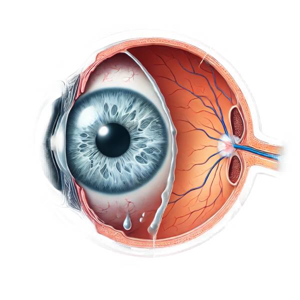
Post-cataract surgery glaucoma, also known as secondary glaucoma, is a condition characterized by an increase in intraocular pressure (IOP) following cataract surgery. Cataract surgery, which involves removing the eye’s natural lens and replacing it with an artificial intraocular lens (IOL), is generally considered safe and effective. However, complications can occur, with one of the most serious being the development of glaucoma.
Understanding Cataract Surgery
Cataract surgery is one of the most common procedures performed worldwide, with a high success rate in restoring vision. The procedure usually entails phacoemulsification, which involves breaking up the cloudy lens with ultrasound waves before removing and replacing it with an IOL. This surgery is typically performed as an outpatient under local anesthesia, and patients can often return to normal activities shortly after the procedure.
The Pathophysiology of Glaucoma Following Cataract Surgery
Several mechanisms can influence the pathogenesis of post-cataract surgery glaucoma:
- Inflammatory Response: Surgery causes natural inflammation. However, excessive inflammation can cause an increase in IOP. This inflammation can cause trabecular meshwork obstruction, which reduces aqueous humor outflow and results in elevated IOP.
- Trabecular Meshwork Damage: Inadvertent damage to the trabecular meshwork (the eye’s drainage system) can occur during cataract surgery. Damage to this structure can obstruct the drainage of aqueous humor, resulting in elevated IOP.
- Corticosteroid Use: Postoperative corticosteroids are frequently prescribed to reduce inflammation. However, some patients are steroid responders, which means their IOP rises in response to steroid use. This increase may exacerbate the risk of glaucoma.
- Preexisting Conditions: Patients with preexisting ocular conditions, such as narrow angles or pseudoexfoliation syndrome, are more likely to develop glaucoma following surgery. These conditions may predispose the eye to aqueous humor outflow obstruction.
- Iris and Lens Fragment Obstruction: Residual lens fragments or inflammatory debris can clog the trabecular meshwork, preventing normal aqueous outflow and causing elevated IOP.
Clinical Presentation
The clinical presentation of post-cataract surgery glaucoma varies greatly. It can appear shortly after surgery (acute) or months to years later (chronic). The key symptoms and signs are:
- Increased Intraocular Pressure (IOP): Elevated IOP, a characteristic of glaucoma, is frequently detected during routine postoperative follow-ups. Patients may not notice symptoms at first.
- Vision Changes: Patients may notice blurry vision or halos around lights. Severe cases can result in significant vision loss.
- Eye Pain: Although not always present, some patients may experience eye pain or discomfort.
- Redness and swelling: These are signs of inflammation and may be associated with elevated IOP.
- Optic Nerve Damage: Prolonged elevated IOP can cause optic nerve damage, which is visible during an eye exam.
Risk Factors
Several risk factors can predispose patients to develop glaucoma following cataract surgery:
- Preexisting Glaucoma: Patients who have a history of glaucoma are more likely to experience IOP spikes after cataract surgery.
- Inflammatory Eye Conditions: Conditions such as uveitis can increase the risk of developing glaucoma following surgery due to inflammation.
- Advanced Age: Older patients are more likely to experience complications after cataract surgery, including glaucoma.
- Complex Surgery: Complicated cataract surgeries, such as those with longer operative times or surgical complications, can raise the risk.
- Genetic Predisposition: Some people may have a genetic predisposition to high IOP or glaucoma.
Pathologic Findings
Histopathological examination of eyes with post-cataract surgery glaucoma frequently reveals alterations in the trabecular meshwork and surrounding tissues. Inflammatory cells, fibrotic changes, and cellular debris can clog the trabecular meshwork. Furthermore, changes in the structure and function of the trabecular meshwork can impair aqueous humor outflow, resulting in elevated IOP.
Epidemiology
The risk of post-cataract surgery glaucoma varies depending on a number of factors, including patient demographics, surgical techniques, and the presence of preexisting conditions. According to research, the risk of developing glaucoma following cataract surgery ranges between 1% and 4%, with higher rates in patients with predisposing factors. Despite the low incidence, the consequences for affected individuals can be severe, necessitating close monitoring and management.
Different Types of Glaucoma Following Cataract Surgery
The timing and underlying mechanism of post-cataract surgery glaucoma can be
- Acute Postoperative Glaucoma: This type develops within the first few days or weeks after surgery. It is frequently associated with inflammation, corticosteroid use, or mechanical blockage of the trabecular meshwork due to residual lens fragments or viscoelastic material.
- Chronic Postoperative Glaucoma: This type develops several months or years after surgery. Scarring and fibrosis of the trabecular meshwork, as well as progressive damage to the optic nerve as a result of prolonged elevated IOP, are typical causes.
Effects on Quality of Life
The development of glaucoma after cataract surgery can have a significant impact on a patient’s quality of life. Even mild vision loss can have an impact on daily activities and independence. The psychological burden of managing a chronic condition, the need for regular follow-ups, and the potential side effects of medications can all have an impact on patients’ well-being.
Research and Advances
Ongoing research aims to better understand the mechanisms underlying post-cataract surgery glaucoma, as well as develop prevention and management strategies. Advances in surgical techniques, such as micro-invasive glaucoma surgery (MIGS), show promise in lowering the incidence and severity of glaucoma following cataract surgery. Furthermore, research into genetic markers and personalized medicine approaches seeks to identify high-risk patients and tailor interventions accordingly.
Diagnostic methods
Clinical evaluation, diagnostic tests, and imaging studies are all required to diagnose post-cataract surgery glaucoma. A timely and accurate diagnosis is critical for effective management and avoiding optic nerve damage.
Clinical Evaluation
The first step in diagnosing post-cataract surgery glaucoma is a comprehensive eye examination that includes:
- Visual Acuity Test: Assessing vision clarity can help determine the severity of visual impairment.
- Intraocular Pressure (IOP) Measurement: An eye care professional uses tonometry to measure the pressure inside the eye. Elevated IOP is a strong indicator of glaucoma.
- Slit-Lamp Examination: This examination gives a detailed view of the anterior segment of the eye, including the cornea, iris, and lens, allowing for the detection of any abnormalities or inflammation.
Gonioscopy
Gonioscopy is a critical diagnostic tool that visualizes the angle between the iris and the cornea. This procedure aids in assessing the trabecular meshwork’s condition and identifying any angle abnormalities or blockages that may contribute to elevated IOP.
Visual Field Testing
Visual field testing assesses peripheral vision and aids in detecting vision loss due to optic nerve damage. This test is critical for tracking the progression of glaucoma and determining the effectiveness of treatment.
Optical Coherence Tomography(OCT)
OCT is a non-invasive imaging technique for obtaining high-resolution cross-sectional images of the retina and optic nerve head. It is useful in determining the thickness of the retinal nerve fiber layer and detecting early signs of optic nerve damage. OCT is useful for diagnosing glaucoma and tracking its progress over time.
Pachymetry
Pachymetry measures corneal thickness. Corneal thickness can influence IOP readings; thus, this measurement aids in accurately interpreting IOP readings and determining the risk of glaucoma.
Anterior Segment Imaging
Advanced imaging techniques, such as anterior segment OCT and ultrasound biomicroscopy (UBM), yield detailed images of the anterior segment, including the trabecular meshwork and angle structures. These imaging techniques aid in detecting structural abnormalities that may contribute to elevated IOP.
Fundus Photography
Fundus photography provides detailed images of the retina and optic nerve head. It is useful for documenting the appearance of the optic nerve and tracking changes over time, which is critical for determining glaucoma progression.
Lab Tests
In some cases, laboratory tests may be required to identify underlying systemic conditions causing elevated IOP. For example, blood tests may be performed to assess blood glucose levels in diabetic patients or to screen for inflammatory markers in cases of suspected uveitis.
Post-Cataract Surgery Glaucoma Treatment
Managing post-cataract surgery glaucoma necessitates a multifaceted approach that includes medical, surgical, and occasionally laser interventions. The severity of the condition, the underlying cause, and the patient’s overall health all influence the management strategy chosen.
Medical Management
- Topical Medications: The first line of treatment is usually topical eye drops that lower intraocular pressure (IOP). These medications can be divided into several categories:
- Prostaglandin Analogues: These increase the outflow of aqueous humor, which lowers IOP. Examples include bimatoprost and latanoprost.
- Beta-blockers: These inhibit the production of aqueous humor. Timolol and betaxolol are two commonly used examples.
- Alpha Agonists: These reduce aqueous humor production while increasing its outflow. Examples include brimonidine and apraclonidine.
- Carbonic Anhydrase Inhibitors: They reduce aqueous humor production. They are available as eye drops (dorzolamide, brinzolamide) and oral medications (acetazolamide).
- Rho Kinase Inhibitors: A novel class of drugs that lower IOP by increasing aqueous outflow via the trabecular meshwork.
- Oral Medications: If topical medications are ineffective, oral carbonic anhydrase inhibitors such as acetazolamide may be prescribed to further reduce IOP.
- Anti-Inflammatory Agents: Nonsteroidal anti-inflammatory drugs (NSAIDs) or corticosteroids can be used to treat inflammation, which is a common cause of increased IOP after cataract surgery.
Laser Therapy
- Laser Trabeculoplasty: This procedure is commonly used when medications alone do not work. It involves using a laser to improve aqueous humor drainage through the trabecular meshwork. Types include:
- Argon Laser Trabeculoplasty (ALT): An argon laser is used to create small burns in the trabecular meshwork, which increases fluid outflow.
- Selective Laser Trabeculoplasty (SLT): A lower-energy laser targets pigmented cells in the trabecular meshwork, reducing tissue damage and potentially providing longer-lasting results.
- Laser Iridotomy: In angle-closure glaucoma, a laser is used to create a small opening in the iris, which allows aqueous humor to flow more freely and lowers IOP.
Surgical Management
When medications and laser therapies fail to control IOP, surgical intervention may be required:
- Trabeculectomy is a common surgical procedure for glaucoma. It entails making a small flap in the sclera (the white part of the eye) and removing some of the trabecular meshwork. This allows aqueous humor to drain into an external reservoir (bleb) beneath the conjunctiva, reducing IOP.
- Glaucoma Drainage Devices: These devices, also known as tube shunts or aqueous shunts, help drain excess aqueous humor from the eye and into an external reservoir. Examples include the Ahmed valve, the Baerveldt implant, and the Molteno implant.
- Minimally Invasive Glaucoma Surgery (MIGS): MIGS procedures are less invasive than traditional surgeries and frequently used in conjunction with cataract surgery. Examples include the iStent, Hydrus Microstent, and Xen Gel Stent. These devices improve aqueous outflow by reducing complications and shortening recovery time.
- Cyclophotocoagulation: This procedure employs a laser to inhibit the ciliary body’s ability to produce aqueous humor. It is possible to perform it either externally (transscleral cyclophotocoagulation) or internally (endocyclophotocoagulation).
Post-operative Care and Monitoring
Effective management of post-cataract surgery glaucoma goes beyond initial treatment.
- Regular Follow-Up: Continuous monitoring of IOP and optic nerve health is critical. Follow-up visits allow for timely changes to treatment plans and early detection of complications.
- Adherence to Treatment: In order to effectively manage their condition, patients must follow the prescribed medication regimens and follow-up schedules.
- Patient Education: Educating patients on the importance of medication adherence, potential side effects, and signs of worsening glaucoma is critical for long-term success.
Lifestyle and Supportive Measures
- Diet and Exercise: A healthy lifestyle, which includes a well-balanced diet and regular exercise, can help to improve overall eye health. Patients should avoid activities that cause significant increases in IOP, such as heavy lifting and certain yoga positions.
- Protective Eyewear: Wearing protective eyewear during activities that could injure the eye is critical, especially after surgery.
Trusted Resources and Support
Books
- “Cataract Surgery: A Patient’s Guide to Treatment” by Robert S. Feder, MD
- This book provides comprehensive information on cataract surgery, postoperative care, and managing complications such as glaucoma.
- “Glaucoma: A Patient’s Guide to the Disease” by Graham E. Trope, MD
- An informative resource that covers various types of glaucoma, including secondary glaucoma, and offers insights into diagnosis and treatment options.
Organizations
- American Academy of Ophthalmology (AAO)
- Website: www.aao.org
- The AAO offers extensive resources on glaucoma, including patient education materials, research updates, and professional guidelines.
- Glaucoma Research Foundation (GRF)
- Website: www.glaucoma.org
- The GRF provides valuable information on glaucoma types, treatment options, and ongoing research, along with support resources for patients and caregivers.


