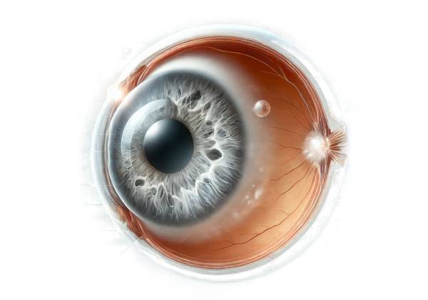
Posterior subcapsular cataract (PSC) is a type of cataract characterized by cloudiness or opacity in the back of the lens, specifically beneath the lens capsule. This condition has a significant impact on vision and typically progresses faster than other types of cataracts, such as nuclear or cortical cataracts. PSCs are particularly troublesome because they can impair reading, reduce vision in bright light, and cause glare or halos around lights, all of which can have a negative impact on daily activities and overall quality of life.
Anatomy and Function of the Lens
The human lens is a transparent, biconvex structure that sits behind the iris and pupil. It is essential for focusing light onto the retina and providing clear vision. The lens has three major components: the lens capsule (a thin, elastic outer covering), the lens epithelium (a layer of cells beneath the capsule), and the lens fibers. The transparency and proper operation of these components are critical to maintaining good vision.
Pathophysiology of Posterior Subcapsular Cataracts
In a PSC, the opacity appears at the back of the lens, just in front of the posterior capsule. This particular location is critical because it is directly in the path of light entering the eye, causing significant disruption in vision. The exact pathogenesis is the migration of lens epithelial cells to the posterior pole, where they proliferate and form abnormal, balloon-like structures known as bladder cells. These cells disrupt the normal arrangement of lens fibers, resulting in the characteristic cloudiness of a PSC.
Risk Factors and Causes
Several factors contribute to the development of PSCs, and understanding these can aid in identifying at-risk individuals and implementing preventive measures.
- Age: Age is the leading risk factor for all types of cataracts, including PSC. As the lens ages, its proteins and fibers may degrade and clump together, resulting in opacity.
- Steroid Use: Long-term use of corticosteroids, whether systemic (oral or injectable) or topical (including inhaled steroids for asthma), is strongly associated with the development of PSCs. Steroids can change the metabolism of lens epithelial cells, promoting migration and proliferation.
- Diabetes: People with diabetes are more likely to develop PSCs. High blood sugar levels can accumulate sorbitol in the lens, resulting in osmotic stress and lens opacity.
- Radiation Exposure: Ionizing radiation, such as X-rays or radiation therapy for cancer, can harm lens epithelial cells and raise the risk of PSC.
- Inflammatory Eye Diseases: Chronic inflammation in the eye, such as uveitis, can contribute to the development of PSCs by causing cellular stress and damage within the lens.
- Genetic Predisposition: Some genetic factors and family histories of cataracts can predispose people to developing PSCs at a younger age.
- Lifestyle Factors: Smoking, excessive alcohol consumption, and poor nutrition can all lead to the development of PSCs. These factors can cause oxidative stress in the lens, which can damage proteins and fibers.
Symptoms and Clinical Presentation
The symptoms of PSCs vary according to the severity and progression of the opacity. Common symptoms include:
- Blurred Vision: One of the most common symptoms, as the opacity directly blocks the passage of light to the retina.
- Glare and Halos: Patients frequently report significant glare issues, especially in bright light or while driving at night. Halos around lights are a common complaint.
- Difficulty Reading: Near vision may be more affected than distance vision, making reading difficult.
- Decreased Visual Acuity: Progressive clouding can cause a significant reduction in visual acuity over time.
Impact on Daily Life
PSC can have a significant impact on daily life, particularly given the rapid progression of this type of cataract. Activities that require clear near vision, such as reading, sewing, or computer use, can become difficult. Furthermore, increased glare and halos can impair driving safety, especially at night or in bright sunlight. This loss of visual function can lead to decreased independence and quality of life, making early diagnosis and treatment critical.
Pathologic Findings
Histopathological examination of PSCs reveals distinct changes in the lens fibers and epithelial cells. The posterior subcapsular region exhibits migration and abnormal proliferation of lens epithelial cells, resulting in clusters of swollen, bladder-like cells. These cells have abnormal protein and cellular debris accumulations that scatter light and cause the opacity observed in PSC.
Difference from Other Cataracts
PSC is one of several types of cataracts, but it can be distinguished from others by its location and specific symptoms:
- Nuclear Cataracts: These form in the center of the lens and frequently cause gradual hardening and yellowing of the lens. They primarily affect distance vision and make seeing difficult in low light.
- Cortical Cataracts: These affect the lens’s outer edges and are characterized by spoke-like opacities that progress toward the center. They can interfere with glare and depth perception.
- Anterior Subcapsular Cataracts: These occur beneath the front capsule of the lens and can also be caused by trauma or inflammation.
Epidemiology
PSC is a fairly common type of cataract, especially in older adults. The exact prevalence varies according to the population studied and the diagnostic criteria used. However, it is widely acknowledged that PSCs are responsible for a significant proportion of visually impairing cataracts, particularly in patients with a history of steroid use or diabetes.
Complications
If left untreated, PSCs can cause significant visual impairment and blindness. PSC can progress faster than other types of cataracts, necessitating early intervention. Furthermore, the presence of a PSC can complicate the surgical removal of the cataract, potentially increasing the risk of postsurgical complications.
Research and Advances
Ongoing research into the pathogenesis and risk factors for PSC aims to improve prevention and treatment strategies. Advances in surgical techniques and intraocular lens (IOL) technology continue to improve patient outcomes following cataract surgery. Furthermore, efforts to understand the molecular mechanisms underlying PSC may lead to novel pharmacological interventions that slow or prevent the progression of the cataracts.
Diagnostic methods
To confirm the presence and extent of the cataract and distinguish it from other ocular conditions, a clinical examination, imaging studies, and, in some cases, laboratory tests are used.
Clinical Examination
A comprehensive clinical examination by an ophthalmologist is the first step in diagnosing PSC. This usually includes:
- Visual Acuity Test: This test determines the sharpness of vision at different distances. Patients with PSC frequently have reduced visual acuity, especially for near tasks.
- Slit-Lamp Examination: Using a slit-lamp microscope, the ophthalmologist can examine the lens thoroughly. This allows for the identification of the typical posterior subcapsular opacities, which appear as granular or plaque-like deposits on the lens’s back surface.
- Dilated Eye Exam: Eye drops can dilate the pupil, allowing for a more thorough examination of the lens and other structures within the eye. This can help determine the size of the cataract and identify any other ocular abnormalities.
Imaging Studies
While the clinical examination is often sufficient to diagnose PSC, imaging studies can provide more detail and confirm the diagnosis:
- Optical Coherence Tomography (OCT): OCT is a non-invasive imaging technique for obtaining high-resolution cross-sectional images of the eye. It can help assess the cataract’s thickness and density, as well as the impact on the posterior segment, which includes the retina and optic nerve.
- Ultrasound Biomicroscopy: This imaging technique uses high-frequency ultrasound to examine the anterior segment of the eye, including the lens. It can be useful in situations where other conditions, such as corneal opacities or dense cataracts, obscure the lens’ view.
Additional Tests
In some cases, additional tests may be required to assess the overall health of the eye and identify any underlying conditions contributing to the development of PSC.
- Glare Testing: Specialized tests can determine the impact of glare on visual function, which is common in PSC patients.
- Contrast Sensitivity Testing: This test assesses the ability to distinguish between different shades of gray in PSC.
- Blood Tests: In patients with risk factors such as diabetes or autoimmune diseases, blood tests may be used to effectively evaluate and manage these conditions.
Posterior Subcapsular Cataract Management
Managing posterior subcapsular cataract (PSC) necessitates a comprehensive approach tailored to the severity of the cataract and the patient’s specific needs. The primary goal of management is to restore and maintain optimal vision, which frequently requires surgical intervention. Here are the main strategies for managing the PSC:
Non-surgical Management
In the early stages of PSC, non-surgical interventions may help manage symptoms and slow cataract progression:
- Prescription Glasses or Contact Lenses: Changing the prescription for glasses or contact lenses can temporarily improve vision by compensating for changes in the lens.
- Anti-Glare Lenses: Applying anti-glare coatings to glasses can help reduce glare and halos, which are common symptoms of PSC. Sunglasses with UV protection can also help reduce light sensitivity and glare.
- Magnifying Aids: For patients who struggle with near tasks, magnifying lenses or reading aids can improve vision for reading and other close-up activities.
- Lifestyle Modifications: Patients are encouraged to modify their daily activities to reduce the impact of vision changes. This includes having adequate lighting at home and work, avoiding night driving, and using larger print materials.
Surgical Management
When PSC significantly impairs vision and interferes with daily activities, cataract surgery becomes necessary. The surgical removal of the cloudy lens is the most effective way to restore vision. The primary surgical procedure is:
- Phacoemulsification is the most commonly used technique for cataract surgery. It entails making a small incision in the cornea, using ultrasound waves to break up the cloudy lens, and then suctioning out the fragments. An intraocular lens (IOL) is then implanted to replace the missing lens. Phacoemulsification is popular because of its low invasiveness, quick recovery time, and high success rate.
- Extracapsular Cataract Extraction (ECCE): When the cataract is too dense or difficult to phacoemulsify, ECCE may be used. This entails making a larger incision to remove the lens in one piece before implanting an IOL. While effective, ECCE requires a longer recovery time and carries a higher risk of complications than phacoemulsification.
Different types of intraocular lenses (IOLs)
The selection of IOL is an important aspect of cataract surgery and can be tailored to the patient’s visual needs and lifestyle.
- Monofocal IOLs: These lenses provide clear vision at a single distance, usually set for distance vision. Patients may still need glasses for reading and close-up tasks.
- Multifocal IOLs: These lenses have multiple focal points, providing better vision at close, intermediate, and far distances. Multifocal IOLs can reduce the need for glasses for most activities.
- Toric IOLs: Toric IOLs can correct astigmatism and improve vision.
- Accommodative IOLs are lenses that move or change shape within the eye, mimicking the lens’s natural focusing ability and providing a range of vision.
Post-operative Care
Effective postoperative care is critical for ensuring a successful outcome and avoiding complications.
- Medications: Typically, patients are given antibiotic and anti-inflammatory eye drops to prevent infection and reduce inflammation. It is critical to adhere to the prescribed regimen to ensure proper healing.
- Follow-Up Visits: Regular follow-up visits are scheduled to monitor the healing process, check the IOL’s positioning, and evaluate visual acuity. These visits aid in the early detection and management of any complications, such as infection or elevated intraocular pressure.
- Activity Restrictions: Patients should avoid strenuous activities, heavy lifting, and rubbing their eyes during the initial healing period. Protective eyewear may be prescribed to protect the eye from injury and bright light.
Managing Complications
While cataract surgery is generally safe, complications may arise. Effective management of these complications is essential.
- Posterior Capsular Opacification (PCO): The most common postoperative complication occurs when the posterior capsule becomes cloudy. PCO can be treated with a quick and painless laser procedure known as YAG laser capsulotomy, which makes a clear opening in the capsule.
- Cystoid Macular Edema (CME): The accumulation of fluid in the macula can impair vision. Anti-inflammatory medications are typically used to treat the condition, either as eye drops or injections.
- Infections and Inflammation: Prompt medical attention is critical for treating infections and chronic inflammation. This may necessitate prolonged use of antibiotics and anti-inflammatory medications.
Trusted Resources and Support
Books
- “Cataract Surgery: A Patient’s Guide to Treatment” by Robert S. Feder, MD
- This book provides comprehensive information on cataract surgery, including the types of cataracts, treatment options, and postoperative care.
- “Ophthalmology” by Myron Yanoff and Jay S. Duker
- A detailed textbook that offers in-depth coverage of various eye conditions, including cataracts, with the latest research and treatment methodologies.
Organizations
- American Academy of Ophthalmology (AAO)
- Website: www.aao.org
- The AAO offers extensive resources on cataracts, including patient education materials, research updates, and professional guidelines.
- National Eye Institute (NEI)
- Website: www.nei.nih.gov
- The NEI provides comprehensive information on eye health, research on eye diseases, and support for patients and families affected by cataracts.


