What is Retinoschisis?
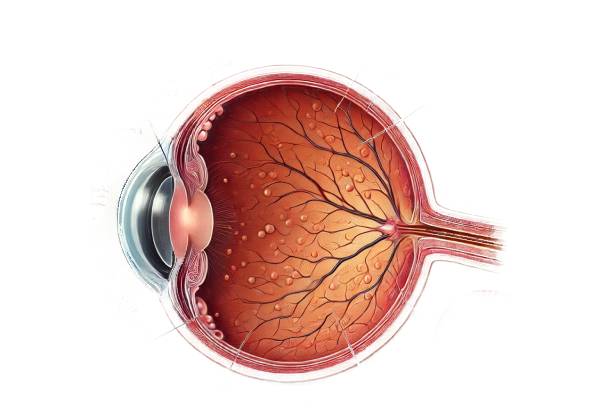
Retinoschisis is an ocular condition characterized by the splitting of the retina, the light-sensitive layer of tissue located at the back of the eye. The retina is essential for vision, as it captures visual information and transmits it to the brain via the optic nerve. Retinoschisis involves the separation of the retinal layers, particularly within the macula, which is responsible for central vision, or the peripheral retina. This separation can lead to visual impairment, which may vary depending on the extent and location of the schisis (splitting). Retinoschisis can occur in different forms, each with its own underlying causes, clinical presentations, and implications for vision.
Types of Retinoschisis
Retinoschisis is broadly categorized into two main types: juvenile (X-linked) retinoschisis and senile (degenerative) retinoschisis. Each type has distinct features and affects different populations.
- Juvenile Retinoschisis (X-Linked Retinoschisis): This form of retinoschisis is a hereditary condition primarily affecting males, as it is linked to mutations in the RS1 gene on the X chromosome. The RS1 gene is responsible for encoding retinoschisin, a protein crucial for maintaining the structural integrity of the retina. When the RS1 gene is mutated, retinoschisin is either absent or dysfunctional, leading to the separation of retinal layers. Juvenile retinoschisis typically presents in early childhood, often between the ages of 5 and 10, and predominantly affects the macula. The condition can lead to a significant reduction in central vision, though peripheral vision is often preserved. In more severe cases, peripheral retinoschisis can occur, leading to complications such as retinal detachment.
- Senile Retinoschisis (Degenerative Retinoschisis): This form of retinoschisis is more common in older adults and is generally associated with age-related degenerative changes in the retina. Unlike juvenile retinoschisis, senile retinoschisis is not hereditary and does not involve mutations in the RS1 gene. Instead, it is thought to result from the weakening of the retinal layers due to the natural aging process. Senile retinoschisis usually affects the peripheral retina and is often discovered incidentally during routine eye examinations, as it may not cause noticeable symptoms. However, in some cases, it can lead to peripheral visual field defects or, rarely, complications such as retinal detachment.
Pathophysiology of Retinoschisis
The retina is composed of multiple layers, each with specific functions crucial for processing visual information. In retinoschisis, there is a splitting or separation between the layers of the retina, most commonly between the outer plexiform layer (where photoreceptors connect with the retinal interneurons) and the inner nuclear layer (which contains the cell bodies of these interneurons). This separation can disrupt the normal transmission of visual signals from the photoreceptors to the brain, leading to visual impairment.
In juvenile retinoschisis, the underlying mutation in the RS1 gene leads to a deficiency or dysfunction of retinoschisin, a protein that helps to maintain the cohesion between the retinal layers. Without adequate retinoschisin, the structural integrity of the retina is compromised, resulting in schisis. The macula is particularly vulnerable, which is why central vision loss is a prominent feature of this condition. Peripheral retinoschisis can also occur, especially in advanced stages, and may progress to more serious complications if not managed appropriately.
Senile retinoschisis, on the other hand, is thought to result from degenerative changes within the retina due to aging. The vitreous humor, the gel-like substance that fills the eye, undergoes changes over time, leading to the liquefaction and posterior vitreous detachment. These changes can create traction on the retinal layers, particularly in the peripheral retina, causing the layers to separate. This form of retinoschisis is typically less severe than juvenile retinoschisis and may remain stable over time, though monitoring is essential to detect any progression or complications.
Clinical Presentation and Symptoms
The symptoms of retinoschisis can vary widely depending on the type of retinoschisis, its location within the retina, and the extent of the retinal splitting. In some cases, retinoschisis may be asymptomatic and discovered incidentally during an eye examination. However, when symptoms do occur, they can significantly impact vision.
- Juvenile Retinoschisis Symptoms: Individuals with juvenile retinoschisis often present with visual difficulties during childhood. The most common symptom is a reduction in central vision, which may manifest as difficulty reading, recognizing faces, or seeing fine details. This reduction in vision is typically due to the involvement of the macula. Children with juvenile retinoschisis may also experience strabismus (misalignment of the eyes) or nystagmus (involuntary eye movements) due to poor central vision. Peripheral vision is usually preserved initially, but in advanced cases, peripheral retinoschisis can develop, leading to peripheral visual field defects or even retinal detachment, which can cause a sudden loss of vision.
- Senile Retinoschisis Symptoms: Senile retinoschisis often presents without symptoms, particularly in the early stages. When symptoms do occur, they are usually related to peripheral vision and may include the appearance of visual field defects, such as missing or blurred areas in the peripheral vision. In rare cases, if the schisis progresses and causes a retinal detachment, patients may experience a sudden onset of floaters, flashes of light, or a shadow or curtain over a part of their vision, which requires immediate medical attention.
Epidemiology and Risk Factors
Retinoschisis is a relatively rare condition, with juvenile retinoschisis estimated to affect approximately 1 in 5,000 to 25,000 males. As it is an X-linked recessive disorder, it predominantly affects males, with female carriers typically remaining asymptomatic, though they may pass the condition on to their sons. The condition is usually diagnosed in early childhood, and the severity of vision loss can vary widely among individuals.
Senile retinoschisis, on the other hand, is more common in the elderly population, with an estimated prevalence of 3-8% among individuals over the age of 40. The condition is equally distributed between males and females and is often associated with other age-related ocular conditions, such as posterior vitreous detachment or cataracts. The exact cause of senile retinoschisis is not well understood, but it is believed to result from age-related degenerative changes in the retina.
Risk factors for retinoschisis include a family history of the condition, particularly in cases of juvenile retinoschisis, as well as advancing age for senile retinoschisis. High myopia (nearsightedness) has also been identified as a potential risk factor for the development of senile retinoschisis, as myopic eyes tend to have longer axial lengths, which may predispose the retina to splitting.
Complications of Retinoschisis
While retinoschisis itself may be stable and not cause significant vision loss, there are several potential complications that can arise, particularly if the condition progresses. These complications can significantly impact vision and require prompt medical intervention.
- Retinal Detachment: One of the most serious complications of retinoschisis is retinal detachment, which occurs when the retina separates from the underlying supportive tissue. Retinal detachment can lead to a sudden and severe loss of vision and requires urgent surgical intervention to prevent permanent blindness. Retinal detachment is more likely to occur in cases of juvenile retinoschisis, particularly when the peripheral retina is involved.
- Vitreous Hemorrhage: In some cases, the abnormal blood vessels associated with retinoschisis may rupture, leading to bleeding into the vitreous humor. This can cause the sudden appearance of floaters or cloudy vision and may require surgical intervention if the bleeding is severe.
- Cystic Macular Changes: In juvenile retinoschisis, the macula may develop cystic changes, leading to further deterioration of central vision. These cysts can be challenging to treat and may contribute to progressive vision loss.
Understanding the various aspects of retinoschisis, including its types, pathophysiology, and potential complications, is essential for early diagnosis and effective management of the condition.
Diagnostic Methods
Diagnosing retinoschisis involves a combination of clinical evaluation, imaging studies, and, in some cases, genetic testing. Early detection is crucial for monitoring the condition and preventing complications such as retinal detachment.
Clinical Examination
The initial step in diagnosing retinoschisis is a thorough clinical examination by an ophthalmologist. During the examination, the patient’s visual acuity is assessed to determine the extent of vision loss, particularly in the central vision for those with juvenile retinoschisis. A detailed patient history is also important to identify any family history of the condition, which can be particularly relevant in cases of juvenile retinoschisis.
- Fundoscopy: The most critical part of the clinical examination is fundoscopy, where the ophthalmologist uses an ophthalmoscope to examine the back of the eye provides high-resolution cross-sectional images of the retina, allowing for detailed visualization of the retinal layers. This imaging modality is particularly useful for detecting the separation of retinal layers, which is the hallmark of retinoschisis.
- Macular OCT: In cases of juvenile retinoschisis, macular OCT can reveal the characteristic schisis cavities within the macula, often showing multiple cystic spaces between the retinal layers. This allows the ophthalmologist to assess the extent of the schisis and monitor any changes over time. Macular OCT is also helpful in identifying associated complications, such as cystic changes or macular holes.
- Peripheral OCT: For patients with suspected senile retinoschisis, peripheral OCT can be used to visualize the peripheral retina, where the schisis is more likely to occur. This imaging can show the splitting of the retinal layers and help differentiate retinoschisis from other conditions that may affect the peripheral retina, such as retinal tears or detachments.
Ultrasound B-Scan
Ultrasound B-scan is another valuable diagnostic tool, especially when the view of the retina is obscured due to media opacities, such as vitreous hemorrhage or cataracts. B-scan ultrasonography provides a cross-sectional image of the eye, allowing the ophthalmologist to assess the retina’s overall structure.
- Differentiating Retinoschisis from Retinal Detachment: One of the key roles of B-scan ultrasound is to differentiate retinoschisis from retinal detachment. In retinoschisis, the schisis cavity typically appears as a dome-shaped elevation with smooth, non-tapering edges, and the split retinal layers usually remain attached at the periphery. In contrast, retinal detachment often presents with a more irregular elevation, with the retina appearing free-floating and mobile within the vitreous cavity. B-scan can also help detect the presence of subretinal fluid, which is more common in retinal detachment.
Fluorescein Angiography
Fluorescein angiography is an imaging technique that involves injecting a fluorescent dye into the bloodstream and capturing images as the dye circulates through the retinal blood vessels. This technique is not routinely used for diagnosing retinoschisis but may be employed in specific cases to assess the retinal vasculature and identify any areas of leakage or neovascularization.
- Assessing Vascular Changes: In juvenile retinoschisis, fluorescein angiography can help detect abnormal blood vessels or areas of capillary non-perfusion, which may be associated with the schisis. In some cases, it can also help in evaluating the extent of vascular changes that could contribute to complications such as vitreous hemorrhage.
Genetic Testing
Genetic testing is particularly relevant in cases of juvenile retinoschisis, where identifying mutations in the RS1 gene can confirm the diagnosis. Genetic testing may be recommended for individuals with a family history of juvenile retinoschisis or for those who present with the clinical features of the condition. Confirming the genetic basis of the disease can provide valuable information for family counseling and can also guide the management and monitoring of affected individuals.
Retinoschisis Management
The management of retinoschisis varies depending on the type of the condition, the severity of the retinal splitting, the presence of symptoms, and the risk of complications such as retinal detachment. Treatment strategies aim to preserve vision, prevent further progression, and manage any complications that arise.
Observation and Monitoring
In many cases, particularly with senile (degenerative) retinoschisis, the condition may be stable and asymptomatic, requiring no immediate intervention. Regular monitoring is crucial to ensure that the retinoschisis does not progress or lead to complications such as retinal detachment. Patients diagnosed with retinoschisis are typically advised to undergo periodic eye examinations, during which an ophthalmologist will assess the retina for any changes in the schisis area, the development of new symptoms, or signs of complications.
Monitoring includes regular visual acuity tests, fundoscopy, and imaging studies like OCT to track any changes in the retinal structure. For asymptomatic patients, this conservative approach is often sufficient, as many cases of senile retinoschisis do not progress significantly over time.
Laser Photocoagulation
Laser photocoagulation is a treatment option that may be considered in cases of retinoschisis where there is a high risk of progression to retinal detachment. This technique involves using a laser to create small burns around the edges of the schisis area to create a barrier and prevent the schisis from extending. The laser seals the retina to the underlying tissue, reducing the likelihood of further splitting or detachment.
Laser treatment is more commonly used in cases where peripheral retinoschisis shows signs of impending detachment, such as the presence of retinal breaks or holes. While laser photocoagulation is not typically used for the macular form of retinoschisis, it can be effective in preventing the progression of peripheral cases.
Vitrectomy
Vitrectomy is a surgical procedure that may be necessary in more severe cases of retinoschisis, particularly when complications such as retinal detachment occur. Vitrectomy involves the removal of the vitreous humor, the gel-like substance inside the eye, to relieve traction on the retina and allow the surgeon to address any retinal breaks or tears that may be contributing to the detachment.
During the procedure, the surgeon may also remove any fibrous tissue that has developed on the retina as a result of the schisis, and reattach the retina to its underlying layers. In some cases, the surgeon may use a gas bubble or silicone oil to help hold the retina in place while it heals. Vitrectomy is generally reserved for cases where conservative treatments have failed, or when the risk of severe vision loss is high due to retinal detachment.
Retinal Detachment Repair
If retinoschisis progresses to retinal detachment, surgical repair is necessary to prevent permanent vision loss. Retinal detachment repair can involve several different techniques, depending on the location and extent of the detachment. Common methods include scleral buckling, pneumatic retinopexy, and vitrectomy.
- Scleral Buckling: This procedure involves placing a silicone band (buckle) around the outside of the eye to create inward pressure, which helps to reattach the detached retina to the underlying tissue. Scleral buckling is often used for peripheral detachments associated with retinoschisis.
- Pneumatic Retinopexy: This is a less invasive procedure where a gas bubble is injected into the eye to press the detached retina back into place. The patient’s head is then positioned in a specific way to keep the bubble in contact with the detachment site until the retina reattaches.
- Vitrectomy: As previously mentioned, vitrectomy may be necessary in more complex cases where other methods are not sufficient to repair the detachment.
Genetic Counseling
For individuals with juvenile (X-linked) retinoschisis, genetic counseling is an important aspect of management. Since this form of retinoschisis is hereditary, understanding the genetic basis of the condition can help in family planning and early diagnosis in at-risk individuals. Genetic counseling provides families with information about the inheritance pattern, the likelihood of passing the condition to offspring, and the implications for other family members.
Visual Rehabilitation
For patients with significant vision loss due to retinoschisis, visual rehabilitation is an essential part of management. This may include the use of low vision aids, such as magnifying glasses, electronic reading devices, or specialized lenses, to enhance remaining vision. Occupational therapy and mobility training can also help individuals adapt to their visual limitations and maintain independence in daily activities.
Overall, the management of retinoschisis requires a tailored approach based on the type of retinoschisis, the severity of the condition, and the patient’s individual needs. Early detection and regular monitoring are key to preventing complications and preserving vision.
Trusted Resources and Support
Books
- “Inherited Retinal Disease: A Diagnosis and Management Guide” by Stephen H. Tsang and Theodore L. Smith: This book provides an in-depth overview of inherited retinal conditions, including retinoschisis, and offers guidance on diagnosis and management.
- “Retina: Medical and Surgical Management” by Stephen J. Ryan: A comprehensive resource on retinal diseases, this book covers the clinical aspects of retinoschisis, including diagnosis, treatment options, and surgical interventions.
Organizations
- Foundation Fighting Blindness (FFB): A leading organization dedicated to finding treatments and cures for inherited retinal diseases, including retinoschisis. FFB provides educational resources, research updates, and support networks for patients and families.
- American Academy of Ophthalmology (AAO): Offers a wealth of information on eye conditions, including retinoschisis, with resources for both patients and healthcare professionals.
- The National Eye Institute (NEI): A government organization that provides comprehensive information on eye diseases, including retinoschisis, and supports research aimed at improving treatment and outcomes for individuals with retinal conditions.


