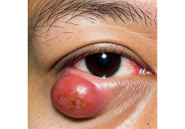
Sebaceous gland carcinoma (SGC) is a rare and aggressive malignancy that originates in the sebaceous glands and most commonly affects the periocular region. While sebaceous glands exist throughout the body, they are most concentrated around the eyes, where they play an important role in the health and function of the eyelids and ocular surface. SGC is notorious for its ability to masquerade as more benign conditions, resulting in diagnostic delays that can have a significant impact on patient outcomes.
Structure and Function of Sebaceous Glands
Sebaceous glands are small oil-producing glands that reside in the skin’s dermis layer. They are most visible on the scalp, face, and upper torso, but they are also essential components of the eyelids, particularly in the tarsal plate. These glands produce sebum, a lipid-rich substance that lubricates the skin and hair, acts as a barrier against external environmental factors, and contributes to the maintenance of the tear film on the ocular surface.
The Meibomian glands, which are modified sebaceous glands, are the most common type of sebaceous gland in the eyelid. These glands produce meibum, a vital component of the tear film that prevents the aqueous layer from evaporating, thereby maintaining ocular surface stability and comfort. Dysfunction of these glands, such as Meibomian gland dysfunction, can result in dry eye disease. Sebaceous glands include Zeis glands, which are associated with eyelash follicles, as well as skin sebaceous glands.
Pathology of Sebaceous Gland Carcinoma
SGC develops as a result of the malignant transformation of sebaceous gland cells. The exact cause of SGC is unknown, despite the identification of several risk factors. These include advanced age, female gender, Asian ethnicity, prior radiation therapy to the head and neck, and genetic predispositions, particularly mutations associated with the Muir-Torre syndrome, a subset of Lynch syndrome.
The pathogenesis of SGC is thought to be a combination of genetic mutations, chronic inflammation, and immune dysregulation. For example, in Muir-Torre syndrome, patients have mutations in DNA mismatch repair genes, which causes microsatellite instability and an increased risk of malignancies, including SGC.
SGC typically appears between the sixth and seventh decades of life, with a slight female predilection. However, it can occur in younger people, especially those with predisposing genetic conditions. The periocular region is the most common site of SGC, accounting for roughly 40% of all cases. The upper eyelid is more commonly affected in this region due to the higher density of Meibomian glands.
Clinical Features of Sebaceous Gland Carcinoma
One of the most difficult aspects of SGC is its ability to mimic a wide range of benign and malignant conditions, frequently leading to misdiagnosis. Clinically, SGC can manifest in several ways:
- Nodular Form: The most common manifestation of SGC is a firm, painless, yellowish nodule that is frequently mistaken for a chalazion or hordeolum (stye). This form may be slow-growing and can last for months or years before a proper diagnosis is made.
- Pagetoid Spread: SGC can also cause a diffuse thickening of the eyelid margin, which often resembles blepharitis. This presentation is particularly insidious because it can result in widespread involvement of the eyelid, conjunctiva, and cornea. The term “pagetoid spread” refers to the intraepithelial spread of malignant cells, which is a characteristic feature of SGC.
- Mimicking Other Conditions: SGC can resemble other more common eyelid cancers, such as basal cell carcinoma or squamous cell carcinoma. It is also possible to confuse it with benign lesions such as seborrheic or actinic keratosis. Furthermore, it can mimic inflammatory conditions such as chronic blepharitis, making early diagnosis difficult.
- Systemic Manifestations: Although rare, SGC can spread to regional lymph nodes and distant organs such as the lungs, liver, and brain. Patients with metastatic disease may experience systemic symptoms like weight loss, fatigue, or neurological deficits.
- Ocular Symptoms: Patients with SGC may feel ocular discomfort, irritation, tearing, or a foreign body sensation. If the tumor invades the conjunctiva, cornea, or intraocular structures, it can impair vision.
- Multifocality and Recurrence: SGC is known for its multifocality, which means that multiple independent tumors can develop concurrently or sequentially within the same or different eyelids. Recurrence following surgical excision is common, especially if the tumor is not completely removed, emphasizing the importance of comprehensive and precise treatment.
Histology of Sebaceous Gland Carcinoma
SGC has atypical sebaceous cells with large, pleomorphic nuclei and abundant vacuolated cytoplasm due to lipid content. These cells have the ability to invade surrounding tissues such as the dermis, conjunctiva, and, in some cases, the orbit. The tumor can have both solid and cystic patterns, and pagetoid spread of malignant cells within the epithelium is a distinguishing feature.
Immunohistochemistry is frequently used to aid in diagnosis, with SGC displaying positive markers such as epithelial membrane antigen (EMA), androgen receptor (AR), and adipophilin. Staining for lipid content with Oil Red O can also help distinguish SGC from other tumors, but it requires fresh or frozen tissue, limiting its utility.
Prognosis and Challenges in Management
The prognosis of SGC is largely determined by the stage at diagnosis and the effectiveness of initial treatment. Early-stage SGC confined to the eyelid has a better prognosis, with an estimated 5-year survival rate of 80-90%. However, the prognosis worsens significantly in cases of orbital invasion, regional lymph node involvement, or distant metastasis, with a 5-year survival rate of less than 50%.
SGC’s aggressive nature, the possibility of local recurrence, and the risk of metastasis make early diagnosis and treatment critical. Unfortunately, because of its ability to mimic benign conditions, SGC is frequently diagnosed late, which can lead to a more complicated clinical outcome.
The challenge in managing SGC stems from its rarity, which has resulted in a lack of widespread awareness among clinicians, as well as its diverse clinical presentations. This emphasizes the importance of maintaining a high index of suspicion for SGC in patients with atypical or recurring eyelid lesions, especially those with known risk factors.
Differential Diagnosis
SGC’s differential diagnosis is broad because it can mimic a variety of benign and malignant conditions. Conditions to consider include:
- Chalazion: A benign, granulomatous inflammation of the Meibomian gland that frequently appears as a painless eyelid nodule. Unlike SGC, chalazia are usually self-limiting and respond to simple treatments such as warm compresses.
- Basal Cell Carcinoma (BCC): The most common malignant eyelid tumor, BCC usually appears as a pearly nodule with telangiectasia and can ulcerate. Unlike SGC, BCC rarely metastasizes but can cause extensive local tissue damage.
- Squamous Cell Carcinoma (SCC): Another common eyelid cancer, SCC often appears as a scaly, ulcerated lesion. It has a higher metastasis potential than BCC, but lacks the sebaceous differentiation found in SGC.
- Blepharitis: Blepharitis is a chronic inflammation of the eyelid margins that can cause diffuse eyelid thickening, erythema, and crusting, similar to the spread of SGC through the pagetoids. However, blepharitis typically responds to lid hygiene measures and lacks the malignant characteristics of SGC.
- Other Benign Lesions: Seborrheic keratosis, actinic keratosis, and papillomas can all mimic SGC. These benign lesions are typically well-defined and lack the invasive features of SGC.
Given the wide range of conditions that SGC can mimic, an accurate diagnosis requires a thorough clinical evaluation, as well as appropriate diagnostic tests.
Methods to Diagnose Sebaceous Gland Carcinoma
Diagnosing sebaceous gland carcinoma (SGC) can be difficult due to its diverse clinical presentation and ability to mimic other ocular diseases. A combination of clinical evaluation, histopathological examination, and imaging studies is frequently required to make a definitive diagnosis.
Clinical Evaluation
The first step in diagnosing SGC is a thorough clinical examination of the affected eyelid and its surrounding structures. Key features that may raise suspicion for SGC are:
- Persistent or recurrent eyelid lesions: Any eyelid lesion that does not respond to standard treatments or returns after excision should be investigated for malignancy, including SGC.
- Nodular or diffuse thickening: Firm, yellowish, and slow-growing nodules, as well as diffuse thickening of the eyelid margin, require further investigation.
- Unilateral blepharitis: Persistent unilateral blepharitis that does not respond to treatment may indicate underlying SGC, especially if it is associated with lash loss (madarosis) or focal ulceration.
- Pagetoid spread: The presence of intraepithelial lesions on the conjunctiva, similar to chronic conjunctivitis, may indicate SGC spread.
Biopsy & Histopathology
A biopsy of the lesion is typically used to make a definitive diagnosis of SGC, which is then examined histopathologically. Various biopsy techniques may be used:
- Incisional Biopsy: If the lesion is large or affects a significant portion of the eyelid, an incisional biopsy may be performed. This entails removing a small portion of the lesion for analysis while leaving the majority intact. Incisional biopsies are especially useful when the lesion’s malignancy is unknown, allowing for histopathological confirmation before moving forward with more extensive surgical interventions.
- Excisional Biopsy: If the lesion is small or well-defined, an excisional biopsy, which removes the entire lesion, may be preferable. This method not only provides a definitive diagnosis, but it can also be curative if the lesion is completely removed with clear margins. However, excisional biopsy should be approached with caution in the periocular region to reduce the risk of incomplete removal, which could result in recurrence.
- Conjunctival Mapping Biopsy: If there is suspected pagetoid spread, a conjunctival mapping biopsy may be performed. Multiple small biopsies are taken from different areas of the conjunctiva to determine the extent of intraepithelial spread. This approach aids in surgical planning and ensures that all affected areas receive adequate treatment.
Histopathological examination of the biopsy specimen is critical for confirming the diagnosis of SGC. The pathologist will look for SGC-specific features, such as atypical sebaceous cells with large, pleomorphic nuclei and abundant vacuolated cytoplasm. The presence of pagetoid spread, in which malignant cells infiltrate the overlying epithelium, is a characteristic of SGC that distinguishes it from other cancers.
Immunohistochemistry
Immunohistochemical (IHC) staining is frequently used to confirm the diagnosis of SGC, especially when the histological features are ambiguous or when SGC is distinguished from other malignancies. Key IHC markers used to diagnose SGC include:
- Epithelial Membrane Antigen (EMA): Sebaceous gland carcinoma frequently expresses EMA, which can help distinguish it from other tumors that do not.
- Androgen Receptor (AR): AR is another marker that frequently appears in SGC. Its presence confirms the diagnosis, especially when other markers are inconclusive.
- Adipophilin: This marker detects lipid droplets within sebaceous cells, assisting in the identification of sebaceous differentiation, a key feature of SGC.
- Oil Red O Staining: While not an IHC marker, Oil Red O staining detects intracellular lipid content in fresh or frozen tissue specimens. It is especially useful in distinguishing SGC from other non-sebaceous malignancies, but its use is limited by the need for fresh tissue.
Imaging Studies
Imaging aids in the diagnosis of SGC, particularly in determining the extent of local invasion and detecting potential metastatic disease. Common imaging modalities used in the evaluation of SGC are:
- High-Resolution Ultrasonography: Ultrasonography is useful for determining the extent of eyelid lesions, particularly their depth and involvement of adjacent structures. It is a non-invasive, quick, and user-friendly imaging modality that provides real-time results.
- Magnetic Resonance Imaging (MRI): MRI is especially useful for determining the extent of orbital invasion and the involvement of nearby soft tissues. It provides excellent soft tissue contrast and can aid in the differentiation of various types of tissue involvement. MRI with contrast enhancement can help delineate tumor margins and detect perineural spread.
- Computed Tomography (CT) Scan: While less common than MRI, CT scans can be useful in assessing bony involvement, especially in cases of suspected orbital or skull base invasion. CT imaging is also useful for detecting calcifications within the tumor, which may occur in SGC.
- Positron Emission Tomography (PET) Scan: PET scans are not commonly used in the diagnosis of SGC, but they may be considered if there is a strong suspicion of metastatic disease. PET scans can detect metabolically active tumor cells throughout the body, giving a complete picture of disease spread.
Sentinel Lymph Node Biopsy
In some cases, especially when there is no clinical evidence of lymph node involvement but a high risk of metastasis, a sentinel lymph node biopsy (SLNB) may be performed. This method entails injecting a tracer near the primary tumor site and determining the first lymph nodes that drain the area. The sentinel lymph node is then surgically removed and checked for metastatic cells. A positive SLNB results in the need for additional treatment, such as lymph node dissection or adjuvant therapy.
Sebaceous Gland Carcinoma Management
To achieve the best possible results, sebaceous gland carcinoma (SGC) management must be multidisciplinary. The primary goals of treatment are to completely eradicate the tumor, preserve visual function, and prevent local recurrence or metastasis. Given the aggressive nature of SGC, timely and appropriate intervention is critical. Surgery, radiotherapy, and systemic therapy are all possible treatment options.
Surgical Management
Surgery remains the cornerstone of treatment for sebaceous gland carcinoma, with the primary goal of completely removing the tumor with clear margins. The size, location, and extent of the tumor determine the appropriate surgical approach:
- Excisional Surgery with Wide Margins: For small, well-defined tumors, complete excision with wide margins (usually 4-5 mm) is recommended. This entails removing the tumor along with a margin of healthy tissue to ensure that all malignant cells are removed. Frozen section control or Mohs micrographic surgery may be used during surgery to ensure clear margins, significantly lowering the risk of recurrence.
- Mohs Micrographic Surgery: Because of its ability to provide immediate, precise microscopic evaluation of excised tissue margins during surgery, Mohs surgery is especially useful in the management of SGC. This technique preserves as much healthy tissue as possible while completely removing the tumor, making it ideal for tumors located in cosmetically and functionally sensitive areas such as the eyelid.
- Orbit Exenteration: If the tumor has invaded the orbit, a major surgical procedure known as orbit exenteration may be required. This involves removing the eye, adnexal structures, and, in some cases, a portion of the surrounding orbital bones and tissues. While orbit exenteration is a radical procedure, it may be necessary in severe cases to achieve disease control on a local level. Exenteration frequently necessitates orbit reconstruction with grafts or flaps.
- Sentinel Lymph Node Biopsy and Lymphadenectomy: Due to the risk of regional lymph node metastasis in SGC, sentinel lymph node biopsy (SLNB) is occasionally performed to detect microscopic metastases in the regional lymph nodes. If metastasis is confirmed, a more extensive lymphadenectomy (removal of affected lymph nodes) may be necessary.
Radiotherapy
Radiotherapy is important in the treatment of sebaceous gland carcinoma, especially when surgery is not an option or when the patient refuses to have surgery. It can be used as a primary treatment or as an adjuvant therapy after surgery.
- Primary Radiotherapy: For patients who are not candidates for surgery due to medical comorbidities or for tumors that cannot be completely removed surgically, radiotherapy may be used as the primary treatment modality. It is especially effective on smaller tumors or those with clear but narrow surgical margins.
- Adjuvant Radiotherapy: Postoperative radiotherapy is frequently recommended for patients with high-risk characteristics, such as positive surgical margins, orbital invasion, or regional lymph node involvement. Adjuvant radiotherapy reduces the risk of local recurrence by focusing on microscopic disease.
- Palliative Radiotherapy: When SGC is advanced and not curable, palliative radiotherapy can be used to relieve symptoms like pain or bleeding while also controlling local tumor growth.
Systemic Therapy
Systemic therapy, including chemotherapy and immunotherapy, is generally not the primary treatment for SGC, but it may be considered in the following scenarios:
- Chemotherapy: Systemic chemotherapy is generally reserved for patients with metastatic disease or inoperable locally advanced tumors. Platinum-based agents like cisplatin or carboplatin are frequently used in combination with other drugs in regimens. However, chemotherapy has a low response rate and is frequently used as a palliative treatment.
- Immunotherapy: While there is limited evidence on the efficacy of immunotherapy in SGC, recent advances in cancer immunotherapy point to potential benefits in some cases, particularly those with mismatch repair deficiency or a high mutational burden. Immune checkpoint inhibitors, for example, could be studied in clinical trials or as compassionate use scenarios.
Follow-up and Surveillance
Given the high risk of recurrence and metastasis, long-term monitoring and surveillance are critical components of SGC management. Patients should have regular ophthalmic exams, which include visual acuity testing, slit-lamp examination, and dilated funduscopy. Imaging studies, such as MRI or PET scans, may be required to monitor regional or distant metastasis.
Patients with a history of SGC should be educated on the symptoms of recurrence and the significance of regular follow-up. Any new or recurring lesions should be biopsyed as soon as possible to rule out recurrence. Due to the risk of late recurrence, lifelong surveillance is frequently recommended.
Trusted Resources and Support
Books
- “Ocular Oncology” by Jerry A. Shields and Carol L. Shields: This comprehensive text covers the spectrum of ocular tumors, including sebaceous gland carcinoma, and provides detailed insights into diagnosis and management.
- “Eye Pathology: An Atlas and Text” by Ralph C. Eagle Jr.: This book offers an in-depth look at the histopathology of ocular conditions, including sebaceous gland carcinoma, with high-quality images and explanations.
Organizations
- American Academy of Ophthalmology (AAO): The AAO provides extensive resources and guidelines on the diagnosis and management of various ocular conditions, including SGC.
- Ocular Melanoma Foundation: While primarily focused on melanoma, this foundation offers resources and support for patients with rare ocular cancers, including sebaceous gland carcinoma.
- National Cancer Institute (NCI): The NCI offers up-to-date information on the latest research, clinical trials, and treatment options for various cancers, including sebaceous gland carcinoma.


