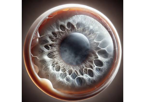
What is secondary cataract?
Secondary cataract, also known as posterior capsule opacification (PCO), is a common complication that can develop following cataract surgery. While the term “secondary cataract” may imply the formation of a new cataract, it actually refers to the clouding of the lens’s posterior capsule, which remains intact following the removal of the primary cataract. As with the original cataract, this opacification can impair vision, resulting in decreased visual acuity and contrast sensitivity.
Anatomy of the Eye and Cataract Surgery
To understand secondary cataracts, you must first understand the anatomy of the eye and how cataract surgery works. The eye is a complex organ, with the lens focusing light onto the retina, where it is processed and transmitted to the brain. The lens is typically transparent, allowing light to pass through and be precisely focused.
A cataract is a condition in which the eye’s natural lens becomes cloudy, resulting in blurred vision, glare, and difficulty seeing in low light conditions. The most common cause of cataracts is aging, but trauma, radiation, and certain medications can all contribute to their development.
Cataract surgery is one of the most common surgical procedures worldwide. During surgery, the cloudy lens is removed and replaced by an artificial intraocular lens (IOL). This procedure is very effective for restoring vision. However, the surgery leaves the lens’s posterior capsule intact, which serves as support for the new IOL. The remaining capsule can become opacified over time, resulting in secondary cataracts.
Pathology of Secondary Cataract
Secondary cataract occurs when lens epithelial cells that remain after cataract surgery start to proliferate and migrate across the posterior capsule. These cells can thicken and cloud the capsule, obstructing the passage of light and causing vision impairment. The process is gradual and can take months or even years following the initial cataract surgery.
There are two primary types of posterior capsule opacification:
- Fibrous PCO: This type of secondary cataract develops when epithelial cells undergo fibrous metaplasia, which results in the formation of fibrotic tissue on the posterior capsule. The fibrous tissue can contract and wrinkle the capsule, resulting in significant visual distortion.
- Elschnig’s Pearls: In this type of PCO, lens epithelial cells proliferate and cluster into small, pearl-like structures on the posterior capsule. These clusters can scatter light, resulting in glare or halos that impair vision.
Several factors influence the development of secondary cataract. These include:
- Age: Younger patients are more likely to develop secondary cataracts because they have a higher regenerative capacity and more active lens epithelial cells.
- Surgical Technique: The technique used during cataract surgery can influence the risk of developing secondary cataracts. For example, leaving fewer residual lens epithelial cells and ensuring proper IOL placement can help reduce the risk.
- Type of Intraocular Lens (IOL): The design and material of the IOL can also influence the development of PCO. Hydrophobic acrylic IOLs, for example, have been associated with a lower risk of secondary cataract than hydrophilic lenses.
- Patient Factors: Diabetes, uveitis, and a history of previous eye surgeries all increase the risk of secondary cataract. Additionally, patients with specific genetic predispositions may be more vulnerable.
Clinical Features of Secondary Cataract
Secondary cataract symptoms are similar to those of primary cataract, and their severity varies depending on the extent of posterior capsule opacification. Common symptoms include:
- Blurred Vision: The most common symptom of secondary cataract is a gradual decrease in visual clarity, with patients frequently reporting that their vision has become hazy or cloudy. This blurriness could be similar to what they had before their original cataract surgery.
- Glare and Halos: Many patients with secondary cataracts have increased sensitivity to light, especially bright lights or headlights at night. This can manifest as glare or the formation of halos around lights, making night driving especially difficult.
- Decreased Contrast Sensitivity: Patients may experience a decrease in their ability to distinguish between different shades of color or subtle differences in lighting, making everyday tasks such as reading or recognizing faces more difficult.
- Difficulty with Near Vision: Although secondary cataract affects both near and distance vision, some patients may experience greater difficulty with near tasks like reading or using a smartphone.
- Double Vision or Ghosting: In some cases, secondary cataract-related irregularities in the posterior capsule can cause visual distortions such as seeing double images or ghosting, in which a faint duplicate of an object appears adjacent to it.
The progression of symptoms varies from patient to patient. Some people may experience a gradual and steady decline in vision, while others may notice symptoms appear suddenly. It is important to note that secondary cataracts do not cause pain or redness, and the eye appears normal from the outside.
Effects on Quality of Life
Secondary cataract can have a significant impact on a patient’s quality of life, especially if they have previously undergone cataract surgery with the expectation of having their vision restored. The recurrence of vision problems can cause frustration, anxiety, and disappointment, especially if the secondary cataract appears soon after surgery.
Patients may struggle with everyday tasks such as reading, driving, or recognizing faces, reducing their independence and confidence. Furthermore, the glare and halos caused by secondary cataracts can make night driving dangerous, limiting a patient’s ability to perform daily activities.
Differential Diagnosis
When a patient presents with symptoms of decreased vision following cataract surgery, it is critical to rule out secondary cataract in the differential diagnosis, but other conditions must also be considered. This includes:
- Cystoid Macular Edema (CME): CME is a condition in which fluid accumulates in the macula, or central part of the retina, causing swelling and vision distortion. It can occur as a complication of cataract surgery and should be distinguished from secondary cataract, which requires different treatment.
- Retinal Detachment: Although uncommon, retinal detachment can occur following cataract surgery and cause symptoms similar to secondary cataract, such as blurred vision and floaters. Prompt diagnosis and treatment are critical for avoiding permanent vision loss.
- Glaucoma: Patients with glaucoma may experience gradual vision loss due to optic nerve damage, which can be misdiagnosed as secondary cataract. However, glaucoma usually causes peripheral vision loss first, whereas secondary cataracts primarily affect central vision.
- Intraocular Lens (IOL) Dislocation: Displacement or dislocation of the IOL can cause visual disturbances similar to those associated with secondary cataract. This condition necessitates surgical intervention and should be detected immediately.
- Corneal Edema: Swelling of the cornea can result in hazy vision and glare, similar to secondary cataract. Endothelial cell loss during cataract surgery, as well as other conditions like Fuchs’ dystrophy, can cause corneal edema.
A thorough clinical examination and appropriate diagnostic tests are required to correctly diagnose secondary cataract and distinguish it from the other conditions.
Diagnostic Techniques for Secondary Cataract
Secondary cataract diagnosis requires a combination of clinical evaluation, specialized imaging techniques, and, in some cases, additional tests to confirm the diagnosis and rule out other potential causes of visual impairment.
Clinical Evaluation
The initial step in diagnosing secondary cataract is a thorough clinical examination by an ophthalmologist. This includes:
- Visual Acuity Test: This standard eye test assesses a patient’s ability to see at different distances. A decrease in visual acuity following cataract surgery may indicate the development of a secondary cataract.
- Slit-Lamp Examination: The slit-lamp, a specialized microscope used in eye exams, allows the ophthalmologist to closely examine the eye’s structures, such as the cornea, lens and retina. During this examination, the doctor can directly observe the posterior capsule and detect any opacification or abnormalities that could indicate a secondary cataract.
- Fundoscopy: Also known as ophthalmoscopy, this test examines the retina and optic nerve in the back of the eye. While the primary goal is to detect retinal issues, a hazy image of the retina may indicate posterior capsule opacification.
- Patient History and Symptom Review: The ophthalmologist will go over the patient’s history of symptoms, focusing on the onset, duration, and progression of vision changes. They will also inquire about any previous eye surgeries, underlying conditions, or medications that may be causing the symptoms.
Imaging Techniques
In some cases, additional imaging techniques may be used to assess the extent of posterior capsule opacification or to rule out other causes of vision problems.
- Ocular Coherence Tomography (OCT): OCT is a non-invasive imaging technique for obtaining detailed cross-sectional images of the retina and optic nerve. While OCT is primarily used to diagnose retinal conditions, it can also aid in distinguishing secondary cataract from other causes of visual impairment, such as cystoid macular edema.
- Specular Microscopy: This imaging technique examines the corneal endothelium and can help rule out corneal edema as a cause of vision problems. It may also provide information about the condition of the cornea after cataract surgery.
- B-Scan Ultrasonography: When the posterior segment of the eye cannot be seen clearly due to dense opacification, B-scan ultrasonography may be used. This ultrasound technique produces detailed images of the eye’s internal structures, allowing the ophthalmologist to examine the retina, choroid, and vitreous body even when a secondary cataract obscures the view through the pupil. This is especially useful for ruling out retinal detachment and other serious posterior segment conditions that may present with similar symptoms.
Additional Tests
Depending on the clinical findings and the patient’s history, additional tests may be required to confirm the diagnosis of secondary cataract or to rule out other potential causes of visual impairment.
- Glare Testing: Glare testing determines how light scatter from the posterior capsule’s opacification affects visual function. This test can help quantify the effects of secondary cataract on the patient’s vision, especially in situations with bright lights or high contrast.
- Contrast Sensitivity Testing: Contrast sensitivity tests assess the ability to distinguish between various shades of light and dark. Secondary cataracts frequently cause a decrease in contrast sensitivity, making this test useful for determining the functional impact of the condition on a patient’s daily activities.
- Refraction Test: A refraction test can help determine whether the patient’s visual symptoms are the result of changes in their eyeglass prescription or a secondary cataract. However, if the patient’s vision does not improve with a new prescription, the visual decline is most likely due to secondary cataract or another underlying condition.
Effective Management of Secondary Cataract
Secondary cataract, also known as posterior capsule opacification (PCO), is typically easy to treat and very effective. The primary goal of treatment is to restore clear vision by removing the opaque posterior capsule. The most common and effective treatment for secondary cataract is YAG laser capsulotomy. While this procedure is the standard of care, other management options are considered based on the patient’s unique needs and circumstances.
YAG Laser Capsulotomy
YAG laser capsulotomy is the most effective treatment for secondary cataracts. This non-invasive, outpatient procedure uses a specialized laser to create a small opening in the opacified posterior capsule, allowing light to pass through and restore clear vision. The procedure is quick, lasting only a few minutes, and causes little discomfort.
Procedure Overview:
- Preparation: Prior to the procedure, the ophthalmologist uses dilating drops to widen the pupil and a topical anesthetic to numb the eye. This ensures that the patient is comfortable throughout the procedure.
- Laser Application: The patient sits in a slit-lamp microscope with a YAG laser. The ophthalmologist uses a laser to make a precise opening in the center of the opacified posterior capsule. The laser energy is carefully calibrated to remove the opacification without causing damage to the surrounding tissues.
- Post-Procedure: Following the procedure, the patient is typically able to resume normal activities almost immediately. Some patients may experience mild floaters or blurred vision, but these usually go away after a few days.
Effectiveness and safety:
YAG laser capsulotomy has a high success rate, with the majority of patients reporting significant vision improvement shortly after the procedure. Although the risk of complications is low, potential risks include increased intraocular pressure, retinal detachment, and intraocular lens (IOL) damage. However, these complications are uncommon, and the benefits of the procedure frequently outweigh the risks.
Alternative Management Options
Alternative management strategies may be considered in some cases, especially if YAG laser capsulotomy is not available right away or if the patient has specific contraindications.
- Observation: If the secondary cataract is mild and does not significantly impair the patient’s quality of life, observation may be a viable option. Patients can be monitored with regular follow-up visits to assess the progression of the opacification. If the vision deteriorates, YAG laser capsulotomy can be performed at a later date.
- Pharmacological Approaches: While not a standard treatment, some studies are looking into the use of pharmacological agents to prevent or delay the development of posterior capsule opacification. These agents seek to prevent the proliferation of lens epithelial cells that cause opacification. However, these approaches are still in the experimental stage and are not widely available for clinical application.
- Surgical Capsulectomy: In rare cases, especially when YAG laser capsulotomy is not possible, a surgical capsulectomy may be performed. This procedure entails manually removing the opacified capsule via a small incision. Surgical capsulectomy is more invasive than laser surgery and has a higher risk of complications, so it is typically reserved for cases where other treatments are not possible.
Post-treatment care and follow-up
Following YAG laser capsulotomy or any alternative treatment, patients must schedule follow-up appointments with their ophthalmologist to monitor their recovery and ensure there are no complications. Most patients notice an immediate improvement in their vision, but any persistent symptoms should be evaluated right away.
Patients should be aware of potential complications, such as a sudden increase in floaters, light flashes, or a curtain-like shadow across their vision, which may indicate retinal detachment. In such cases, immediate medical attention is required.
Prevention of Secondary Cataracts
While there is no guaranteed method for preventing secondary cataracts, several strategies during the initial cataract surgery can help to reduce the risk. This includes:
- Careful Removal of Lens Epithelial Cells: During cataract surgery, the surgeon can take extra precautions to remove as many lens epithelial cells as possible, reducing the risk of PCO development.
- Use of Advanced IOL Designs: Intraocular lenses with sharp-edged designs or made of cell proliferation-inhibiting materials, such as hydrophobic acrylic, can reduce the risk of secondary cataract.
- Optimized Surgical Techniques: Techniques such as meticulous cortical clean-up and the use of capsular tension rings can help keep the posterior capsule intact and reduce the risk of opacification.
Despite these precautions, secondary cataract remains a common complication of cataract surgery, but it can be effectively managed with appropriate treatment.
Trusted Resources and Support
Books
- “Cataract Surgery: Technique, Complications, and Management” by Roger F. Steinert: This comprehensive book covers all aspects of cataract surgery, including the management of secondary cataract, with detailed insights into surgical techniques and patient care.
- “Lens and Cataract” by David F. Chang: Part of the Essentials in Ophthalmology series, this book provides a detailed discussion of cataract and related conditions, including secondary cataract, with a focus on practical management strategies.
Organizations
- American Academy of Ophthalmology (AAO): The AAO offers extensive resources on cataract and secondary cataract, including clinical guidelines, patient education materials, and access to the latest research.
- National Eye Institute (NEI): The NEI provides valuable information on eye conditions, including secondary cataract, with resources for both patients and healthcare professionals, as well as updates on current research and clinical trials.
- Prevent Blindness: This organization focuses on public education about eye health and offers resources on cataract and secondary cataract, including information on prevention, treatment options, and patient support services.


