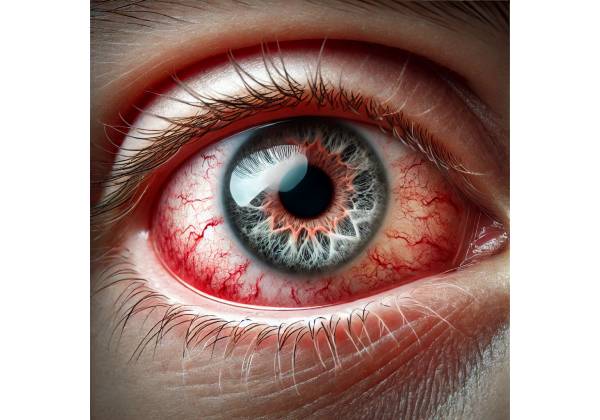
What is secondary glaucoma?
Secondary glaucoma is a broad category of eye conditions distinguished by elevated intraocular pressure (IOP) due to a known cause or underlying pathology. Secondary glaucoma, as opposed to primary glaucoma, usually develops as a result of other ocular or systemic conditions, trauma, inflammation, or specific medications. Secondary glaucoma’s elevated IOP can damage the optic nerve, resulting in vision loss and possibly blindness if not treated.
The anatomy and physiology of intraocular pressure
Understanding secondary glaucoma requires a basic understanding of the eye’s anatomy and physiology, particularly the mechanisms that regulate intraocular pressure. The anterior chamber, which is the space between the cornea and the iris, maintains the eye’s shape and optical properties by balancing fluid production and drainage.
The ciliary body, which is located behind the iris, produces the fluid known as aqueous humor. Aqueous humor flows from the posterior chamber, between the iris and the lens, to the anterior chamber, where it nourishes the avascular structures of the eye, such as the cornea and lens, and aids in the maintenance of proper eye pressure. The fluid then exits the eye primarily via the trabecular meshwork, a porous tissue located at the angle where the iris meets the cornea. The fluid eventually exits the eye through Schlemm’s canal and enters the bloodstream.
In a healthy eye, aqueous humor production and drainage are balanced, resulting in a stable intraocular pressure (typically 10 to 21 mmHg). When this balance is upset—whether due to increased fluid production, decreased drainage, or both—IOP rises, resulting in glaucoma.
Types of Secondary Glaucoma
The underlying cause of the elevated intraocular pressure determines the classification of secondary glaucoma. These categories include:
- Neovascular Glaucoma: Neovascular glaucoma is a severe and difficult type of secondary glaucoma that develops when abnormal blood vessels (neovascularization) form on the iris and over the trabecular meshwork. These vessels can obstruct the flow of aqueous humor, resulting in elevated IOP. Neovascular glaucoma is frequently associated with ischemic retinal conditions, including diabetic retinopathy, central retinal vein occlusion (CRVO), and ocular ischemic syndrome. The prognosis is generally poor due to the disease’s aggressive nature.
- Traumatic Glaucoma: Blunt or penetrating ocular injuries can both cause traumatic glaucoma. Trauma can cause angle recession, hyphema (bleeding in the anterior chamber), or lens dislocation, all of which disrupt the normal aqueous humor outflow and raise IOP. Traumatic glaucoma can develop quickly or gradually, even years after the initial injury.
- Uveitic Glaucoma: Uveitis is a type of inflammation that affects the uveal tract, which includes the iris, ciliary body, and choroid. Inflammation can cause the formation of synechiae (adhesions between the iris and the lens or cornea), which obstruct the trabecular meshwork and cause elevated IOP. Corticosteroids for uveitis can increase the risk of secondary glaucoma.
- Steroid-Induced Glaucoma: Prolonged use of corticosteroids, either topical, systemic, or intraocular, can result in steroid-induced glaucoma. Steroids can raise IOP by decreasing the flow of aqueous humor through the trabecular meshwork. Individuals are at varying risk of developing this type of glaucoma, with some being more susceptible to steroid-induced IOP elevation.
- Pigmentary Glaucoma: Pigment granules from the iris enter the anterior chamber and accumulate in the trabecular meshwork, preventing fluid outflow. This type of glaucoma is more common in younger, myopic people and can result in fluctuating IOP levels, which can cause optic nerve damage over time.
- Pseudoexfoliative Glaucoma: Pseudoexfoliative glaucoma is associated with pseudoexfoliation syndrome, which is characterized by the accumulation of fibrillary material on the lens, iris, and trabecular meshwork. This material can obstruct aqueous outflow, resulting in increased IOP. Pseudoexfoliative glaucoma is more common in older people and frequently more aggressive than primary open-angle glaucoma.
- Phacolytic Glaucoma: This condition develops in the presence of a hypermature (overripe) cataract. Lens proteins can leak into the anterior chamber, triggering an inflammatory response that obstructs the trabecular meshwork and raises IOP. This condition is most common in elderly patients who have delayed cataract surgery.
- Angle-Closure Glaucoma: Secondary angle-closure glaucoma develops when the angle between the iris and cornea becomes clogged, either due to an abnormality in the eye’s anatomy or due to other factors such as inflammation or swollen lens. This type of glaucoma can be acute or chronic, and it often necessitates prompt treatment to avoid vision loss.
Pathophysiology of Secondary Glaucoma
The pathophysiology of secondary glaucoma is complex and varies according to the underlying cause. However, an increase in intraocular pressure due to impaired aqueous humor outflow is the common denominator in all forms of secondary glaucoma.
In conditions such as neovascular glaucoma, the formation of new blood vessels on the iris and trabecular meshwork causes physical obstruction of the outflow channels, resulting in an increase in IOP. Similarly, in uveitic glaucoma, inflammatory cells and debris can clog the trabecular meshwork, reducing outflow and increasing pressure.
Trauma can cause structural damage to the eye’s drainage system, such as angle recession, which widens the angle while damaging the trabecular meshwork, resulting in inefficient fluid drainage. Steroid-induced glaucoma, on the other hand, is caused by the drug’s effect on trabecular meshwork cells, which reduces their efficiency in allowing aqueous humor to exit the eye.
In pigmentary glaucoma, pigment particles shed from the back of the iris can accumulate in the trabecular meshwork, causing mechanical blockage and increased resistance to outflow. Pseudoexfoliative glaucoma is characterized by the deposition of pseudoexfoliative material, which obstructs the trabecular meshwork and impedes fluid drainage.
Regardless of the cause, secondary glaucoma can cause progressive optic nerve damage due to persistently elevated intraocular pressure. The optic nerve, which sends visual information from the retina to the brain, is extremely sensitive to pressure. When IOP remains high, it compresses nerve fibers, reducing blood flow and causing cell death. This damage causes characteristic visual field loss over time, beginning with peripheral vision and progressing to central vision if the condition is not properly managed.
Symptoms and Clinical Presentation
Secondary glaucoma symptoms can vary greatly depending on the underlying cause, the rate of IOP increase, and the degree of optic nerve damage. Some types of secondary glaucoma, such as steroid-induced or pigmentary glaucoma, may develop slowly and asymptomatically in the early stages, whereas others, such as neovascular or traumatic glaucoma, can present with a sudden onset and severe symptoms.
Symptoms of secondary glaucoma include:
- Blurred Vision: Patients may experience blurred or hazy vision, which can vary depending on their IOP. This symptom is often more noticeable during activities that raise IOP, such as bending over or straining.
- Eye Pain: Eye pain is a common symptom of acute secondary glaucoma, including angle-closure and traumatic glaucoma. The pain can range from mild discomfort to severe, throbbing pain, which is frequently accompanied by redness and a sensation of pressure in the eye.
- Headache: Elevated IOP can cause referred pain, which appears as a headache, particularly around the brow or temple area. Angle-closure glaucoma can cause severe headaches, nausea, and vomiting.
- Halos Around Lights: Patients with secondary glaucoma may report seeing halos around lights, particularly in low-light conditions. This occurs as a result of elevated IOP-induced corneal edema, which scatters light entering the eye.
- Visual Field Loss: As secondary glaucoma progresses, patients may experience gradual loss of peripheral vision. This can manifest as difficulty navigating in low-light conditions or frequent collisions with objects. Without treatment, visual field loss can progress to tunnel vision and then blindness.
- Redness and Tearing: Inflammatory forms of secondary glaucoma, such as uveitic or phacolytic glaucoma, frequently cause redness, tearing, and light sensitivity as a result of the underlying inflammation.
Secondary glaucoma symptoms can be subtle or overlap with other ocular conditions, so patients at risk should have regular eye exams. Early detection and treatment are critical for preventing irreversible optic nerve damage and maintaining vision.
Diagnostic Techniques for Secondary Glaucoma
Secondary glaucoma diagnosis necessitates a thorough clinical evaluation that includes a detailed patient history, comprehensive eye examination, and specialized diagnostic testing. The goal is to identify the underlying cause of elevated intraocular pressure, determine the extent of optic nerve damage, and recommend appropriate treatment.
Comprehensive Eye Examination
A thorough eye examination is the first step in detecting secondary glaucoma. This includes:
- Visual Acuity Test: This test determines the sharpness of a patient’s vision at different distances. Although not specific to glaucoma, decreased visual acuity may indicate advanced optic nerve damage or other ocular conditions.
- Tonometry is a critical test for measuring intraocular pressure. Several methods are available, including Goldmann applanation tonometry, which is considered the gold standard. Elevated IOP is a hallmark of glaucoma, and multiple measurements are frequently required to confirm consistent IOP elevation over time. Tonometry helps to quantify IOP levels, which are important in determining the severity of secondary glaucoma.
- Gonioscopy: A special lens is used to examine the angle between the iris and the cornea (the anterior chamber angle). This test is critical for distinguishing between open-angle and angle-closure types of secondary glaucoma. It enables the ophthalmologist to examine the structure of the trabecular meshwork and detect any abnormalities, such as neovascularization, angle recession due to trauma, or synechiae (adhesions), that may contribute to IOP elevation.
- Slit-Lamp Examination: A slit-lamp microscope is used to thoroughly examine the anterior segment of the eye, which includes the cornea, iris, lens, and anterior chamber. This examination may reveal signs of inflammation, pigment dispersion, or pseudoexfoliation material, all of which can lead to secondary glaucoma. Furthermore, the slit-lamp examination allows the ophthalmologist to detect corneal edema, hyphema, and other abnormalities that could indicate high IOP.
- Fundoscopy (Ophthalmoscopy): Fundoscopy is an examination of the optic nerve head (optic disc) in the back of the eye. The optic disc in secondary glaucoma may exhibit cupping, a characteristic change caused by nerve fiber loss from elevated IOP. The degree of optic disc cupping, as well as any hemorrhages or pallor, can reveal important information about the extent of glaucoma damage.
Imaging Techniques
Advanced imaging techniques are frequently used to assess optic nerve damage and track the progression of glaucoma.
- Optical Coherence Tomography (OCT) is a non-invasive imaging technique that generates detailed cross-sectional images of the retina and optic nerve head. OCT is especially useful for detecting and quantifying RNFL thinning, which is a key indicator of glaucoma damage. OCT can also detect macular edema, which is sometimes associated with uveitic or neovascular glaucoma.
- Scanning Laser Ophthalmoscopy (HRT): The Heidelberg Retina Tomograph (HRT) is another imaging technique for examining the optic nerve head. It generates a 3D topographic map of the optic disc, which allows for the evaluation of optic nerve cupping and the tracking of structural changes over time.
- Visual Field Testing (Perimetry): Visual field testing is an important diagnostic tool for detecting functional vision loss in glaucoma. This test maps the patient’s visual field and identifies areas of visual field loss. Visual field defects in secondary glaucoma can begin in the peripheral vision and progress to the central vision if not treated. Automated perimetry is a common method for tracking the progression of visual field loss.
Additional Diagnostic Tests
Additional tests may be required to further evaluate the condition based on the type of secondary glaucoma suspected.
- Fluorescein Angiography: This procedure involves injecting a fluorescent dye into the bloodstream and photographing the retinal blood vessels. It is especially useful in neovascular glaucoma to detect abnormal blood vessel growth on the retina and iris.
- Ultrasound Biomicroscopy (UBM): UBM generates high-resolution images of the anterior segment structures, such as the ciliary body, iris, and trabecular meshwork. It is useful for diagnosing angle-closure glaucoma, phacolytic glaucoma, and other structural abnormalities that cause elevated IOP.
- Anterior Segment Optical Coherence Tomography (AS-OCT): AS-OCT can produce detailed images of the anterior chamber angle, cornea, and iris. It is especially useful for diagnosing angle-closure glaucoma and assessing the efficacy of interventions like laser peripheral iridotomy.
- Blood Tests and Systemic Evaluation: If secondary glaucoma is suspected of being caused by a systemic condition, such as sarcoidosis or juvenile idiopathic arthritis, blood tests and a systemic evaluation may be required. These tests can aid in detecting underlying systemic diseases that necessitate concurrent treatment.
- Genetic Testing: In some cases, particularly when pseudoexfoliative or pigmentary glaucoma is suspected, genetic testing may be recommended. While not a routine procedure, it can provide additional information about a patient’s risk of developing secondary glaucoma.
Secondary Glaucoma: Best Management Practices
Management of secondary glaucoma is complex and must
be specific to the condition’s underlying cause. The main objectives are to reduce intraocular pressure (IOP), prevent further optic nerve damage, and address the underlying
Cause contributing
to the elevated IOP. Management strategies range from medical therapy to surgical intervention, depending on the disease’s severity and progression.
Medical Management
Medical management is frequently the first line of treatment for secondary glaucoma, and it aims to lower IOP through the use of topical and systemic medications. The medication prescribed is determined by the type of secondary glaucoma, as well as the patient’s overall health and response to treatment.
- Topical Medications: There are several types of eye drops available to lower IOP, including:
- Prostaglandin Analogues: Drugs like latanoprost and bimatoprost increase the outflow of aqueous humor via the uveoscleral pathway, lowering IOP. They are frequently first-line agents due to their efficacy and once-daily dosing.
- Beta-Blockers: Medications such as timolol inhibit beta-adrenergic receptors in the ciliary body, reducing aqueous humor production. They are often used in conjunction with other medications.
- Alpha Agonists: Brimonidine and apraclonidine decrease aqueous humor production while increasing uveoscleral outflow. They are frequently used as an adjunctive therapy.
- Carbonic Anhydrase Inhibitors: These drugs, available as both topical (e.g., dorzolamide) and systemic (e.g., acetazolamide), inhibit the enzyme carbonic anhydrase, which reduces aqueous humor production.
- Miotics: Pilocarpine, a cholinergic agonist, stimulates aqueous outflow by contracting the ciliary muscle and expanding the trabecular meshwork. However, because of the negative side effects, it is less commonly used.
- Systemic Medications: In cases of acute IOP elevation or when topical therapy is ineffective, systemic medications such as oral carbonic anhydrase inhibitors (e.g., acetazolamide) or hyperosmotic agents (e.g., mannitol) may be used to rapidly lower IOP.
- Corticosteroids: In uveitic glaucoma, corticosteroids are used to reduce inflammation. However, because corticosteroids can raise IOP, their use must be closely monitored, and adjunctive IOP-lowering medications are frequently required.
Laser Therapy
Laser therapy is frequently used when medical management is insufficient or when IOP needs to be reduced permanently.
- Laser Trabeculoplasty: Laser trabeculoplasty, also known as argon laser trabeculoplasty (ALT) or selective laser trabeculoplasty (SLT), improves aqueous outflow through the trabecular meshwork. This procedure is especially useful in open-angle secondary glaucoma, such as pigmentary or pseudoexfoliative glaucoma.
- Laser Peripheral Iridotomy: In cases of angle-closure glaucoma, a small hole is created in the peripheral iris, allowing aqueous humor to flow more freely into the anterior chamber and preventing angle closure.
- Cyclophotocoagulation: This laser procedure targets the ciliary body and reduces aqueous humor production. It is commonly used in cases of refractory glaucoma or when other surgical options are ineffective.
Surgical Management
When medical and laser therapies fail to control IOP, or when the glaucoma has progressed, surgical intervention may be required.
- Trabeculectomy: One of the most common glaucoma surgeries entails creating a drainage fistula to allow aqueous humor to bypass the trabecular meshwork and flow directly out of the eye, lowering IOP. This surgery is effective in a variety of secondary glaucomas, but there are risks such as infection, scarring, and hypotony (low IOP).
- Glaucoma Drainage Devices: Patients who are not candidates for trabeculectomy or who have failed previous surgeries may benefit from glaucoma drainage devices (e.g., Ahmed, Baerveldt, or Molteno implants). These devices drain aqueous humor from the anterior chamber into an external reservoir, lowering IOP.
- Cyclodestructive Procedures: When other surgeries fail to treat advanced or refractory glaucoma, cyclodestructive procedures, such as cyclocryotherapy or cyclophotocoagulation, may be used to destroy a portion of the ciliary body in order to reduce aqueous humor production.
- Lens Extraction: In phacolytic or phacomorphic glaucoma, where a hypermature cataract is causing the IOP to rise, cataract surgery to remove the lens may be required to treat the condition.
Management of Underlying Conditions
In secondary glaucoma, it is critical to treat the underlying cause of elevated IOP. This could include treating systemic diseases like diabetes or uveitis, managing retinal conditions associated with neovascular glaucoma, or discontinuing medications like corticosteroids if they are contributing to elevated IOP.
Follow-Up and Monitoring
Regular follow-up is essential in secondary glaucoma management to monitor IOP, assess optic nerve health, and adjust treatment as needed. Visual field testing, optic nerve imaging, and IOP measurements are critical components of ongoing monitoring to ensure that the condition is under control and prevent further vision loss.
Trusted Resources and Support
Books
- “Glaucoma: A Patient’s Guide to the Disease” by Graham E. Trope: This book provides an accessible yet comprehensive overview of glaucoma, including secondary glaucoma, with practical advice for patients on managing the condition and understanding treatment options.
- “The Glaucoma Book: A Practical, Evidence-Based Approach to Patient Care” by Paul N. Schacknow and John R. Samples: This book offers in-depth information on the diagnosis, management, and treatment of various forms of glaucoma, making it a valuable resource for both healthcare professionals and patients.
Organizations
- Glaucoma Research Foundation: This organization provides extensive resources for patients with glaucoma, including information on secondary glaucoma, treatment options, and ongoing research. They also offer patient support services and education.
- American Academy of Ophthalmology (AAO): The AAO offers a wealth of information on glaucoma, including clinical guidelines, educational materials for patients, and updates on the latest research and treatment options.
- World Glaucoma Association (WGA): The WGA is dedicated to advancing knowledge and care of glaucoma worldwide. They provide resources for both professionals and patients, including information on secondary glaucoma and its global impact.


