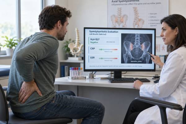
A spondyloarthritis blood test panel usually combines HLA-B27 with general inflammation markers such as CRP and ESR. The panel can strengthen or weaken clinical suspicion, but it cannot confirm or exclude spondyloarthritis by itself. HLA-B27 is an inherited immune marker associated most strongly with axial spondyloarthritis. CRP and ESR may show active systemic inflammation, yet both are normal in many people with genuine disease.
The result has meaning only when matched to the pattern of symptoms. Inflammatory back pain beginning before age 45, sacroiliac pain, heel enthesitis, swollen fingers or toes, recurrent uveitis, psoriasis, inflammatory bowel disease, or arthritis after infection can all point toward the spondyloarthritis family. Imaging often provides the decisive objective evidence: X-rays may show established sacroiliitis, MRI can detect earlier active inflammation, and ultrasound can assess peripheral joints and tendon attachments. The panel is therefore a probability tool and a disease-activity aid—not a stand-alone screening or diagnostic package.
- HLA-B27 positivity increases spondyloarthritis likelihood when symptoms fit, but many healthy carriers never develop disease.
- A negative HLA-B27 result does not rule out axial or peripheral spondyloarthritis.
- Normal CRP and ESR are common and should not stop evaluation when the clinical pattern is convincing.
- HLA-B27 generally needs testing only once; CRP and ESR may be repeated to monitor activity.
- Sudden painful red-eye symptoms, a hot swollen joint with fever, or new neurologic deficits require urgent assessment.
Table of Contents
- What the Panel Can and Cannot Do
- HLA-B27: Genetic Risk, Not a Diagnosis
- CRP, ESR, and Other Inflammation Tests
- How Results Differ Across Spondyloarthritis Patterns
- Why Imaging and Clinical Features Matter More
- Testing Process and Interpretation Traps
- Follow-Up, Monitoring, and Urgent Symptoms
What the Panel Can and Cannot Do
Spondyloarthritis is a family of inflammatory conditions rather than one laboratory-defined disease. It includes axial spondyloarthritis, psoriatic arthritis, reactive arthritis, inflammatory bowel disease–associated arthritis, and undifferentiated forms. Some people have mainly spinal and sacroiliac symptoms; others have peripheral joints, tendon attachments, digits, skin, bowel, or eyes involved.
A blood panel can help with three tasks:
- Estimate diagnostic probability. HLA-B27 adds weight when the history is compatible.
- Look for current systemic inflammation. CRP and ESR may support active disease.
- Establish a broader baseline. Blood count, kidney, liver, and infection tests can identify mimics or prepare for treatment.
The panel cannot show exactly which joint or enthesis is inflamed. It cannot distinguish axial spondyloarthritis from psoriatic arthritis with certainty, and it cannot replace an eye examination, skin examination, bowel evaluation, or imaging.
A panel result is also a snapshot. CRP can change over days, ESR over a longer interval, while HLA-B27 does not change at all. Symptoms may fluctuate independently of systemic markers, and pain may persist from structural damage or mechanical strain after inflammation improves. For that reason, ordering the three tests together does not make them interchangeable. Each should be tied to a separate question before the blood is drawn.
A useful panel may contain:
| Test | Main question | Key limitation |
|---|---|---|
| HLA-B27 | Is an important inherited susceptibility marker present? | Positive in healthy people; negative in many affected patients |
| CRP | Is there a measurable acute-phase response? | May be normal; rises with infection and many other conditions |
| ESR | Is there a broader inflammatory effect on blood proteins? | Affected by age, anemia, pregnancy, kidney disease, and immunoglobulins |
| Complete blood count | Is there anemia, infection evidence, or another blood abnormality? | Nonspecific |
| Kidney and liver panel | Are there organ issues or a treatment baseline? | Does not diagnose spondyloarthritis |
RF, anti-CCP, and ANA may be added when rheumatoid arthritis, lupus, or another connective tissue disease is a realistic alternative. Negative RF does not define spondyloarthritis, and positive RF does not automatically exclude it. Multiple diseases and incidental antibodies can coexist.
HLA-B27: Genetic Risk, Not a Diagnosis
HLA-B27 is a form of the human leukocyte antigen B protein found on cell surfaces. It helps the immune system present protein fragments to immune cells. The marker is inherited and remains stable throughout life, so repeat testing is rarely useful.
The laboratory usually reports HLA-B27 as present or absent. It is not a concentration with a normal, optimal, or toxic range. Testing may use molecular methods that detect the allele or flow cytometry that detects the antigen.
HLA-B27 has its strongest clinical association with axial spondyloarthritis, including radiographic axial spondyloarthritis, historically called ankylosing spondylitis. In many European-ancestry populations, most patients with ankylosing spondylitis carry HLA-B27. The proportion is lower in many other ancestral groups, and background carriage in healthy people also varies widely.
That population variation changes interpretation. A positive result in a symptomatic person from a population where HLA-B27 is uncommon may shift probability differently from the same result where the marker is common. Yet no ancestry-based percentage can replace the individual clinical picture.
A positive result is most useful when several compatible features are present:
- Chronic back pain beginning before age 45
- Improvement with movement rather than rest
- Night pain or alternating buttock pain
- Sacroiliitis on MRI or X-ray
- Acute anterior uveitis
- Psoriasis or inflammatory bowel disease
- Heel enthesitis or dactylitis
- First-degree family history of spondyloarthritis
A positive result without symptoms does not justify imaging, treatment, or repeated surveillance by itself. Most carriers never develop ankylosing spondylitis. Routine testing of healthy family members or children usually creates uncertainty rather than actionable prediction.
A negative result cannot exclude disease. HLA-B27-negative axial spondyloarthritis is well recognized, especially in populations with lower allele frequency and among patients with psoriasis- or bowel-associated disease. The dedicated HLA-B27 test guide explains positive and negative results in more detail.
CRP, ESR, and Other Inflammation Tests
CRP is made by the liver in response to inflammatory signaling. It can rise within hours and fall relatively quickly when inflammation improves. ESR measures how quickly red blood cells settle in a tube over one hour and usually changes more slowly.
In axial spondyloarthritis, an elevated CRP can support active inflammation and is used in disease-activity scores such as ASDAS-CRP. Elevated CRP may also be associated with greater average risk of radiographic progression and can help document objective inflammation when certain advanced therapies are considered.
However, many patients with active axial disease have normal CRP. Inflammation may be localized to the sacroiliac joints or entheses without generating a strong systemic response. Some people simply have a low-CRP biological pattern. A normal value should not outweigh inflammatory symptoms or MRI evidence.
ESR may add complementary information, particularly in peripheral disease or when CRP is unavailable. It is less direct and more vulnerable to confounding. Anemia can raise ESR, while unusual red-cell shapes can lower it. Age and pregnancy also affect reference expectations.
Other tests are chosen for specific questions:
- Fecal calprotectin may support evaluation for intestinal inflammation when bowel symptoms suggest inflammatory bowel disease; it is not a routine spondyloarthritis blood marker.
- Creatine kinase may be checked if muscle disease rather than joint or enthesis pain is possible.
- Uric acid provides gout context but cannot diagnose an acute crystal attack.
- Ferritin may rise with systemic inflammation but is too nonspecific for routine spondyloarthritis diagnosis.
- Cytokine panels and research biomarkers are not established clinical diagnostic tests.
A 2024 biomarker guideline for axial spondyloarthritis gave strongest support to HLA-B27 for suspected disease and CRP/ESR for activity assessment. This reflects a practical reality: many proposed molecules have interesting research associations, but none has replaced these established tests plus imaging and clinical evaluation.
How Results Differ Across Spondyloarthritis Patterns
The same panel can mean different things depending on the disease pattern.
Axial spondyloarthritis
HLA-B27 is most informative here. CRP may support active disease, but normal CRP and ESR are common. MRI of the sacroiliac joints is often central when X-rays are normal and suspicion remains high. A positive HLA-B27 plus inflammatory back pain is supportive, not conclusive.
Psoriatic arthritis
HLA-B27 is more relevant when axial involvement is present and less useful for purely peripheral disease. CRP and ESR may be high with active polyarthritis but can remain normal. Skin and nail examination, dactylitis, enthesitis, and imaging patterns often carry more diagnostic weight. A negative RF can support the differential but is not required.
Inflammatory bowel disease–associated arthritis
Blood markers may reflect bowel inflammation, joint inflammation, anemia, infection, or all of these. HLA-B27 is associated more with axial disease than with short-lived peripheral arthritis that parallels bowel activity. Coordination with gastroenterology is often needed because some medications help both systems while others do not.
Reactive arthritis
Reactive arthritis follows certain gastrointestinal or genitourinary infections. HLA-B27 is not required for diagnosis and does not identify the triggering organism. It may be associated with a greater tendency toward axial or persistent disease in some patients. Infection testing depends on timing and exposure; antibody panels are not routinely useful for proving the trigger.
Peripheral undifferentiated spondyloarthritis
Enthesitis, dactylitis, asymmetric lower-limb arthritis, uveitis, family history, and related conditions shape the diagnosis. HLA-B27 and CRP can add support, but ultrasound and examination may be more informative than sacroiliac imaging when spinal symptoms are absent.
| Pattern | HLA-B27 value | CRP/ESR value |
|---|---|---|
| Predominantly axial | Often important diagnostic support | Activity/prognosis support; frequently normal |
| Peripheral psoriatic | Limited unless axial features exist | May reflect polyarticular activity |
| IBD-associated | Most relevant to axial disease | May reflect bowel or joint inflammation |
| Reactive | Not diagnostic; may affect phenotype | Can rise during acute inflammation |
Why Imaging and Clinical Features Matter More
The diagnosis begins with recognizing a characteristic pattern. Inflammatory back pain often starts before age 45, lasts at least three months, improves with activity, worsens with rest, and may wake the person during the second half of the night. No single feature is specific, so combinations matter.
Spondyloarthritis can also announce itself outside the spine. Heel pain at the Achilles tendon or plantar fascia, a whole swollen digit, recurrent one-eye uveitis, psoriasis, chronic bloody diarrhea, or arthritis after infection may be more revealing than the back symptoms.
For suspected axial disease, plain pelvic X-rays can show structural sacroiliitis. X-rays may remain normal for years, so MRI is used to detect active bone marrow edema and other inflammatory or structural lesions. MRI interpretation requires expertise because athletes, postpartum patients, mechanical stress, infection, and degenerative change can mimic sacroiliitis.
For peripheral disease, ultrasound can show synovitis, tenosynovitis, enthesitis, bursitis, and increased Doppler blood flow. It can also help distinguish active inflammation from mechanical tendon disease and guide aspiration of a swollen joint.
Classification criteria, including ASAS frameworks, organize features for research but should not be treated as automatic diagnostic algorithms. A patient can have spondyloarthritis without meeting criteria, and someone can meet a feature combination while another diagnosis fits better.
The ankylosing spondylitis blood test panel is most relevant when axial symptoms dominate. A broader evaluation is needed when skin, bowel, peripheral joints, or entheses are the central problem.
Testing Process and Interpretation Traps
The panel uses routine venous blood. Fasting is generally unnecessary unless other bundled tests require it. HLA-B27 results may take several days if the sample goes to a reference laboratory; CRP and ESR often return sooner.
Common interpretation errors include:
- Ordering HLA-B27 for ordinary mechanical back pain without inflammatory features
- Treating a positive HLA-B27 as proof of ankylosing spondylitis
- Treating normal CRP and ESR as proof that inflammation is absent
- Repeating HLA-B27 to monitor a flare
- Assuming all heel pain is enthesitis or all MRI edema is spondyloarthritis
- Using a negative RF result as a diagnostic criterion for spondyloarthritis
- Ignoring infection, crystal arthritis, or mechanical disease because the panel looks supportive
Pretest probability is the hidden variable. A positive HLA-B27 result is much more useful in a young adult with inflammatory back pain and uveitis than in an older adult with localized pain after lifting. Likewise, a high CRP has more relevance when it rises with swollen joints than when fever and cough suggest infection.
Assay differences matter less for HLA-B27 because the result is usually binary, but rare subtypes and method limitations can occasionally create discrepancies. CRP and ESR should be interpreted using the laboratory’s own units and reference range. Standard CRP, not high-sensitivity cardiovascular CRP, is generally used for inflammatory disease activity.
Direct-to-consumer genetic data may infer HLA-related variants without performing full clinical typing. A medical laboratory may need to confirm the result before it influences diagnosis.
Follow-Up, Monitoring, and Urgent Symptoms
After testing, the next step depends on the symptom pattern and objective evidence.
A positive HLA-B27 with convincing axial symptoms may lead to rheumatology referral and sacroiliac imaging. A negative result with the same symptoms may still lead to imaging because the disease remains possible. High CRP or ESR can support active inflammation, while normal values shift emphasis toward examination and MRI rather than ending the evaluation.
Once spondyloarthritis is diagnosed, HLA-B27 is not repeated. Monitoring may include CRP, ESR, validated scores, function, spinal mobility, swollen joints, enthesitis, skin and bowel activity, and treatment-safety tests. ASDAS incorporates back pain, morning stiffness, patient global assessment, peripheral pain/swelling, and CRP or ESR. Other scores are used for psoriatic or peripheral disease.
A CRP decline can support response but should agree with symptoms and function. Some treatments alter CRP strongly, and some patients never have elevated markers. Imaging is repeated selectively when it will change management rather than on a fixed schedule for everyone.
Prepare for follow-up by recording:
- Age when back or joint symptoms began
- Whether movement or rest changes pain
- Night waking and alternating buttock pain
- Heel, digit, skin, nail, eye, or bowel symptoms
- Recent gastrointestinal or genitourinary infection
- Family history of spondyloarthritis, psoriasis, uveitis, or inflammatory bowel disease
- Response to anti-inflammatory medication
Sudden eye pain, redness, light sensitivity, or blurred vision needs same-day eye assessment for possible acute anterior uveitis. A hot swollen joint with fever requires urgent evaluation and often aspiration for infection. New leg weakness, saddle numbness, loss of bladder or bowel control, or severe pain after trauma requires emergency care.
The panel is most powerful when it directs the right next test. It is least useful when its three results are treated as a diagnostic verdict.
References
- ASAS-EULAR recommendations for the management of axial spondyloarthritis: 2022 update 2023 (Guideline)
- A guideline on biomarkers in the diagnosis and evaluation in axial spondyloarthritis 2024 (Guideline)
- Recommendations | Spondyloarthritis in over 16s: diagnosis and management | Guidance | NICE 2024 (Guideline)
- Diagnosis and Treatment of Ankylosing Spondylitis 2024 (Review)
- Exploring the Pathogenesis of Spondylarthritis beyond HLA-B27 2024 (Review)
- Tailoring Inflammatory Biomarker Assessment in Axial Spondyloarthritis: The Potential of ESR in Different Clinical Settings 2025 (Clinical Study)
Disclaimer
This article is for general education and cannot diagnose spondyloarthritis. HLA-B27, CRP, and ESR must be interpreted with symptoms, examination, imaging, ancestry, and possible alternative conditions by a qualified clinician. Seek urgent care for a hot swollen joint with fever, new neurologic deficits, or sudden painful red-eye symptoms with light sensitivity or vision change.