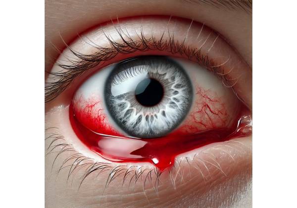
A subconjunctival hemorrhage is a common and usually harmless ocular condition characterized by the sudden appearance of a bright red or dark red patch on the white part of the eye (sclera). A small blood vessel bursts, leaking blood into the space between the conjunctiva and the sclera. The conjunctiva is a thin, transparent membrane that protects the sclera and lines the inside of the eyelids. While subconjunctival hemorrhages may appear alarming, they rarely cause pain, vision changes, or serious injury, and they frequently resolve on their own without the need for medical intervention.
Anatomy and Pathophysiology
Understanding a subconjunctival hemorrhage requires knowledge of basic eye anatomy. The sclera is the eye’s tough, white outer layer, which provides protection and structure. The conjunctiva is a mucous membrane containing small blood vessels that are usually invisible to the naked eye. These blood vessels are delicate and may rupture under certain conditions, resulting in a subconjunctival hemorrhage.
When one of these blood vessels ruptures, blood seeps into the area beneath the conjunctiva. Because the conjunctiva is clear, the blood stands out against the white background of the sclera, forming a red patch that varies in size. The conjunctiva serves as a barrier, preventing blood from spreading into the cornea or other parts of the eye.
The blood that accumulates beneath the conjunctiva does not drain; rather, it is gradually absorbed by the body over time. This process can take anywhere from a few days to a couple of weeks, depending on the size of the hemorrhage. During this time, the color of the hemorrhage may shift from bright red to darker red or even yellow as the blood is broken down and reabsorbed.
The causes of subconjunctival hemorrhage
Subconjunctival hemorrhages can occur spontaneously or as a result of other factors that put pressure on the small blood vessels in the eye. Some of the common causes are:
- Sudden Increase in Venous Pressure: One of the most common causes of subconjunctival hemorrhage is a sudden rise in venous pressure, which can rupture the delicate blood vessels. Activities that can cause this increase in pressure include:
- Coughing: Forceful coughing can increase blood vessel pressure and cause rupture.
- Sneezing: A strong sneeze can produce a similar effect, causing blood vessels to burst.
- Vomiting: The strain of vomiting can raise venous pressure in the head and neck, contributing to a hemorrhage.
- Heavy Lifting or Straining: Activities requiring strenuous physical exertion, such as lifting heavy objects or straining during bowel movements, can raise venous pressure and cause a hemorrhage.
- Eye Trauma: Direct trauma to the eye or surrounding area can result in a subconjunctival hemorrhage. This trauma can be minor, such as rubbing the eye too vigorously, or more severe, such as being struck by an object or having eye surgery.
- Systemic Health Conditions: Some systemic health conditions can increase the risk of subconjunctival hemorrhages. This includes:
- Hypertension (High Blood Pressure): High blood pressure can weaken blood vessel walls, making them more likely to rupture.
- Diabetes: Poorly controlled diabetes can damage blood vessels throughout the body, including those in the eyes, raising the risk of hemorrhage.
- Blood Clotting Disorders: Conditions that impair the blood’s ability to clot, such as hemophilia or thrombocytopenia, can cause spontaneous bleeding, including subconjunctival hemorrhages.
- Medications: Taking blood-thinning medications like aspirin, warfarin, or anticoagulants can increase the risk of bleeding, including in the small blood vessels of the eye.
- Infections: Ocular infections, such as conjunctivitis, can occasionally result in subconjunctival hemorrhage due to inflammation and increased fragility of the blood vessels.
- Aging: As people age, their blood vessels become more fragile, increasing their susceptibility to rupture. This can result in an increased risk of subconjunctival hemorrhage in older adults.
- Post-Surgical Complications: Subconjunctival hemorrhage can occur as a result of eye surgeries such as cataract surgery, LASIK, or other procedures that manipulate the eye. This is usually a temporary and harmless occurrence, but it can appear dramatic.
- Contact Lens Use: Improper or excessive use of contact lenses can irritate the eyes, resulting in a subconjunctival hemorrhage. This is especially true if the contact lenses are not properly cleaned or if they are worn for an extended time.
- Idiopathic: In many cases, a subconjunctival hemorrhage occurs with no known cause. These idiopathic cases are typically harmless and resolve on their own.
Symptoms of Subconjunctival Hemorrhage
The primary and most visible symptom of a subconjunctival hemorrhage is the appearance of a bright red or dark red patch on the sclera. This red patch is usually well-defined and can cover a small or large portion of the white of the eye. The appearance of the hemorrhage can be startling, especially if it occurs unexpectedly, but it is usually painless.
Other important aspects of a subconjunctival hemorrhage include:
- No Pain: Unlike many other eye conditions, a subconjunctival hemorrhage is not painful. The individual may be unaware of the hemorrhage until they see it in a mirror or someone else notices it.
- No Vision Changes: A subconjunctival hemorrhage does not impair vision. Individuals with this condition will not have blurry vision, double vision, or other visual disturbances. The hemorrhage occurs on the eye’s outer surface and does not affect the cornea, lens, or retina.
- No Discharge or Tearing: Unlike conjunctivitis or other infections, a subconjunctival hemorrhage does not result in eye discharge, excessive tearing, or other secretions. Aside from the red patch, the eye appears normal.
- No Light Sensitivity: A subconjunctival hemorrhage does not cause photophobia, or light sensitivity. The condition does not impair the eye’s ability to tolerate light.
- No Swelling: Although the red patch appears alarming, there is no swelling of the eyelids or surrounding tissues. The condition is limited to the surface of the sclera.
Duration and Treatment of a Subconjunctival Hemorrhage
A subconjunctival hemorrhage usually heals on its own without treatment. Over time, the body gradually absorbs the blood, and the red patch fades. The duration of the hemorrhage varies according to its size, but it typically resolves within one to two weeks. During this time, the hemorrhage’s appearance may change, with the red patch becoming darker and then yellowish as the blood breaks down.
In most cases, a subconjunctival hemorrhage does not recur, and people can resume their normal activities with no long-term consequences. However, recurrent hemorrhages may necessitate further investigation to determine whether an underlying systemic condition requires treatment.
Potential Complications
A subconjunctival hemorrhage is usually harmless and self-limiting, but in some cases, it can be associated with more serious underlying issues. For example, frequent or recurring hemorrhages may indicate a bleeding disorder or uncontrolled hypertension. In rare cases, a subconjunctival hemorrhage may indicate a more serious trauma or eye injury requiring immediate medical attention.
Subconjunctival hemorrhage after eye surgery is usually a minor complication. However, if it is accompanied by pain, vision changes, or other troubling symptoms, additional testing may be required to rule out complications.
Psychological and Social Impact
Although a subconjunctival hemorrhage is not life-threatening, its dramatic and unsightly appearance can cause psychological stress. Individuals may be self-conscious about the red patch on their eye, especially if it is large or visible. People may avoid social interactions or feel embarrassed about their appearance until the hemorrhage heals. Reassurance and education about the condition’s benign nature can help to alleviate these concerns.
Diagnostic Approaches for Subconjunctival Hemorrhage
A subconjunctival hemorrhage is usually easy to diagnose, and a visual examination of the eye is often sufficient. However, in some cases, additional diagnostic tests may be required to rule out underlying conditions or complications.
Visual Examination
The primary method for diagnosing a subconjunctival hemorrhage is a visual examination by an eye care professional. During this examination, the ophthalmologist or optometrist will examine the eye, concentrating on the sclera to detect the characteristic red patch associated with a hemorrhage. The red patch is usually well defined and does not extend beyond the conjunctiva. The healthcare provider will evaluate the size, shape, and location of the hemorrhage and may inquire about the onset of symptoms and any possible causes, such as recent trauma, coughing, or sneezing.
In most cases, a visual examination is sufficient to diagnose a subconjunctival hemorrhage, particularly if the condition is isolated and simple. The absence of pain, vision changes, or discharge strengthens the diagnosis.
Patient History
A thorough patient history is essential in the diagnosis of a subconjunctival hemorrhage. The eye care professional will inquire about the patient’s medical history, recent activities, and any possible risk factors that contributed to the hemorrhage. Key questions could include:
- Recent Trauma: Has there been any recent injury or trauma to the eye or surrounding area, even if minor, such as vigorous rubbing of the eye or being struck by an object?
- Coughing or Sneezing: Has the patient ever had a severe cough or sneeze that resulted in a sudden increase in venous pressure?
- Vomiting or Straining: Has the patient recently vomited or engaged in activities that cause significant strain, such as lifting heavy objects or exerting extreme physical effort?
- Medication Use: Does the patient take any medications that may increase the risk of bleeding, such as blood thinners (e.g., aspirin, warfarin) or other anticoagulants?
- Systemic Health Conditions: Does the patient have a history of systemic conditions such as hypertension, diabetes, or blood clotting disorders that could put them at risk for a subconjunctival hemorrhage?
- Infections: Has the patient recently had any eye infections or inflammation, such as conjunctivitis, which could be associated with a hemorrhage?
This information assists the healthcare provider in identifying any underlying factors that may have contributed to the hemorrhage and determining whether additional investigation or treatment is required.
Slit Lamp Examination
The eye care professional may perform a slit-lamp examination in some cases, particularly if there is concern about other underlying eye conditions or if the hemorrhage occurs repeatedly. A slit lamp is a specialized microscope that provides a magnified view of the eye’s structures, such as the conjunctiva, sclera, cornea, and anterior chamber.
A slit-lamp examination allows the eye care professional to closely inspect the hemorrhage and assess the overall health of the eye. This examination can help rule out other possible causes of the redness, such as conjunctivitis, corneal abrasions, or uveitis, which may necessitate different treatment options. The slit-lamp examination is especially useful when the diagnosis is not immediately clear from visual inspection alone.
Blood Pressure Measurement
Because hypertension is a known risk factor for subconjunctival hemorrhage, taking the patient’s blood pressure is an essential part of the diagnostic process. Elevated blood pressure raises the risk of blood vessel rupture, resulting in a hemorrhage. If high blood pressure is detected, the patient may be referred to a primary care physician or a cardiologist for further evaluation and treatment.
Blood Tests
Blood tests may be ordered if the hemorrhage occurs again or if an underlying systemic condition is suspected. Blood tests can aid in detecting issues such as:
- Coagulation Disorders: Tests like prothrombin time (PT), activated partial thromboplastin time (aPTT), and platelet count can evaluate a patient’s blood clotting ability and identify any clotting disorders that may predispose them to bleeding.
- Diabetes: A blood glucose or hemoglobin A1c test can help determine whether the patient’s hemorrhage is due to poorly controlled diabetes.
- Complete Blood Count (CBC): A CBC can reveal information about the patient’s overall health, such as their red and white blood cell counts, which may indicate anemia or infection.
Imaging Studies
Imaging studies, such as an orbital CT scan or MRI, may be recommended in rare cases, especially if there is a concern about trauma or other serious underlying conditions. These imaging techniques can provide detailed views of the eye and surrounding structures, assisting in the identification of any fractures, tumors, or other abnormalities that may be causing the hemorrhage.
Subconjunctival Hemorrhage: Treatment and Management
Subconjunctival hemorrhage is typically a harmless condition that resolves on its own without the need for medical intervention. However, management focuses on symptom relief, underlying causes, and preventing recurrence. The treatment for a subconjunctival hemorrhage varies depending on the severity of the condition, the presence of any underlying health issues, and the frequency of recurrence.
Self-Care & Home Remedies
In most cases of subconjunctival hemorrhage, self-care measures are adequate to manage the condition while it heals. This includes:
- Observation and Reassurance: Because subconjunctival hemorrhage is usually harmless and self-limiting, the primary management strategy is to simply monitor the situation and reassure the patient. The red patch usually fades within one to two weeks as the body reabsorbs blood. Patients should be informed that the hemorrhage may change color as it heals, progressing from bright red to darker red and finally yellowish.
- Avoiding Eye Rubbing: Patients should be advised not to rub their eyes, as this can aggravate the hemorrhage or cause further trauma to the delicate blood vessels.
- Cold Compresses: Applying a cold compress to the affected eye within 24 to 48 hours of the hemorrhage’s onset may help reduce any swelling or discomfort. Wrap ice in a clean cloth and gently press it against the closed eyelid for short periods of time to create a cold compress.
- Warm Compresses: After the first 48 hours, using warm compresses can improve blood circulation and help the body absorb blood more effectively. To apply a warm compress, soak a clean cloth in warm water, wring it out, and place it on the closed eyelid for 10-15 minutes several times per day.
- Lubricating Eye Drops: While subconjunctival hemorrhage rarely causes dryness or irritation, some patients may experience a mild foreign body sensation or discomfort. To soothe and relieve the eye, use over-the-counter lubricating eye drops (artificial tears).
Medical Treatment
Most subconjunctival hemorrhages resolve on their own, so no specific medical treatment is required. However, in some cases, medical intervention may be required:
- Addressing Underlying Causes: If the hemorrhage is caused by an underlying systemic condition, such as hypertension or a bleeding disorder, it is critical to treat that condition to avoid recurrence. For example, if high blood pressure is identified as a contributing factor, the patient may need to consult with their primary care physician about managing their blood pressure through lifestyle changes, medication, or both.
- Medication Review: Patients taking blood thinners, such as aspirin, warfarin, or other anticoagulants, may require a medication review if they have recurring hemorrhages. In some cases, dosage adjustments may be required, or alternative medications may be considered. However, any medication changes should be made only after consulting with a healthcare professional.
- Treatment for Associated Infections: If a subconjunctival hemorrhage is caused by an eye infection, such as conjunctivitis, the underlying infection should be treated accordingly. Depending on the cause of the infection, you may need to use antibiotic or antiviral eye drops.
- Surgical Intervention: In very rare cases, particularly if the hemorrhage is large, persistent, or caused by significant trauma, surgical intervention may be required. This could include procedures to repair damaged blood vessels or treat any resulting ocular injuries. However, such cases are uncommon, and surgery is rarely necessary for a simple subconjunctival hemorrhage.
Preventive Strategies
Subconjunctival hemorrhage prevention is primarily concerned with addressing underlying risk factors and maintaining good eye health. Prevention strategies include:
- Control of Systemic Conditions: Proper management of systemic conditions such as hypertension and diabetes is critical in preventing subconjunctival hemorrhage from occurring again. Patients should take their medications as prescribed, eat a healthy diet, exercise regularly, and schedule regular check-ups with their doctors.
- Eye Protection: Keeping the eyes safe from trauma is critical for preventing injury-related hemorrhages. This can include wearing protective eyewear while engaging in activities that endanger the eyes, such as sports, woodworking, or chemical handling.
- Proper Use of Contact Lenses: Contact lens wearers can reduce the risk of irritation and subsequent hemorrhage by following proper hygiene practices, such as washing their hands before handling lenses and using appropriate cleaning solutions.
- Avoiding Valsalva Maneuver: The Valsalva maneuver, which involves forcefully exhaling against a closed airway (as in coughing, sneezing, or straining), can raise venous pressure and cause a subconjunctival hemorrhage. Patients should be encouraged to avoid or carefully manage activities requiring excessive straining.
Patients can effectively manage a subconjunctival hemorrhage and lower their risk of recurrence by combining self-care, medical management, and preventive strategies. It is critical for patients to understand that, while the appearance of the hemorrhage may be alarming, it is usually a harmless condition that will resolve with time.
Trusted Resources and Support
Books
- “The Eye Book: A Complete Guide to Eye Disorders and Health” by Gary H. Cassel: This comprehensive guide provides valuable information on a wide range of eye conditions, including subconjunctival hemorrhage, with practical advice on maintaining eye health and preventing common issues.
- “Your Eyes: An Owner’s Guide” by Paul J. Ruggieri: This book offers an accessible overview of various eye conditions, including subconjunctival hemorrhage, and provides tips on how to protect and care for your eyes.
Organizations
- American Academy of Ophthalmology (AAO): The AAO provides extensive resources on eye health and various ocular conditions, including subconjunctival hemorrhage. Their website offers patient education materials, guidelines for treatment, and access to professional eye care services.
- National Eye Institute (NEI): The NEI, part of the U.S. National Institutes of Health, offers research-based information on eye conditions, treatments, and preventive care. Their resources are valuable for patients seeking to learn more about eye health and the management of conditions like subconjunctival hemorrhage.


