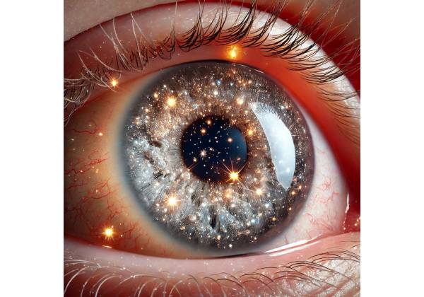
Synchysis scintillans is a rare but distinct ocular condition defined by the presence of freely floating, refractile crystals or cholesterol deposits in the eye’s vitreous humor. The condition is typically associated with degenerative changes in the vitreous body and is frequently observed in eyes that have previously experienced trauma, inflammation, or are in the advanced stages of certain ocular diseases. Synchysis scintillans is usually bilateral, meaning it affects both eyes, but it can sometimes appear unilaterally. The condition is usually benign and does not cause significant visual impairment, but its presence can indicate underlying ocular pathology.
Anatomy and Physiology of Vitreous Humor
To understand synchysis scintillans, you must first understand the anatomy and physiology of the vitreous humor. The vitreous humor is a clear gel-like substance that fills the space between the lens and the retina in the eye’s posterior segment. It accounts for roughly two-thirds of the eye’s volume and serves a variety of important functions, including maintaining the eye’s shape, allowing light to pass through to the retina, and acting as a shock absorber to protect the retina from mechanical damage.
Water makes up approximately 99% of the vitreous humor, with collagen fibers, hyaluronic acid, and other proteins accounting for the remaining 1%. In a healthy eye, the vitreous is clear and free of opacities, allowing light to reach the retina without interference. However, with age or as a result of various pathological conditions, the vitreous can degenerate, resulting in the appearance of floaters, opacities, or other abnormalities such as synchysis scintillans.
Pathogenesis of Synchysis Scintillans
The accumulation of cholesterol crystals or calcium-lipid complexes in the vitreous humor is the hallmark of synchysis scintillans. These crystals are highly refractile, which means they reflect and scatter light, giving them a sparkling, glittering appearance under an ophthalmic examination. The term “scintillans” comes from the Latin word for “sparkling,” which accurately describes the visual effect of these crystals.
The exact mechanism for the formation of these crystals is unknown, but it is thought to be linked to degenerative changes in the vitreous body. Several factors may contribute to the development of synchysis scintillans, including:
- Vitreous Liquefaction (Syneresis): As we age, the vitreous humor gradually undergoes a process known as liquefaction or syneresis, in which the gel-like structure breaks down, resulting in the formation of liquid pockets within it. As the vitreous liquefies, it loses its structural integrity, causing cholesterol and other lipids to crystallize.
- Cholesterol Metabolism: Abnormalities in cholesterol metabolism or high cholesterol levels in the blood can contribute to the formation of cholesterol crystals in the vitreous. This is more common in people who have systemic illnesses like hyperlipidemia or diabetes.
- Ocular Trauma: Previous trauma to the eye can disrupt the vitreous structure, allowing cellular debris and lipids to enter the vitreous cavity. This debris may act as a nidus for the formation of synchysis scintillans.
- Chronic Inflammation: Chronic uveitis or other inflammatory conditions of the eye can cause ocular tissue breakdown and lipid release into the vitreous. Inflammatory cells and mediators can also affect the vitreous environment, promoting crystal formation.
- Degenerative Ocular Diseases: Conditions like proliferative diabetic retinopathy, retinal detachment, and advanced cataracts can cause significant changes in vitreous structure and composition, raising the risk of synchysis scintillans.
Clinical Features of Synchysis Scintillans
Patients with synchysis scintillans typically exhibit symptoms related to visual disturbances caused by floating crystals in the vitreous. The most common symptom is the appearance of “floaters,” which are small moving spots or shapes in the visual field. The movement of the crystals within the vitreous as the eye moves causes these floaters, which are often more visible against a bright background, such as a blue sky or a white wall.
The appearance of these floaters varies, but they are typically described as shimmering, sparkling, or glittering, which is due to the refractile nature of the cholesterol crystals. Synchysis scintillans floaters are distinct and shiny, as opposed to typical vitreous floaters, which can appear as dark spots or cobweb-like shapes.
Despite the presence of these floaters, the majority of patients with synchysis scintillans have no significant visual impairment. Although floaters can be annoying or distracting, they rarely interfere with central vision or cause significant visual loss. However, in rare cases, if the crystals become densely packed or are associated with other ocular conditions, they can cause more severe visual disturbances.
Differential Diagnosis
Other conditions that can cause floaters or opacities in the vitreous humor should be considered when diagnosing synchysis scintillans. Some of these conditions are:
- Asteroid Hyalosis: Asteroid hyalosis is another condition characterized by the presence of small, white, calcium-containing opacities in the vitreous fluid. Unlike synchysis scintillans, the opacities in asteroid hyalosis are usually more numerous, smaller, and less mobile. They are usually firmly attached to the vitreous framework and do not cause any significant visual symptoms.
- Vitreous Hemorrhage: A vitreous hemorrhage occurs when blood leaks into the vitreous cavity, which can be caused by trauma, diabetic retinopathy, or retinal vein occlusion. The blood can cause floaters or a sudden loss of vision, depending on the severity of the hemorrhage. In contrast to the sparkling crystals seen in synchysis scintillans, vitreous hemorrhage is characterized by darker, more diffuse opacities.
- Posterior Vitreous Detachment (PVD): PVD is a common age-related condition in which the vitreous gel detaches from the retina. This process can result in the formation of floaters, which are commonly characterized as dark spots or cobweb-like shapes. PVD, unlike synchysis scintillans, does not involve the presence of refractile crystals.
- Uveitis: Uveitis, or inflammation of the uveal tract, can cause an accumulation of inflammatory cells and debris in the vitreous humor, resulting in floaters and vision problems. Uveitis floaters are typically less refractile and more diffuse than synchysis scintillans floaters.
- Retinal Detachment: In cases of retinal detachment, the vitreous may contain pigmented cells or hemorrhagic debris, which can result in floaters. Retinal detachment is a serious condition that frequently causes additional symptoms such as flashes of light, a curtain-like shadow over the visual field, and sudden loss of vision.
Prognosis
The prognosis for patients with synchysis scintillans is generally favorable because the condition is usually benign and does not cause significant visual impairment. However, the presence of synchysis scintillans may indicate an underlying ocular or systemic disease, so a thorough evaluation of the patient is required to identify any associated conditions.
Most floaters caused by synchysis scintillans do not require treatment, and patients can become accustomed to their presence over time. In rare cases where the floaters are especially bothersome or interfere with vision, additional management options may be considered, as discussed in the management section of this article.
Diagnosing Synchysis Scintillans: Essential Techniques
Synchysis scintillans is diagnosed using a combination of clinical examination, imaging studies, and, in some cases, laboratory tests to confirm the presence of vitreous crystals and rule out other conditions that cause similar symptoms. Common diagnostic methods include the following:
Clinical Examination
The first step in diagnosing synchysis scintillans is a thorough clinical examination by an ophthalmologist. The exam typically includes:
- Slit-Lamp Biomicroscopy: Slit-lamp biomicroscopy is a valuable tool for studying the anterior segment of the eye, as well as the vitreous and retina. During the examination, the ophthalmologist can see the vitreous humor and recognize the distinctive refractile crystals of synchysis scintillans. The crystals appear to be free-floating, shiny particles that move in response to eye movements. The slit lamp enables close examination of the size, shape, and distribution of these crystals.
- Indirect Ophthalmoscopy: Indirect ophthalmoscopy offers a more comprehensive view of the vitreous cavity and retina. This technique is especially useful for evaluating the posterior segment of the eye and determining any associated retinal pathology, such as retinal detachment or uveitis, that may be contributing to the patient’s symptoms. This examination allows you to see the floating crystals in synchysis scintillans, as well as their characteristic sparkle.
- Visual Acuity and Refraction Tests: While synchysis scintillans rarely causes significant visual impairment, it is critical to evaluate the patient’s visual acuity and refractive status. These tests determine whether the floaters are affecting the patient’s vision and guide future treatment.
Imaging Studies
Imaging studies may be used to confirm the diagnosis of synchysis scintillans and assess the extent of vitreous involvement. Common imaging techniques include the following:
- Ultrasound B-Scan: B-scan ultrasonography is a non-invasive imaging technique that employs sound waves to generate cross-sectional images of the eye. Ultrasound B-scan is especially useful when media opacities, such as cataracts or dense floaters, make it difficult to see the vitreous fluid. In patients with synchysis scintillans, a B-scan can detect highly reflective, mobile particles within the vitreous cavity. These particles are the cholesterol crystals or calcium-lipid complexes that characterize the condition. The B-scan can also help rule out other conditions, such as vitreous hemorrhage or retinal detachment, which can cause similar symptoms.
- Optical Coherence Tomography (OCT): While OCT is most commonly used to evaluate the retina and optic nerve, it can also provide useful information about the vitreous body in certain situations. OCT uses light waves to create detailed cross-sectional images of the retina and vitreous interface. Although OCT is not the primary diagnostic tool for synchysis scintillans, it can aid in assessing the vitreoretinal interface and detecting any abnormalities in the eye’s posterior segment that may be associated with the condition.
- Fluorescein Angiography (FA): Fluorescein angiography is most commonly used to assess retinal circulation and detect vascular abnormalities, but it can also aid in the differential diagnosis of vitreous opacities. FA can help rule out other causes of vitreous opacities, such as diabetic retinopathy or retinal vein occlusion, which can both cause floaters.
Lab Tests
Synchysis scintillans is typically diagnosed through clinical examination and imaging, rather than laboratory tests. However, if an underlying systemic condition, such as hyperlipidemia or diabetes mellitus, is suspected of contributing to the crystal formation, blood tests may be ordered to assess the patient’s lipid profile and blood glucose levels. These tests can aid in identifying systemic risk factors that may require attention as part of the overall management strategy.
Synchysis Scintillans Management
Synchysis scintillans management typically focuses on symptoms and underlying conditions rather than directly treating the vitreous crystals. Because synchysis scintillans is frequently a benign condition, many patients do not require aggressive treatment, especially if their visual function remains largely intact. However, if the floaters severely impair vision or are particularly bothersome, several management strategies can be considered.
Observation and Monitoring
For the majority of patients with synchysis scintillans, the primary treatments are observation and regular monitoring by an ophthalmologist. Because the condition is usually benign and the associated floaters are not vision-threatening, many patients gradually learn to live with the presence of floaters. Regular follow-up visits allow the ophthalmologist to monitor the condition for changes and ensure that no additional complications or underlying conditions, such as retinal detachment or progressive vitreous degeneration, develop. During these visits, the patient’s visual acuity and ocular health are evaluated, and any new symptoms are addressed right away.
Addressing the Underlying Conditions
If synchysis scintillans is associated with an underlying systemic condition, such as hyperlipidemia, diabetes mellitus, or chronic inflammation, managing these conditions can be critical to the overall treatment plan. Controlling systemic factors that contribute to the formation of vitreous crystals can help stabilize the condition and reduce the risk of future complications. This may entail collaborating with a primary care physician, an endocrinologist, or other specialists to control blood lipids, blood glucose, and systemic inflammation.
Vitrectomy
If the floaters associated with synchysis scintillans are particularly dense or interfere with daily activities, surgery may be considered. Vitrectomy is a surgical procedure that removes the vitreous humor and any floating crystals from the eye. This procedure can significantly reduce visual disturbances because it removes the source of the floaters.
Vitrectomy is usually reserved for patients who have severe symptoms that cannot be treated with conservative measures, as the procedure has risks such as retinal detachment, infection, and cataract formation. The patient should consult with their ophthalmologist to weigh the potential benefits of the procedure against the risks. Vitrectomy is typically performed as an outpatient procedure using local or general anesthesia, and patients may require several weeks of recovery time.
Laser Vitreolysis
Laser vitreolysis is a less invasive treatment for floaters caused by synchysis scintillans. This procedure uses a laser to break up or vaporize vitreous opacities, reducing their impact on vision. Laser vitreolysis is an outpatient procedure that does not require surgical incisions, making it an appealing option for patients who are not candidates for vitrectomy or who prefer a less invasive approach.
However, laser vitreolysis is not appropriate for all patients with synchysis scintillans because the procedure’s efficacy is dependent on the size, location, and composition of the vitreous crystals. The procedure also has its own set of risks, including the possibility of retinal or lens damage. Patients should consult with their ophthalmologist about the potential benefits and risks of laser vitreolysis to determine if it is a viable option for their specific situation.
Patient Education and Adaptations
Educating patients about synchysis scintillans and its typically benign course is an important part of management. Patients should be reassured that floaters are harmless and that, in many cases, the brain adjusts to their presence over time, making them less noticeable. Providing patients with floater-coping strategies, such as focusing on distant objects or using proper lighting, can help to reduce their impact on daily activities.
Patients who find the floaters particularly bothersome may benefit from psychological support or counseling, especially if the floaters cause significant anxiety or distress. Learning to manage expectations and develop coping mechanisms can help patients improve their quality of life while reducing the need for more invasive treatments.
Trusted Resources and Support
Books
- “Vitreous: In Health and Disease” by J. Sebag: This comprehensive book provides detailed information on the anatomy, physiology, and pathology of the vitreous humor, including conditions like synchysis scintillans. It is an excellent resource for both clinicians and patients seeking to understand vitreous-related disorders.
- “Clinical Ophthalmology: A Systematic Approach” by Jack J. Kanski and Brad Bowling: This widely respected textbook offers in-depth coverage of various ocular conditions, including synchysis scintillans, with an emphasis on diagnosis, management, and patient care. It is a valuable reference for medical professionals and informed patients alike.
Organizations
- American Academy of Ophthalmology (AAO): The AAO is a leading organization that provides a wealth of resources on eye health and ocular conditions, including vitreous disorders like synchysis scintillans. Their website offers patient education materials, access to the latest research, and directories of certified eye care providers.
- National Eye Institute (NEI): As part of the U.S. National Institutes of Health, the NEI conducts and supports research on eye diseases and offers extensive educational resources for patients and healthcare professionals. The NEI provides up-to-date information on vitreous conditions and promotes eye health through public awareness campaigns.


