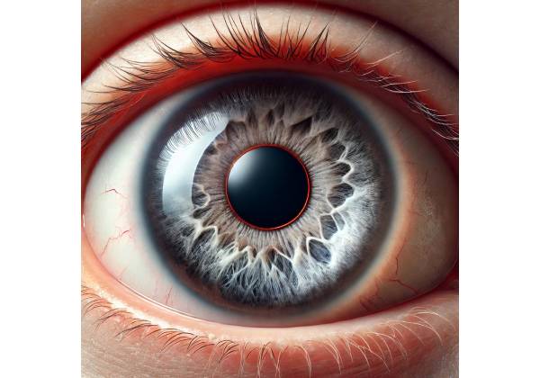
Vossius ring, also known as Vossius ring sign, is an ocular condition in which a circular imprint appears on the anterior surface of the eye’s lens. This ring is made up of pigment or epithelial cells from the iris that are deposited on the lens after blunt trauma to the eye. The condition is named after Adolf Vossius, a German ophthalmologist who first described it in the late 1800s.
Vossius ring is commonly associated with ocular trauma, especially when a significant impact causes the eye to compress and then rapidly decompress. This sudden compression-decompression event may cause the iris to make forceful contact with the lens’s anterior surface. As a result, pigment or epithelial cells from the iris migrate to the lens, leaving the distinctive ring-shaped imprint. While Vossius ring is usually not a vision-threatening condition, it can indicate significant eye trauma, which may lead to other complications that require attention.
Anatomy of the Eye and the Formation of Vossius Ring
To understand how a Vossius ring forms, you must first understand the eye’s relevant structures, specifically the iris, lens, and anterior chamber.
- The Iris: The iris is the colored part of the eye that controls the diameter and size of the pupil, thereby determining how much light reaches the retina. The iris has a layer of pigmented epithelial cells that give it its color. These cells are essential in the formation of a Vossius ring because they are the ones that imprint on the lens surface.
- The Lens: The lens is a transparent, biconvex structure that sits behind the iris and pupil. It works with the cornea to focus light on the retina, providing clear vision. The lens is flexible and can change shape to focus on objects at various distances, a process known as accommodation.
- The Anterior Chamber: The anterior chamber is the fluid-filled space that connects the cornea and the iris. It helps to keep intraocular pressure stable while also nourishing the corneal endothelium and lens.
When the eye receives a blunt force, the following sequence of events usually results in the formation of a Vossius ring:
- Trauma Impact: The eye is subjected to blunt trauma, which can occur from a variety of sources, including being struck by a ball, a fist, or another object.
- Compression and Decompression: The force of the impact causes the eyeball to flatten or compress temporarily. Following this, the eye rapidly decompresses as the force decreases. This sudden change in pressure and shape brings the posterior surface of the iris into contact with the anterior surface of the lens.
- Pigment Transfer: When the iris makes contact with the lens, pigment epithelial cells or pigment granules are transferred to the lens surface. Due to the pupil’s circular shape, this transfer occurs in a ring-like pattern, resulting in the formation of a Vossius ring.
- Ring Persistence: The ring may remain on the lens surface for a variety of time periods. In some cases, it fades or disappears over time, while in others, it remains visible for an extended period of time.
Clinical Features and Symptoms
The Vossius ring does not typically cause symptoms or direct vision impairment. However, its presence indicates previous significant ocular trauma, which may have resulted in other injuries that impair vision.
- Visual Observation: The most distinguishing feature of the Vossius ring is that it is visible during clinical examination, particularly under slit-lamp microscope. It manifests as a well-defined, circular or ring-shaped discoloration on the anterior surface of the lens, which corresponds to the size and shape of the pupil at the time of trauma. The ring’s color can range from light brown to dark brown, depending on the pigmentation of the iris.
- Associated Ocular Injuries: While Vossius ring is generally safe, it is frequently associated with other ocular injuries caused by the same trauma. These injuries may include:
- Hyphema: The presence of blood in the anterior chamber of the eye, often caused by trauma. Hyphema can cause pain, blurred vision, and an increased risk of elevated intraocular pressure, which can progress to glaucoma if not treated properly.
- Lens Dislocation or Subluxation: Trauma can cause the lens to shift out of its normal position, resulting in lens dislocation or subluxation. This can cause significant visual disturbances and may necessitate surgical intervention.
- Traumatic Cataract: A cataract, or clouding of the lens, can form after trauma, especially if the impact was severe enough to damage the lens fibers. Traumatic cataracts can impair vision and require cataract surgery to restore clarity.
- Iridodialysis is the detachment of the iris from its base at the ciliary body, resulting in a D-shaped pupil. It can cause visual disturbances and increased sensitivity to light.
- Corneal Edema: Trauma can cause corneal swelling, resulting in temporary blurred vision and discomfort.
- Retinal Injuries: Trauma severe enough to cause a Vossius ring can also result in retinal injuries, such as commotio retinae (retinal bruising) or retinal detachment, both of which are serious conditions that require immediate medical attention.
Pathophysiology of Vossius Ring
The pathophysiology of Vossius ring is directly related to the dynamics of ocular trauma as well as the physical interaction between the iris and lens. The main processes involved are:
- Mechanical Impact: The initial mechanical impact increases intraocular pressure and deforms the globe. This causes a temporary contact between the iris and the lens, which is necessary for the formation of the Vossius ring.
- Cellular Transfer: When pigment epithelial cells or granules from the iris come into contact with the lens, they are mechanically dislodged and transferred to the lens. The amount of transfer depends on the force of the impact and the duration of the contact.
- Pigment Adhesion: Once applied, the pigment adheres to the lens surface. The natural stickiness of the lens capsule, as well as the properties of the pigment granules, facilitate adhesion. Over time, some of the pigment may be absorbed or dislodged by the eye’s normal movements or the aqueous humor that circulates within the anterior chamber.
- Ring Persistence: Individuals differ in how long the Vossius ring remains on the lens surface. In some cases, the ring fades or disappears as the aqueous humor gradually resorbs or washes away the pigment. In other cases, the ring may be visible for an extended period of time, particularly if the pigment has deeply adhered to the lens capsule.
Epidemiology and Risk Factors
Vossius ring is a common finding in people who have suffered significant blunt trauma to the eye. The condition is most frequently seen in:
- Young People: Young people, particularly those involved in sports or physical activities, are more likely to sustain blunt trauma that results in the formation of a Vossius ring.
- Occupational Hazards: Certain occupations that involve a high risk of ocular injury, such as construction, manufacturing, or jobs requiring the use of heavy machinery, may predispose people to trauma-related conditions like Vossius ring.
- Lack of Protective Eyewear: The absence of appropriate protective eyewear during high-risk activities significantly increases the risk of ocular trauma and the subsequent development of Vossius ring.
Prognosis and Long-Term Implications
Individuals with a Vossius ring typically have a good prognosis because the condition does not cause long-term visual impairment. However, the presence of a Vossius ring indicates significant ocular trauma, which may result in other injuries that impair vision if not managed properly.
Most of the time, the Vossius ring fades or resolves on its own. However, the resulting injuries, such as hyphema, lens dislocation, or retinal damage, may necessitate medical or surgical intervention to avoid complications and preserve vision. Individuals with a Vossius ring should have a comprehensive eye examination to rule out any associated injuries and receive appropriate treatment as needed.
Methods for Diagnosing Vossius Ring
A thorough clinical examination, often supplemented by imaging techniques to assess the extent of ocular injury and identify any associated conditions, is required to diagnose a Vossius ring. Common diagnostic methods include the following:
Slit Lamp Examination
The slit-lamp examination is the foundation for diagnosing a Vossius ring. This specialized microscope allows the ophthalmologist to closely examine the eye’s structures at high magnification. During the slit-lamp examination, the clinician can see the anterior surface of the lens and recognize the distinctive circular or ring-shaped imprint that defines a Vossius ring.
- Visualization: Under the slit-lamp, the Vossius ring appears as a distinct, pigmented ring on the lens, proportional to the pupil size at the time of trauma. The color and intensity of the ring vary depending on the iris’ pigmentation and the severity of the trauma.
- Assessment of Associated Injuries: In addition to detecting the Vossius ring, the slit-lamp examination allows the ophthalmologist to look for other trauma-related injuries like hyphema, corneal edema, or iridodialysis. This comprehensive evaluation is critical for determining the trauma’s overall impact and directing future treatment.
Dilated Fundus Examination
A dilated fundus examination is frequently used to evaluate the posterior segment of the eye, especially if there is concern about potential retinal or optic nerve damage as a result of the trauma that caused the Vossius ring. This examination uses eye drops to dilate the pupil, allowing the ophthalmologist to get a better view of the retina, optic disc, and posterior segment of the eye.
- Retinal Evaluation: During the dilated fundus examination, the ophthalmologist will look for signs of retinal injury, such as commotio retinae (retinal bruising), retinal tears, or retinal detachment. These conditions can coexist with a Vossius ring in cases of severe blunt trauma and may necessitate immediate treatment to avoid vision loss.
- Optic Nerve Assessment: Signs of damage or swelling (papilledema) to the optic nerve may indicate a more severe traumatic injury to the eye or head. This section of the examination is critical in cases where there is concern about long-term visual complications.
Gonioscopy
Gonioscopy is a technique for examining the anterior chamber angle of the eye, which is where the cornea meets the iris. This test is especially important if angle recession is suspected, which occurs when the structures of the anterior chamber angle are damaged as a result of trauma.
- Angle Recession: In the context of a Vossius ring, angle recession may occur, particularly if the trauma was severe. Angle recession can increase the risk of developing secondary glaucoma, which is characterized by increased intraocular pressure. Identifying angle recession early with gonioscopy allows for more frequent monitoring and timely intervention if intraocular pressure rises.
Imaging Techniques
In some cases, additional imaging techniques may be required to fully assess the extent of ocular injury and aid in the diagnosis of associated conditions.
- Optical Coherence Tomography (OCT): OCT is a non-invasive imaging technique for obtaining detailed cross-sectional images of the retina and optic nerve. This imaging modality is especially useful for detecting subtle changes in the retinal layers and identifying macular edema, which can occur after ocular trauma. OCT may be used if there is a suspicion of posterior segment involvement despite the presence of a Vossius ring.
- Ultrasound B-Scan: An ultrasound B-scan may be performed if a lens opacity caused by the Vossius ring or other trauma-related factors obscures the retinal view. This imaging technique employs sound waves to generate cross-sectional images of the eye, allowing the ophthalmologist to examine the vitreous body, retina, and optic nerve for signs of hemorrhage, retinal detachment, or other structural abnormalities.
- Anterior Segment Optical Coherence Tomography (AS-OCT): AS-OCT generates high-resolution images of the anterior segment of the eye, which includes the cornea, anterior chamber, iris, and lens. This imaging technique is useful for determining the exact location and extent of the Vossius ring, as well as assessing any associated anterior segment injuries.
Intraocular Pressure Measurement
Intraocular pressure (IOP) measurement is a standard part of the diagnostic workup after ocular trauma, especially in the presence of a Vossius ring. Elevated IOP can indicate a variety of complications, including traumatic glaucoma and hyphema.
- Tonometry: Tonometry is a technique for measuring IOP. It is possible to perform it using a variety of methods, including Goldmann applanation tonometry, which is considered the gold standard. Monitoring IOP is critical, especially if there is evidence of angle recession or hyphema, as these conditions can lead to increased IOP and subsequent glaucoma if not addressed.
Vossius Ring Treatment: Options and Approaches
The primary goal of managing a Vossius ring is to address any underlying ocular trauma while also preventing or treating potential complications that may arise as a result of the injury. While the Vossius ring is typically benign and does not directly impair vision, its presence indicates that significant blunt trauma has occurred, which may have resulted in other eye injuries that necessitate careful examination and intervention. Management options include observation, medical treatment, and, in some cases, surgical intervention.
Observation and Monitoring
In most cases, the Vossius ring fades over time as pigment or epithelial cells adhered to the lens surface are absorbed or washed away by the eye’s natural aqueous humor circulation. As a result, many cases of Vossius ring do not require specialized treatment. However, close monitoring is required to ensure that no additional ocular complications arise as a result of the trauma.
- Regular Eye Examinations: Patients with a Vossius ring should have regular eye exams to monitor the condition of their eyes and detect any changes in vision or eye health. The ophthalmologist will evaluate the ring’s persistence and look for signs of associated injuries, such as lens dislocation, cataract formation, or retinal damage.
- Patient Education: Patients should be informed about the nature of the Vossius ring and the significance of reporting any new or worsening symptoms, such as increased blurring of vision, floaters, flashes of light, or eye pain. This education is essential for detecting and treating complications as soon as they arise.
Medical Treatment
While the Vossius ring does not typically require medical treatment, the following conditions may necessitate intervention:
- Management of Hyphema: If the trauma that caused the Vossius ring also resulted in hyphema (blood in the anterior chamber), medical intervention may be required to avoid complications such as elevated intraocular pressure or corneal blood staining. This may include the use of topical corticosteroids to reduce inflammation, as well as medications to lower intraocular pressure if it becomes high.
- Treatment of Elevated Intraocular Pressure: If trauma has caused angle recession or hyphema, medications such as beta-blockers, prostaglandin analogs, or carbonic anhydrase inhibitors may be prescribed to reduce pressure and prevent glaucoma development.
- Steroid Therapy for Inflammation: When trauma causes significant inflammation in the eye, topical or systemic corticosteroids may be used to control the inflammatory response. This is especially important if you are concerned about the development of secondary complications like cataracts or uveitis.
Surgical Intervention
Surgical intervention is rarely required for the management of a Vossius ring itself; however, if the trauma has caused significant structural damage to the eye, surgery may be necessary:
- Cataract Surgery: If the trauma that caused the Vossius ring results in the formation of a traumatic cataract (clouding of the lens), cataract surgery may be necessary to restore clear vision. During the procedure, the cloudy lens is removed and replaced by an artificial intraocular lens (IOL).
- Lens Dislocation Surgery: If the trauma has caused the lens to become dislocated or subluxated (partially dislocated), surgery may be required to reposition or replace the lens. This surgery is critical for restoring proper visual function and avoiding future complications.
- Vitrectomy: If the trauma caused retinal detachment or significant hemorrhage in the vitreous body, a vitrectomy may be necessary. This procedure includes removing the vitreous gel and repairing any retinal tears or detachments. Vitrectomy is frequently combined with other procedures, such as laser photocoagulation, to stabilize the retina and prevent further detachment.
Long-term Follow-up
Long-term follow-up is critical for patients who have suffered ocular trauma and developed a Vossius ring. Regular eye examinations enable the early detection of any delayed complications, such as secondary glaucoma, cataract formation, or retinal detachment. Patients should be encouraged to keep these follow-up appointments and to seek immediate medical attention if they notice any vision changes.
Trusted Resources and Support
Books
- “Traumatic Eye Injuries: Diagnosis and Management” by James C. Augsburger
- This comprehensive guide covers the full spectrum of eye injuries, including those that result in conditions like Vossius ring. It provides detailed information on diagnosis, management, and treatment options for various types of ocular trauma.
- “Ocular Trauma: Principles and Practice” by Ferenc Kuhn
- This book offers in-depth coverage of the mechanisms, diagnosis, and management of ocular trauma. It is an excellent resource for understanding the broader context in which a Vossius ring may occur.
Organizations
- American Academy of Ophthalmology (AAO)
- The AAO provides extensive resources on ocular trauma and related conditions, including Vossius ring. Their website offers patient education materials, clinical guidelines, and updates on the latest research in eye trauma and treatment.
- Eye Injury Registry of America (EIRA)
- EIRA is dedicated to the collection and analysis of data related to eye injuries. Their resources help improve understanding and management of ocular trauma, providing valuable information for both healthcare professionals and patients dealing with conditions like Vossius ring.


