
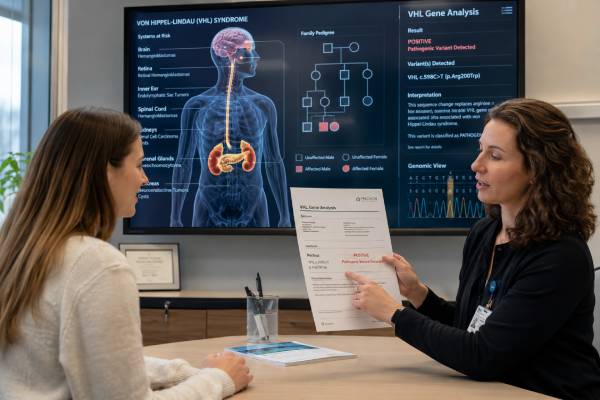
A VHL genetic test looks for an inherited pathogenic variant that causes von Hippel-Lindau syndrome, a lifelong tumor-predisposition condition. Carriers can develop retinal and central nervous system hemangioblastomas, clear cell renal cell carcinoma, pheochromocytoma, pancreatic neuroendocrine tumors, endolymphatic sac tumors, and several benign cysts or tumors. Many lesions are treatable when found early, but they may arise in more than one organ and recur over time.
Testing usually uses blood or saliva and should include sequencing plus deletion-duplication analysis. A positive germline result confirms the syndrome and allows relatives to have focused testing. A negative result does not fully exclude VHL when the clinical pattern is strong, especially if mosaicism is possible. A variant of uncertain significance should not be used alone to diagnose the syndrome or recommend surgery. Results should lead to a written, age-based surveillance plan involving eyes, hearing, blood pressure and metanephrines, brain and spine MRI, and abdominal imaging. The exact schedule depends on age, previous lesions, variant type, family history, and current specialist guidance.
- A pathogenic VHL result confirms a hereditary syndrome that can affect the eyes, brain, spine, kidneys, adrenals, pancreas, and inner ear.
- Surveillance begins in childhood and continues for life, even when the carrier has no symptoms.
- A negative blood test may miss low-level mosaicism or a variant outside the assay’s range.
- A VUS should not direct organ surgery or predictive testing of healthy relatives by itself.
- Each child of a person with a constitutional VHL pathogenic variant has a 50% chance of inheriting it.
Table of Contents
- What VHL Does
- Who May Need VHL Testing
- How Testing Is Performed
- Understanding VHL Results
- Tumors and Other Manifestations
- Surveillance by Organ System
- Treatment, Family Testing, and Pregnancy
- Limitations and Next Steps
What VHL Does
VHL encodes the von Hippel-Lindau protein, part of a complex that marks certain proteins for breakdown. One of its best-known targets is hypoxia-inducible factor, or HIF. When oxygen is adequate, functional VHL helps remove HIF. When VHL function is lost, HIF can accumulate and activate genes involved in blood-vessel growth, glucose metabolism, and cell survival.
A person with von Hippel-Lindau syndrome is usually born with one nonworking VHL copy in every cell. A susceptible cell can form a lesion after the remaining working copy is lost or inactivated. This two-hit mechanism explains why a carrier is predisposed but not born with every tumor.
VHL lesions are often highly vascular. Hemangioblastomas contain abnormal blood-vessel networks and supporting stromal cells. Clear cell renal cell carcinomas are driven in part by HIF signaling. This pathway also explains why HIF-2α inhibitors can be useful for selected VHL-associated tumors, although medication does not replace surveillance.
The syndrome has historically been divided into types. Type 1 families have a low pheochromocytoma risk, while type 2 families have higher pheochromocytoma risk and are further described by renal cancer risk. Certain variant classes correlate with these patterns, but the categories are not perfect. Management should follow the exact variant, family phenotype, and current guidance rather than the label alone.
Most variants found during hereditary evaluation are germline. VHL can also be altered somatically in sporadic clear cell kidney cancer. A VHL change on tumor-only testing does not automatically mean the person has von Hippel-Lindau syndrome. Confirmation in blood or another normal tissue is required before relatives are considered at risk.
The condition is highly variable. Relatives with the same variant may develop different organs, ages, and numbers of lesions. Surveillance is therefore broad and repeated; waiting for one family member’s pattern to repeat can miss disease.
Who May Need VHL Testing
Testing is considered when a person has a VHL-associated lesion, a combination of suggestive findings, or a known familial variant. The threshold is lower when a lesion occurs young, is bilateral or multifocal, or appears with another VHL feature.
Findings that commonly prompt evaluation include:
- More than one central nervous system hemangioblastoma.
- A retinal hemangioblastoma, particularly at a young age or with another VHL feature.
- Clear cell renal cell carcinoma at a young age, in both kidneys, or in multiple tumors.
- Pheochromocytoma that is bilateral, multifocal, early onset, or accompanied by another VHL manifestation.
- Pancreatic neuroendocrine tumor, especially when multiple or accompanied by pancreatic or renal cysts.
- Endolymphatic sac tumor causing hearing or balance symptoms.
- Epididymal or broad-ligament cystadenomas in a suggestive setting.
- A close relative with von Hippel-Lindau syndrome or a pathogenic VHL variant.
A single sporadic lesion at a typical age may have a lower chance of inherited disease, but age cutoffs are not absolute. Multigene testing may be more appropriate for pheochromocytoma or hereditary kidney cancer because several genes overlap.
Clinical diagnosis can sometimes be made from a characteristic combination of lesions, especially in a known VHL family. Molecular confirmation remains valuable because it enables targeted testing of asymptomatic relatives and can help with genotype-informed pheochromocytoma counseling.
Testing children is appropriate after a familial variant is known because eye examinations and other surveillance begin early. Delaying testing until adulthood would miss the period when retinal lesions and pheochromocytoma can first appear.
Before testing, gather ophthalmology records, brain and spine imaging, kidney and pancreas scans, pheochromocytoma biochemistry, operative notes, pathology, audiology, and the exact family laboratory report. A pancreatic “mass” might be a cyst, serous cystadenoma, or neuroendocrine tumor, and each carries different significance.
A broader hereditary cancer panel can be useful when kidney cancer, PPGL, and other lesions do not form a classic VHL pattern. The panel should include genes appropriate to the presenting tumor rather than unrelated genes added without clinical rationale.
How Testing Is Performed
Germline VHL testing usually uses DNA from blood or saliva. No fasting is needed. A complete test should include sequence analysis and deletion-duplication analysis because a substantial minority of disease-causing variants are larger deletions or rearrangements.
Testing options include:
- Targeted familial testing for a known variant.
- Full VHL sequencing for small variants.
- Deletion-duplication analysis for exon or whole-gene copy-number changes.
- A multigene panel for hereditary kidney cancer or PPGL when the phenotype overlaps.
- Mosaicism studies using deeper sequencing or another tissue when blood is negative but suspicion remains high.
Results often take two to six weeks. Targeted testing can be faster. The report should identify the transcript, DNA and protein change, classification, and methods.
Mosaic VHL occurs when the variant arises after fertilization and is present in only some tissues. The allele fraction in blood may be low or undetectable. Mosaicism is more likely to be considered in a person with classic VHL lesions but negative standard testing and no family history. Testing affected tissue, cultured skin fibroblasts, or a higher-depth blood assay can help.
Tumor sequencing can support the evaluation. For example, the same VHL variant in more than one independent lesion may suggest a shared constitutional or mosaic origin. A sporadic clear cell renal carcinoma, however, commonly has somatic VHL loss, so tumor positivity alone is weak evidence for inherited disease.
When a broad panel reports a VHL variant, the laboratory’s classification and allele fraction should be reviewed. A low-level result may need confirmation. Deletions involving VHL and nearby genes can produce a broader phenotype, so the size and boundaries of a copy-number change matter.
Pretest counseling should cover possible uncertain results, implications for children, the lifelong surveillance commitment, and insurance or privacy issues. It should also explain that a positive result may reveal lesions through screening that require observation rather than immediate treatment.
Understanding VHL Results
Laboratories classify variants as pathogenic, likely pathogenic, uncertain significance, likely benign, or benign. The category describes evidence that the variant disrupts VHL; it does not report current tumor burden.
Pathogenic or likely pathogenic
A positive constitutional result confirms von Hippel-Lindau syndrome. The carrier should have a baseline evaluation and age-appropriate lifelong surveillance. Likely pathogenic variants are generally managed like pathogenic variants.
The exact variant can inform whether pheochromocytoma risk is expected to be low or high, but genotype does not predict every lesion. A person from a type 1 family can still need blood pressure and clinical review, and individual plans should follow specialist guidance.
The result should be kept permanently. Relatives need the exact DNA change for focused testing. A report that states only “VHL positive” is insufficient.
Negative
A negative targeted test for the known familial variant is usually a true negative. The person generally does not need VHL-specific surveillance.
A negative full test in a person with multiple classic lesions does not fully exclude VHL. Mosaicism, technical limitations, an undetected structural change, or another syndrome may explain the presentation. Clinical surveillance can remain appropriate while the evaluation continues.
A negative result in an unaffected person is uninformative when no affected relative has had testing. Testing should begin with the relative most likely to carry a pathogenic variant whenever possible.
Variant of uncertain significance
A VUS is not a confirmed diagnosis. It should not be the sole reason for kidney surgery, intensive screening of healthy relatives, or prenatal testing. Management should rest on the person’s lesions and family history.
Evidence for reclassification can include functional studies, tumor loss of the working allele, population frequency, segregation, and reports in unrelated VHL families. Reclassification should be issued by a clinical laboratory. The rules for a VUS result help prevent uncertain evidence from becoming an irreversible medical decision.
Mosaic result
A confirmed mosaic pathogenic variant can cause VHL manifestations, but risk may differ according to tissue distribution. Transmission to a child is possible if reproductive cells carry the variant; the probability may be lower than 50% but cannot always be measured. Surveillance is individualized and often resembles standard VHL care when several organ systems may be involved.
Tumors and Other Manifestations
VHL affects multiple organs, and several lesions can be bilateral or multifocal.
Retinal hemangioblastomas can appear in childhood and threaten vision through leakage, retinal detachment, or location near the optic nerve. Small peripheral lesions can often be treated with laser or cryotherapy. New floaters, vision loss, flashes, or a visual-field change need urgent ophthalmic evaluation.
Central nervous system hemangioblastomas occur in the cerebellum, brainstem, spinal cord, or nerve roots. Symptoms can include headache, vomiting, imbalance, weakness, numbness, pain, bowel or bladder change, or symptoms from a tumor-associated cyst. Imaging growth and symptoms guide surgery; not every small lesion needs immediate treatment.
Clear cell renal cell carcinoma is a major cause of serious illness in VHL. Tumors may arise in both kidneys and recur. Management often uses active surveillance until the largest solid lesion approaches a size threshold, commonly about 3 cm, followed by nephron-sparing treatment when feasible. The strategy balances metastatic prevention with preserving kidney tissue across repeated interventions.
Pheochromocytomas can release catecholamines and cause headaches, sweating, palpitations, tremor, anxiety-like episodes, or high blood pressure. Some are silent. A secretory tumor must be recognized before surgery or pregnancy because catecholamine crises can be life-threatening.
Pancreatic lesions include simple cysts, serous cystadenomas, and pancreatic neuroendocrine tumors. Cysts are often benign and need no treatment unless they cause problems. Neuroendocrine tumors are assessed using size, growth rate, location, metastatic risk, and sometimes genotype. Surgery must be balanced against pancreatic insufficiency and diabetes.
Endolymphatic sac tumors can cause sudden or progressive hearing loss, tinnitus, vertigo, or ear fullness. Hearing loss can become permanent, so new symptoms need prompt audiologic and imaging evaluation.
Epididymal cystadenomas and broad-ligament cystadenomas are usually benign. They can support the diagnosis but may not require treatment unless symptomatic or uncertain.
Tumors can arise independently. A normal brain MRI does not reduce kidney risk, and successful kidney surgery does not end eye surveillance. Care must remain organ-specific and lifelong.
Surveillance by Organ System
VHL surveillance begins in childhood and changes with age. Schedules differ somewhat across countries, but common programs include the following components.
| Area | Common timing or method | Purpose |
|---|---|---|
| Eyes | Dilated retinal examination beginning in infancy or early childhood, often every 6–12 months | Find treatable retinal hemangioblastomas before vision loss |
| Blood pressure and pheochromocytoma | Annual blood pressure and plasma or urine metanephrines beginning in childhood, with age adjusted to variant and family history | Detect catecholamine-producing tumors before a crisis |
| Hearing | Periodic audiology beginning in childhood, with MRI for symptoms or protocol-based screening | Identify endolymphatic sac tumors and hearing change |
| Brain and spine | MRI beginning in later childhood or early adolescence and repeated about every two years when normal | Detect CNS hemangioblastomas |
| Kidneys, adrenals, and pancreas | Abdominal MRI beginning in adolescence and repeated about every two years, with shorter intervals for lesions | Monitor renal tumors, pheochromocytomas, pancreatic neuroendocrine tumors, and cysts |
MRI is favored because surveillance is lifelong and repeated CT would add substantial radiation. Ultrasound can be useful in young children or selected follow-up but is less sensitive for some small or complex lesions. CT and nuclear imaging remain appropriate when MRI cannot answer the clinical question.
Surveillance intervals shorten after a lesion is found. A renal mass near an intervention threshold, a growing pancreatic neuroendocrine tumor, or a CNS lesion with a cyst may need imaging every few months. A stable small lesion may be observed longer.
Metanephrine testing requires attention to posture, stress, medications, and laboratory method. Borderline results may be repeated under controlled conditions. A significant elevation leads to imaging and endocrine evaluation.
Pregnancy, anesthesia, and planned surgery are occasions to confirm that pheochromocytoma screening is current. A carrier should inform the surgical and obstetric teams even if prior tests were normal.
A written surveillance calendar should identify the responsible eye specialist, radiology protocol, endocrine testing laboratory, and VHL center. Fragmented care can lead to duplicated scans in one organ and missed screening in another.
Treatment, Family Testing, and Pregnancy
Treatment is lesion specific. Observation is appropriate for many small asymptomatic lesions. Surgery, laser, cryotherapy, ablation, focused radiation, or systemic therapy may be used when growth or symptoms create risk. The aim is not to remove every finding immediately but to prevent irreversible harm while preserving organ function.
Kidney management often favors partial nephrectomy or ablation rather than total nephrectomy. Repeated bilateral disease makes nephron preservation essential. Brain and spinal hemangioblastomas are usually treated when symptomatic, growing, or associated with a threatening cyst. Eye treatment depends on size and location.
Belzutifan, a HIF-2α inhibitor, can shrink selected VHL-associated renal, CNS, and pancreatic neuroendocrine tumors in patients who do not require immediate surgery. Eligibility, availability, duration, anemia, hypoxia, pregnancy risks, and other adverse effects require oncology oversight. Medication does not remove the inherited variant or eliminate surveillance.
VHL is autosomal dominant. Each child of a constitutional carrier has a 50% chance of inheriting the variant. Once the family variant is known, targeted familial testing identifies who needs surveillance. Children who test negative can usually avoid VHL-specific screening.
A de novo variant can occur. If both parents test negative, sibling risk is low but not zero because of possible parental mosaicism. The affected person’s children remain at 50% risk.
Reproductive options include natural conception, prenatal diagnosis, and in vitro fertilization with preimplantation genetic testing for monogenic disease. Prenatal testing shows whether the variant is present but cannot predict the organs or severity involved.
Before pregnancy, a carrier should update eye, CNS, abdominal, blood-pressure, and metanephrine assessments. Pregnancy can complicate management of pheochromocytoma and retinal or CNS lesions. Imaging and delivery planning should involve a VHL team and maternal-fetal medicine when needed.
Limitations and Next Steps
Genetic testing can miss low-level mosaicism, deep intronic variants, complex rearrangements, or changes outside validated regions. A negative result must be interpreted against the clinical pattern and assay. Variant classifications can also change as new families and functional data are reported.
Surveillance itself has limitations. MRI can find benign or indeterminate lesions that lead to anxiety and repeat scans. It may also miss very small lesions. No schedule prevents every complication, so symptom awareness remains essential.
Useful next steps after a pathogenic result are:
- Confirm the result and meet with a VHL-experienced genetics team.
- Complete baseline eye, hearing, biochemical, CNS, and abdominal assessments appropriate for age.
- Create a written calendar with imaging intervals and responsible specialists.
- Establish lesion-specific thresholds for observation or intervention.
- Screen for pheochromocytoma before surgery or pregnancy.
- Offer targeted testing to relatives, including children at the recommended age.
- Keep all imaging on comparable systems when possible so growth can be measured accurately.
- Review treatment and surveillance advances periodically.
Common mistakes include focusing only on kidney cancer, treating every pancreatic cyst as dangerous, relying on ultrasound alone for all adult surveillance, and assuming a negative blood test excludes mosaic VHL. Another error is removing small lesions too early and causing cumulative organ damage. Specialist care aims to time intervention before metastatic spread or irreversible symptoms while avoiding unnecessary procedures.
The value of a VHL result lies in organized lifelong care. With coordinated surveillance, many lesions can be detected at a stage when vision, hearing, kidney function, neurologic function, and cancer outcomes are better protected.
Because VHL surveillance spans several organs, missed handoffs are a common preventable problem. A shared calendar should identify the next retinal examination, blood-pressure and metanephrine assessment, hearing review, and abdominal and central nervous system imaging. The team should also document which prior lesions were observed, treated, or removed, since growth rate and organ function influence the timing of intervention. New visual change, neurologic symptoms, hearing loss, or spells of headache and palpitations should be assessed between scheduled visits.
References
- Von Hippel-Lindau Syndrome 2025 (Review)
- von Hippel-Lindau disease: Updated guideline for diagnosis and surveillance 2022 (Guideline)
- Von Hippel-Lindau Disease: A Comprehensive Review of Diagnosis, Genetics, Clinical Challenges, and Surveillance 2025 (Review)
- Narrative Review of Von Hippel-Lindau Syndrome 2025 (Review)
- Genetic Counseling and Long-Term Surveillance Using a Multidisciplinary Approach in von Hippel-Lindau Disease 2022 (Review)
- Modern management of von Hippel-Lindau disease 2022 (Review)
Disclaimer
VHL results and surveillance schedules should be interpreted by a hereditary tumor team using the exact variant, age, prior lesions, and current guidance. This article is educational and does not replace individualized care or urgent assessment of sudden vision loss, severe headache, neurologic change, chest symptoms, or marked hypertension.





