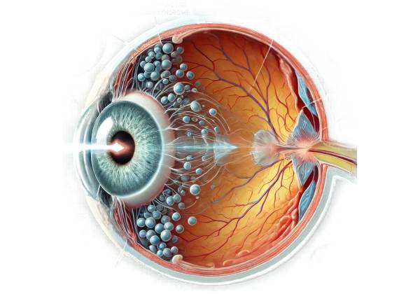
Vitreomacular traction syndrome (VMT) is a condition that affects the eye, specifically the macula, which is the central portion of the retina responsible for detailed and sharp vision. VMT occurs when the vitreous humor, a gel-like substance that fills the eye, attaches abnormally to the macula and exerts traction or pulling forces on it. This abnormal attachment and pulling can cause a variety of visual disturbances and, if untreated, can result in significant visual impairment.
Understanding Anatomy: Vitreous and Retina
To fully comprehend the nature of vitreomacular traction syndrome, one must first understand the basic anatomy of the eye, specifically the vitreous humor and retina.
The vitreous humor:
The vitreous humor is a clear, gel-like substance that fills the space between the lens in the front of the eye and the retina in the back. It is mostly water, collagen fibers, and hyaluronic acid, which contribute to its gel-like consistency. The vitreous connects to the retina at several locations, including the macula, optic nerve, and retinal periphery.
Retina and Macula:
The retina is a thin layer of light-sensitive tissue that covers the back of the eye. It works similarly to the film in a camera, capturing light and converting it into electrical signals that travel to the brain via the optic nerve. The macula is a small but important part of the retina that controls central vision, color vision, and the ability to detect fine details. The fovea, located in the center of the macula, is the area of vision that is most sharp.
The Pathology of Vitreomacular Traction Syndrome
The vitreous humor naturally degenerates with age, a process known as vitreous syneresis. This process involves the liquefaction and shrinkage of the vitreous gel, which eventually leads to posterior vitreous detachment (PVR). During PVD, the vitreous humor separates from the retina, which is a normal and usually harmless process. However, in some cases, the vitreous does not completely detach from the macula, resulting in an abnormal adhesion and VMT.
Vitreomacular traction syndrome occurs when the vitreous humor partially detaches and exerts tractional forces on the macula. These forces can alter the shape of the macula, disrupt its normal function, and cause a variety of visual symptoms. The degree of traction varies; some cases are mild and asymptomatic, while others cause significant macular distortion and visual impairment.
Causes and Risk Factors
Vitreomacular traction syndrome is primarily an age-related condition, but several factors can increase the likelihood of its occurrence:
1. Age:
Age is the most important risk factor for VMT. As people age, their vitreous humor naturally degenerates and becomes more susceptible to detachment. VMT is most commonly diagnosed in people over 50.
2. Gender:
Some studies suggest that women are slightly more likely than men to develop VMT, possibly due to hormonal changes affecting the vitreous and retina.
3. Myopia (nearsightedness):
People with high myopia are at a higher risk for developing VMT. The myopic eye’s elongated shape can cause structural changes in the vitreous humor, increasing the likelihood of incomplete detachment and abnormal adhesion to the macula.
4. Diabetes:
Diabetic retinopathy, a diabetes complication that affects the retina, can raise the risk of VMT. Diabetic retinopathy causes abnormal blood vessel growth and scar tissue, which can exacerbate vitreous traction on the macula.
5. Previous eye surgery:
People who have had cataract surgery or other intraocular procedures may be more likely to develop VMT. Surgical manipulation of the eye can hasten vitreous degeneration and raise the risk of incomplete detachment.
**6. *Inflammatory Eye Conditions*
Chronic inflammation in the eye, such as uveitis, can contribute to the development of VMT by changing the consistency and attachment of the vitreous humor to the retina.
Symptoms of Vitreomacular Traction Syndrome
The symptoms of vitreomacular traction syndrome vary greatly depending on the severity of the traction and macular involvement. Some people with VMT may be asymptomatic, especially in the early stages, while others may have severe visual disturbances. Common symptoms of VMT are:
1. Blurred vision:
One of the most common signs of VMT is a gradual blurring of the central vision. This can make it difficult to perform tasks like reading, driving, and recognizing faces.
2. Distorted vision (metamorphopsia):
Patients with VMT frequently report that straight lines are wavy or distorted, a condition known as metamorphopsia. This symptom results from abnormal pulling on the macula, which disrupts its normal architecture.
3. Reduced Visual Acuity:
Over time, the constant traction on the macula can cause a decrease in visual acuity, making it difficult to see fine details. This can have a significant impact on your ability to engage in daily activities.
4. Central scotoma:
In more severe cases of VMT, patients may develop a central scotoma, which is a dark or blind spot in the center of their vision. This occurs when traction severely impairs the macula’s function.
5. Difficulty with color perception:
The macula is responsible for color vision, so VMT can cause subtle changes in color perception, making it difficult to distinguish between different hues.
Complications Of Vitreomacular Traction Syndrome
If left untreated, vitreomacular traction syndrome can cause a number of serious complications, including permanent vision loss. The complications include:
1. Macular Hole Formation:
One of the most serious complications of VMT is the formation of a macular hole. A macular hole is a full-thickness defect in the macula that forms when tractional forces exceed the macula’s ability to withstand them. This results in a tear, which can cause significant central vision loss and usually necessitates surgical intervention.
**2. Cystoid Macular Edema (CME)
VMT can cause cystoid macular edema, which is the accumulation of fluid in the macula. This fluid buildup can cause further macula distortion, worsening visual symptoms and making the clinical situation more difficult to manage.
3. Retinal detachment:
In rare cases, severe VMT can cause retinal detachment, in which the retina pulls away from the underlying tissue layers. Retinal detachment is a medical emergency that requires immediate surgical intervention to avoid permanent vision loss.
**4. ** Formation of the Epiretinal Membrane (ERM)
VMT can sometimes result in the formation of an epiretinal membrane, also called a macular pucker. This is a thin layer of scar tissue that forms on the surface of the retina, increasing traction and distorting the macula. An ERM can exacerbate vision problems and may necessitate surgical removal.
Effects on Quality of Life
Vitreomacular traction syndrome can significantly impair a person’s quality of life, especially as the condition progresses and symptoms worsen. Visual disturbances caused by VMT, such as blurred vision, metamorphopsia, and decreased visual acuity, can make it difficult to perform daily tasks that require sharp central vision. Reading, driving, and using digital devices can become increasingly difficult, causing frustration and a loss of independence.
Furthermore, the psychological effects of VMT should not be overlooked. The fear of losing vision, as well as the uncertainty about the condition’s progression, can cause anxiety and depression. Individuals with VMT may also feel isolated or self-conscious about their visual impairments, especially if they are unable to participate in social activities or hobbies that they previously enjoyed.
Given the risk of significant visual impairment, individuals with VMT should seek regular eye examinations and appropriate treatment to manage the condition and avoid complications.
Diagnostic methods
Vitreomacular traction syndrome is diagnosed using a combination of clinical evaluation, imaging techniques, and a thorough assessment of the patient’s symptoms. The goal is to detect the presence of VMT, assess the amount of vitreous traction on the macula, and determine the best course of action. Several diagnostic methods are frequently used to assess VMT:
1. Clinical Examination
The first step in diagnosing VMT is a comprehensive clinical examination by an eye care professional. This usually includes:
- Visual Acuity Testing: To determine visual acuity and identify any reductions. This test can help determine how VMT is affecting the patient’s central vision.
- Slit-Lamp Biomicroscopy: A slit-lamp examination allows the clinician to see the anterior segment of the eye in great detail, including the vitreous and retina. Using a special lens, the examiner can assess the vitreous attachment to the retina and look for signs of traction on the macula.
2. Optical Coherence Tomography (OCT
Optical Coherence Tomography (OCT) is the gold standard imaging method for diagnosing vitreomacular traction syndrome. OCT is a non-invasive test that generates high-resolution cross-sectional images of the retina and vitreous. This imaging technique enables clinicians to see the exact location and extent of the vitreomacular adhesion, as well as any associated macular changes like macular edema or the early formation of a macular hole. OCT is especially useful for tracking the progression of VMT over time and determining the need for intervention.
3. B-scan ultrasonography
B-scan ultrasonography is an effective diagnostic tool, especially when the view of the retina is obscured by a dense cataract or vitreous hemorrhage. This ultrasound technique produces cross-sectional images of the eye’s internal structures, such as the vitreous and retina. B-scan ultrasonography can help confirm the presence of vitreomacular traction by demonstrating the relationship between the vitreous and the macula, particularly when other imaging modalities such as OCT are not available.
4. Fluorescein angiography
Fluorescein angiography is a diagnostic test that involves injecting a fluorescent dye into a vein in the patient’s arm and photographing the dye as it circulates through the blood vessels in the retina. The purpose of this test is to evaluate the retinal blood vessels and detect any leakage or abnormal blood flow in the macula. While fluorescein angiography is not the primary diagnostic tool for VMT, it can aid in the identification of associated conditions, such as cystoid macular edema or diabetic retinopathy, which may complicate VMT.
5. Amsler Grid Test
The Amsler Grid test is a simple but effective tool for assessing the central visual field. Patients must concentrate on a central dot within a grid of horizontal and vertical lines. If the patient notices any lines that are wavy, distorted, or missing, it could indicate macular involvement, which is consistent with conditions such as VMT. The Amsler Grid test is especially useful for patients who want to monitor their vision at home and identify any changes that could indicate worsening VMT or progression to more serious complications.
6. Patient History and Symptom Assessment
Gathering a detailed patient history and assessing symptoms are critical steps in the diagnostic process. Understanding when the symptoms first appeared, how they progressed, and any factors that may have exacerbated them can help diagnose VMT. Patients are frequently asked about their visual experience, including any instances of blurred vision, metamorphopsia, or difficulty performing tasks that require detailed vision. A thorough history can help distinguish VMT from other macular conditions and guide diagnostic tests.
Treatment Options for Vitreomacular Traction Syndrome
Managing vitreomacular traction syndrome (VMT) requires a variety of strategies, depending on the severity of the condition, the presence of symptoms, and the risk of complications like macular hole formation or significant vision loss. The management approach is unique to each patient, taking into account their specific symptoms, the progression of the condition, and their overall eye health.
1. Observation and Monitoring
Patients with mild VMT who are asymptomatic or have few symptoms may benefit from a conservative approach of observation. In some cases, VMT resolves spontaneously as the vitreous humor continues to detach from the macula. Regular follow-up visits with an eye care provider are critical for monitoring the condition. During these visits, the patient’s vision will be evaluated, and imaging studies such as Optical Coherence Tomography (OCT) will be used to detect any changes in the vitreomacular interface.
Patients are frequently advised to monitor their vision at home with tools like the Amsler Grid, which can help detect changes in visual distortion. If symptoms worsen or OCT results show progression, more active treatment options may be considered.
2. Pharmaceutical Treatment
Pharmacological intervention is an option for managing VMT, especially when spontaneous resolution is unlikely and there is a risk of progression to more serious conditions such as a macular hole. The main pharmacological treatment for VMT is ocriplasmin (Jetrea).
Ocriplasmin Injection:
Ocriplasmin is a recombinant protease enzyme that, when injected into the vitreous cavity, causes the vitreous to separate from the macula. It works by breaking down the proteins that cause the vitreous to adhere to the macula, thereby reducing traction. Ocriplasmin is especially effective in cases of focal VMT, where adhesion is minimal and spontaneous resolution is unlikely.
The injection is given in a clinical setting, and patients are closely monitored for any potential side effects. While ocriplasmin can be beneficial in some cases, it can cause temporary visual disturbances, eye pain, or inflammation. Not all patients respond to ocriplasmin, so its use should be carefully considered based on individual patient characteristics.
3. Surgical Intervention
Surgical intervention is frequently required for patients with advanced VMT, particularly those who develop a macular hole, significant visual distortion, or decreased visual acuity. Vitrectomies are the most common surgical procedure for VMT.
Vitrectomy:
Vitrectomy is a surgical procedure that removes vitreous gel from the eye to relieve traction on the macula. During surgery, the surgeon makes small incisions in the eye and removes the vitreous with specialized instruments. In some cases, the surgeon may remove any epiretinal membrane (ERM) that has formed on the macula’s surface to further reduce traction.
Vitrectomy is a highly effective procedure for resolving VMT and improving vision. However, there are some risks, such as cataract formation, retinal detachment, infection, and intraocular bleeding. The patient and their eye care provider should have a thorough discussion before deciding to undergo vitrectomy, weighing the potential benefits against the risks.
Surgical Treatment of Macular Hole:
If a macular hole has formed as a result of VMT, the surgical procedure may include additional steps, such as the use of a gas bubble to tamponade the macula and promote hole closure. Patients are frequently required to remain in a face-down position for several days following surgery to ensure that the gas bubble remains in the proper position to promote healing.
4. Lifestyle Changes and Supportive Care
In addition to medical and surgical treatments, lifestyle modifications and supportive care can help manage VMT:
- Visual Aids: Patients with VMT may benefit from visual aids such as magnifiers, better lighting, and adaptive devices to assist with reading and other tasks that require clear vision.
- Regular Eye Examinations: Continued follow-up with an eye care provider is required to monitor the progression of VMT and adjust the treatment plan as necessary. Early detection of complications can result in timely intervention and better visual outcomes.
- Psychological Support: Counseling or support groups can be beneficial to patients who are experiencing anxiety or depression as a result of their fear of losing their vision.
Eye care professionals can effectively manage VMT by combining observation, pharmacological treatments, surgical options, and supportive care to help patients keep their vision and quality of life.
Trusted Resources and Support
Books
- “Retina” by Stephen J. Ryan, MD
This comprehensive textbook offers in-depth information on various retinal conditions, including vitreomacular traction syndrome, and is a valuable resource for both clinicians and patients seeking to understand the condition better. - “Age-Related Macular Degeneration and the Impact of Vitreomacular Adhesion” by Carl D. Regillo, MD
This book provides detailed insights into how vitreomacular conditions like VMT interact with age-related macular changes, offering valuable information for those affected by these conditions.
Organizations
- American Academy of Ophthalmology (AAO)
The AAO offers a wide range of resources, including information on vitreomacular traction syndrome, its diagnosis, and management. The website provides access to patient education materials and the latest research in ophthalmology. - The Macular Society
A UK-based organization that provides support and information to people with macular conditions, including VMT. They offer resources, support groups, and access to the latest developments in treatment and research.


