What is Optical Atrophy?
Optic atrophy is a condition characterized by degeneration or damage to the optic nerve, which transmits visual information from the eye to the brain. This degeneration causes a partial or complete loss of vision, depending on the extent of the damage. Optic atrophy is not a disease, but rather a symptom of a variety of underlying conditions that affect the optic nerve. Understanding the causes, symptoms, and diagnostic methods of optic atrophy is critical for timely detection and treatment.
Comprehensive Analysis of Optic Atrophy
Anatomy and Function of the Optic Nerve
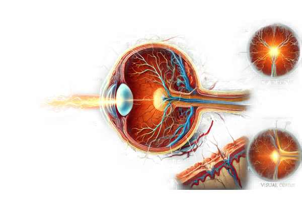
The optic nerve is a crucial part of the visual system. It consists of over a million nerve fibers that carry visual information from the retina to the brain. The retina captures light and converts it into electrical signals, which the optic nerve transports to the visual cortex for processing. Any damage to the optic nerve disrupts the signal transmission, resulting in visual impairment.
Types of Optic Atrophy
Depending on the underlying cause, there are several types of optic atrophy:
- Primary Optic Atrophy: This type develops without any previous inflammation or swelling of the optic nerve. It is frequently associated with glaucoma and hereditary optic neuropathies.
- Secondary Optic Atrophy: This occurs after an episode of optic neuritis or papilledema. It is frequently associated with inflammatory or compressive conditions that cause swelling in the optic nerve.
- Consecutive Optic Atrophy: This occurs as a result of retinal diseases, such as retinitis pigmentosa, in which the primary damage is to the retina, which then affects the optic nerve.
- Glaucomatous Optic Atrophy: Glaucoma-specific, this type is caused by increased intraocular pressure, which gradually damages the optic nerve.
Causes of Optical Atrophy
Optic atrophy has a variety of causes, which can be classified as hereditary or acquired.
Hereditary Causes
- Leber’s Hereditary Optic Neuropathy (LHON): A genetic disorder that causes sudden vision loss, usually in young males. Mutations in mitochondrial DNA are what cause it.
- Dominant Optic Atrophy (DOA) is an autosomal dominant condition that causes gradual vision loss in childhood. It is frequently associated with mutations in the OPA1 gene.
Acquired Causes
- Glaucoma: Chronic high intraocular pressure causes progressive optic nerve damage and atrophy.
- Optic Neuritis: Optic nerve inflammation, which is frequently associated with multiple sclerosis, can cause optic atrophy if it is severe or recurring.
- Ischemic Optic Neuropathy: Optic atrophy can occur when the optic nerve receives insufficient blood supply, which is frequently caused by conditions such as giant cell arteritis or atherosclerosis.
- Trauma: Direct injury to the optic nerve from head trauma or surgical procedures can cause optic atrophy.
- Toxins and Nutritional Deficiencies: Exposure to certain toxins (e.g., methanol) or nutrient deficiencies, such as vitamin B12, can harm the optic nerve.
- Tumors: Intracranial or intraorbital tumors that compress the optic nerve can cause optic atrophy.
- Infections: Serious infections, such as meningitis or encephalitis, can spread to the optic nerve and cause damage.
Symptoms of Optical Atrophy
Symptoms of optic atrophy can vary depending on the extent and location of the optic nerve damage.
- Vision Loss: The main symptom is a progressive or sudden loss of vision. This loss may be partial or complete, affecting one or both eyes.
- Color Vision Deficiency: Patients frequently report a decrease in color vision, which causes colors to appear washed out or less vivid.
- Visual Field Defects: It is common to lose peripheral vision or have blind spots in the visual field.
- Decreased Visual Acuity Patients frequently report blurred vision and difficulty focusing.
- Pupil Abnormalities: The affected eye may have an abnormal pupil response to light.
Pathophysiology of Optic Atrophy.
Optic atrophy occurs when optic nerve fibers degenerate. This degeneration can occur due to a number of mechanisms, including
- Axonal Degeneration: Damage to the axons of the optic nerve disrupts the transmission of visual signals. This could be the result of direct injury, ischemia, or demyelinating diseases.
- Glial Cell Proliferation: After nerve fiber damage, glial cells proliferate and form scar tissue, which further impairs nerve function.
- Mitochondrial Dysfunction: In hereditary optic atrophies, mutations affecting mitochondrial function cause energy deficits in optic nerve cells, resulting in degeneration.
Differential Diagnosis
Several conditions can mimic optic atrophy, requiring a careful differential diagnosis:
- Optic Neuritis: Optic nerve inflammation causes acute vision loss and pain, but if it occurs repeatedly, it can progress to optic atrophy.
- Anterior Ischemic Optic Neuropathy (AION): Sudden vision loss caused by impaired blood flow to the optic nerve, which is frequently associated with systemic vascular diseases.
- Papilledema: Swelling of the optic disc due to increased intracranial pressure can cause visual disturbances, but it is not the same as optic atrophy.
- Retinal Diseases: Conditions such as retinitis pigmentosa primarily affect the retina, but can also cause optic atrophy.
Prognosis
The underlying cause and the extent of nerve damage determine the prognosis of optic atrophy. In some cases, early intervention can halt progression and save remaining vision, whereas in others, vision loss may be permanent.
Methods to Diagnose Optic Atrophy
Optic atrophy diagnosis requires a multifaceted approach that combines clinical evaluation, advanced imaging, and laboratory tests to accurately identify the underlying cause and assess the extent of optic nerve damage.
Clinical Evaluation
- Patient History: A detailed patient history is required to identify potential risk factors and underlying conditions. This includes questions about vision changes, the onset and progression of symptoms, family history of ocular diseases, and any history of systemic diseases or trauma.
- Visual Acuity Testing: This standard eye test assesses the clarity of vision and helps determine the extent of vision loss.
- Pupil Examination: Examining for abnormal pupillary responses, such as a relative afferent pupillary defect (RAPD), can reveal optic nerve dysfunction.
Imaging Studies
- Ophthalmoscopy: A direct examination of the optic disc with an ophthalmoscope is the primary diagnostic tool. Optic atrophy manifests as pallor of the optic disc, with possible changes to the disc margins and blood vessels.
- Optical Coherence Tomography (OCT): OCT can produce high-resolution images of the optic nerve head and retinal nerve fiber layer. It aids in quantifying nerve fiber loss and determining the structural integrity of the optic nerves.
- Magnetic Resonance Imaging (MRI): MRI of the brain and orbits is critical for detecting compressive lesions, demyelinating diseases, and other intracranial pathologies that can lead to optic atrophy. Contrast-enhanced MRI can reveal inflammatory or neoplastic processes involving the optic nerve.
Lab Tests
- Blood Tests: A thorough blood examination can help identify systemic causes of optic atrophy, such as infections, nutritional deficiencies (e.g., vitamin B12), autoimmune disorders, and inflammatory markers.
- Cerebrospinal Fluid (CSF) Analysis: When inflammatory or infectious causes are suspected, CSF analysis can provide useful diagnostic information. Elevated protein levels or the presence of specific antibodies may indicate a condition such as multiple sclerosis or neurosyphilis.
Functional Testing
- Visual Field Testing: Automated perimetry maps the visual field and detects defects like scotomas or peripheral vision loss. This helps to determine the functional impact of optic nerve damage.
- Electrophysiological Tests: Visual evoked potentials (VEP) are tests that measure electrical activity in the brain in response to visual stimulation. Abnormal VEP results can indicate optic nerve dysfunction and aid in determining the location of damage.
Optical Atrophy Treatment
Treatment for optic atrophy focuses on addressing the underlying cause to prevent further damage while also managing symptoms to improve quality of life. While optic nerve damage is often irreversible, early treatment can help preserve remaining vision and prevent progression.
Addressing Underlying Causes
- Glaucoma: Lowering intraocular pressure is critical in treating glaucoma-induced optic atrophy. Prostaglandin analogs, beta-blockers, and carbonic anhydrase inhibitors can all help with this. Advanced cases may necessitate surgical interventions such as trabeculectomy or laser therapy.
- Inflammatory Conditions: Conditions such as optic neuritis or multiple sclerosis may necessitate corticosteroids or immunomodulatory treatments to reduce inflammation and prevent further damage to the optic nerve.
- Ischemic Optic Neuropathy: Managing underlying vascular conditions like hypertension, diabetes, and hyperlipidemia is critical. Antiplatelet therapy or anticoagulants may be used to improve blood flow and prevent future ischemic episodes.
- Nutritional Deficits: Vitamin B12 deficiency can cause optic atrophy. Vitamin B12 supplementation can slow or even reverse the progression of vision loss.
- Toxins and Infections: Eliminating toxins like methanol and treating infections quickly with appropriate antibiotics or antiviral medications can help prevent further optic nerve damage.
Symptom Management
- Visual Aids: Low vision aids like magnifying glasses, telescopic lenses, and electronic devices can help patients make the most of their remaining vision. Vision rehabilitation programs can also provide training on how to use these aids effectively.
- Occupational Therapy: Occupational therapists can assist patients in adjusting to vision loss by teaching strategies for daily living activities and suggesting changes to the home environment.
Innovative and Emerging Therapies
- Neuroprotective Agents: Researchers are looking into neuroprotective drugs that can protect optic nerve cells from damage. Agents such as citicoline and brimonidine are being investigated for their ability to slow the progression of optic atrophy.
- Stem Cell Therapy: Experimental treatments utilizing stem cells seek to regenerate damaged optic nerve fibers. While still in its early stages, this method shows promise for restoring vision in patients with optic atrophy.
- Gene Therapy: Gene therapy is being investigated as a method of correcting genetic mutations that cause hereditary optic neuropathy. Early trials have demonstrated potential for treating conditions such as Leber’s Hereditary Optic Neuropathy (LHON).
- Electrical Stimulation: Techniques such as transcorneal electrical stimulation (TES) and transorbital alternating current stimulation (tACS) are under investigation for their ability to stimulate optic nerve cells and improve visual function.
- Antioxidant Therapy: Researchers are investigating the potential of antioxidants such as alpha-lipoic acid and coenzyme Q10 to reduce oxidative stress and protect optic nerve cells from damage.
Monitoring and Follow-up
Regular follow-up with an ophthalmologist or neurologist is essential for monitoring the condition and adjusting treatment as necessary. Visual field testing, OCT imaging, and other assessments aid in monitoring disease progression and intervention efficacy.
Effective Methods for Improving and Avoiding Optic Atrophy
- Regular Eye Examination:
- Schedule regular eye exams to detect early signs of optic nerve damage and treat underlying conditions promptly.
- Control glaucoma:
- Control intraocular pressure with medications or surgery to avoid glaucomatous optic atrophy.
- Maintain Vascular Health
- To avoid ischemic optic neuropathy, keep risk factors like hypertension, diabetes, and hyperlipidemia under control.
- Avoid Toxicants:
- When working with chemicals, limit your exposure to toxins such as methanol and ensure adequate ventilation.
- Nutritional support:
- Maintain an adequate intake of essential nutrients, particularly vitamin B12, to avoid nutritional optic neuropathy.
- Protect From Infections:
- Treat infections affecting the optic nerve, such as meningitis or encephalitis, as soon as possible with appropriate medications.
- Use Protective Eyewear.
- To avoid traumatic optic neuropathy, wear protective eyewear during activities that are likely to cause eye injury.
- Educate and Increase Awareness:
- Raise awareness of optic atrophy and its risk factors through educational campaigns and support groups.
- Early intervention:*
- Seek immediate medical attention if you are experiencing vision loss to ensure early diagnosis and treatment of any potential causes of optic atrophy.
Books
- “Clinical Neuro-Ophthalmology: A Practical Guide” by Ambar Chakravarty
- “Walsh & Hoyt’s Clinical Neuro-Ophthalmology” by Neil R. Miller and Nancy J. Newman
- “Optic Nerve Disorders: Diagnosis and Management” by Jane W. Chan


