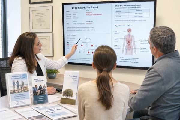
A TP53 genetic test looks for an inherited pathogenic variant associated with Li-Fraumeni syndrome and related heritable TP53 cancer-predisposition syndromes. TP53 is a major tumor-suppressor gene, and constitutional loss of one copy can lead to a very high lifetime risk of cancer, often beginning in childhood or young adulthood. The tumor spectrum includes soft-tissue and bone sarcomas, premenopausal breast cancer, brain tumors, adrenocortical carcinoma, leukemia, and many other cancers. Some carriers develop more than one primary cancer.
Testing usually uses blood or saliva, but interpretation can be unusually complex. A TP53 variant detected at a low level in blood may come from clonal hematopoiesis rather than an inherited syndrome. Mosaicism is another possibility. Confirmation with a non-blood tissue may be needed before labeling the result germline. A true positive result supports intensive lifelong surveillance, often including annual whole-body MRI and brain MRI, while minimizing unnecessary ionizing radiation. A negative or uncertain result must be considered alongside the cancer pattern, tumor findings, family history, and the test’s technical limits.
- A confirmed germline TP53 pathogenic variant can indicate very high, lifelong risk for multiple primary cancers.
- A TP53 variant in blood is not always inherited; clonal hematopoiesis and mosaicism must be considered.
- Surveillance commonly uses frequent clinical review plus annual whole-body and brain MRI, with age-specific organ screening.
- A VUS should not be used alone to diagnose Li-Fraumeni syndrome or recommend preventive surgery.
- Each child of a person with a constitutional TP53 pathogenic variant usually has a 50% chance of inheriting it.
Table of Contents
- What TP53 Does
- Who May Need TP53 Testing
- How Testing Is Performed
- Understanding TP53 Results
- Germline, Mosaic, and Blood-Clone Findings
- Cancer Risks and Surveillance
- Treatment, Family Testing, and Pregnancy
- Limitations and Next Steps
What TP53 Does
TP53 encodes the p53 protein, sometimes called the guardian of the genome. When DNA is damaged or a cell is under stress, p53 can pause cell division, help repair damage, trigger senescence, or direct the cell to die. These actions reduce the chance that a damaged cell becomes malignant.
A person with a germline pathogenic variant usually has one altered TP53 copy in every cell. Cancer can arise after the remaining working copy is lost or other oncogenic changes accumulate. Because p53 protects many tissues, the resulting cancer spectrum is broad and can span the entire lifespan.
Li-Fraumeni syndrome was originally recognized through families with childhood sarcomas and multiple early cancers. Modern testing shows that heritable TP53-related disease is more diverse. Some variants confer classic very high risk, while others are associated with later onset or a narrower pattern. Even with an attenuated variant, management should be based on expert variant interpretation, personal history, and current guidance rather than assumptions from one relative.
The syndrome is not defined by a single cancer. Core tumors include soft-tissue sarcoma, osteosarcoma, premenopausal breast cancer, brain tumors, and adrenocortical carcinoma. Leukemia, lung cancer, gastrointestinal cancers, melanoma, prostate cancer, and other malignancies also occur. A carrier may develop multiple independent primary tumors rather than one cancer repeatedly spreading.
TP53 mutations are also extremely common as acquired changes within tumors. A TP53 mutation on somatic tumor testing usually reflects the cancer’s biology and does not automatically establish Li-Fraumeni syndrome. Germline confirmation is guided by age, cancer type, variant allele fraction, family history, and whether the exact alteration is known to occur constitutionally.
The distinction matters because a true germline result affects lifelong surveillance and relatives, while a tumor-only result generally does not. It also matters for treatment planning: people with Li-Fraumeni syndrome may be more susceptible to radiation-associated second cancers, so clinicians try to avoid unnecessary therapeutic and diagnostic radiation without compromising cancer control.
Who May Need TP53 Testing
Testing is considered when a personal or family cancer pattern meets established criteria such as the modified Chompret criteria, when a highly suggestive tumor occurs, or when a broader hereditary cancer panel identifies TP53. Criteria evolve, and genetics professionals apply them using exact ages, cancer types, and family relationships.
Situations that commonly prompt testing include:
- A tumor in the classic Li-Fraumeni spectrum before age 46 plus an early cancer in a close relative or multiple primary tumors.
- Multiple primary cancers in one person, including at least two core Li-Fraumeni tumors, with the first occurring young.
- Adrenocortical carcinoma, choroid plexus carcinoma, or embryonal anaplastic subtype rhabdomyosarcoma, even without family history.
- Very early breast cancer, particularly before age 31.
- Childhood osteosarcoma or soft-tissue sarcoma with a suggestive family pattern.
- A family with cancers across several generations at unusually young ages.
- A known constitutional TP53 pathogenic variant in a relative.
- A possible germline TP53 finding from tumor sequencing.
A negative family history does not exclude the syndrome. A variant may be de novo, relatives may be young, family size may be small, or medical information may be missing. Some families show reduced penetrance or an attenuated phenotype.
Testing may be ordered as a single-gene assay when suspicion is high or as part of a hereditary cancer panel. Panels can be useful when breast cancer, sarcoma, brain tumors, and other cancers overlap several syndromes. They also increase the chance of uncertain and unexpected findings.
Testing a person who currently has cancer can influence surveillance, surgical planning, radiation discussions, and family care. Testing a healthy relative is most informative after the familial pathogenic variant has been established. Predictive testing in children is appropriate because childhood surveillance can find tumors earlier.
Before testing, gather pathology reports, ages at diagnosis, treatment records, and genetic reports from relatives. “Bone cancer” may mean osteosarcoma, metastatic carcinoma, or another diagnosis; the distinction affects criteria. Similarly, a new breast cancer in a prior breast-cancer survivor may be a second primary or recurrence.
A genetics team should also discuss the possibility that a blood TP53 finding is not constitutional. Older age, prior chemotherapy or radiation, and a low variant allele fraction increase concern for clonal hematopoiesis, although none of these features alone is definitive.
How Testing Is Performed
Initial germline testing commonly uses blood or saliva. The laboratory sequences TP53 and assesses deletions or duplications. Some panels also evaluate promoter or structural changes. Results often take two to six weeks.
The testing process may involve several stages:
- Sequence and copy-number analysis of blood or saliva.
- Review of variant allele fraction and quality metrics.
- Comparison with tumor results, if available.
- Testing of relatives to assess segregation and parent of origin.
- Confirmation in nonhematopoietic tissue when clonal hematopoiesis or mosaicism is possible.
Saliva and buccal swabs contain white blood cells, so they are not fully independent of blood. They may reproduce a clonal hematopoiesis variant. Cultured skin fibroblasts from a punch biopsy are often used as a constitutional source because the culture reduces blood-cell contamination. Eyebrow follicles, nails, or other tissues may be used by specialized laboratories, but validation and contamination risks vary.
A result near 50% variant allele fraction is compatible with a heterozygous germline variant but can also occur when a blood clone is large. A low fraction may indicate mosaicism, clonal hematopoiesis, circulating tumor DNA, recent transfusion, transplant donor cells, or technical artifact. The percentage is a clue, not proof of origin.
People who have received an allogeneic stem-cell transplant require special planning because their blood DNA comes largely from the donor. A skin-fibroblast sample or stored pretransplant material may be needed to assess the patient’s germline.
Tumor testing can provide supportive evidence. The same TP53 variant in more than one independent tumor may support constitutional or mosaic origin. A tumor may also show loss of the working allele. However, TP53 is so commonly mutated in sporadic tumors that tumor presence alone is not enough.
The laboratory should use TP53-specific classification standards when available. Some variants retain partial function or have population-specific evidence that changes their risk interpretation. Expert review is particularly important before intensive surveillance or risk-reducing surgery.
Understanding TP53 Results
A clinical report generally classifies variants as pathogenic, likely pathogenic, uncertain significance, likely benign, or benign. For TP53, the report should also state the observed allele fraction when relevant and note concern for mosaicism or clonal hematopoiesis.
Pathogenic or likely pathogenic
A confirmed constitutional pathogenic or likely pathogenic variant supports a diagnosis of Li-Fraumeni syndrome or a heritable TP53-related cancer syndrome. The person should enter a specialized surveillance program and relatives should be offered targeted testing.
The word “pathogenic” describes the variant, not a guarantee that cancer is present today. It also does not define the exact age or cancer type that will occur. Variant-specific and family data may refine risk, but no result gives certainty.
A positive result found only in blood is not fully confirmed as constitutional when the clinical context suggests a blood clone. Origin should be resolved before relatives are tested or lifelong screening is assigned.
Negative
A negative targeted test for a known familial variant is usually a true negative. The person did not inherit that familial TP53 risk and generally does not need Li-Fraumeni surveillance.
A negative full-gene test in someone with a strong cancer pattern is less definitive. The family may have another syndrome, an undetected TP53 mechanism, mosaicism, or a combination of sporadic cancers. Management can still be influenced by personal and family history.
A tumor that lacks the familial variant does not necessarily rule out cancer in a carrier; tumors can lose the variant-containing region or have technical limitations. Germline status should be determined from an appropriate normal tissue.
Variant of uncertain significance
A VUS should not be used alone to diagnose Li-Fraumeni syndrome, start intensive whole-body MRI in healthy relatives, recommend mastectomy, or alter radiation treatment. Care should follow the clinical history while the variant remains uncertain.
TP53 VUS interpretation can use functional assays, tumor characteristics, population data, computational evidence, and segregation. Functional abnormality alone may not be enough if other evidence conflicts. Reclassification should come from a qualified laboratory or expert panel.
The principles in a genetic variant classification are especially important because the consequences of overcalling TP53 are substantial.
Benign or likely benign
These variants do not explain a hereditary cancer syndrome and do not justify TP53-specific surveillance.
Germline, Mosaic, and Blood-Clone Findings
Three biological explanations can produce a pathogenic TP53 variant in blood.
Constitutional germline means the variant was present from conception and is expected in most tissues. It can be transmitted to children and supports full syndrome management.
Post-zygotic mosaicism means the variant arose during early development and is present in a subset of tissues. Cancer risk and transmission depend on which tissues carry it. A low fraction in blood with detection in another non-neoplastic tissue supports mosaicism.
Clonal hematopoiesis means the variant arose in a blood-forming stem cell later in life and expanded. It may be associated with age, smoking, prior chemotherapy, or radiation. It generally does not mean the person has Li-Fraumeni syndrome, though it can carry hematologic risk.
Distinguishing them can require a stepwise evaluation:
- Review the allele fraction and sequencing quality.
- Ask about prior cancer therapy and hematologic abnormalities.
- Check whether the variant appears in tumor tissue.
- Test cultured skin fibroblasts or another validated non-blood tissue.
- Test parents or children when appropriate and with counseling.
- Review blood counts and consider hematology input for a suspected clone.
A variant detected in a child with a classic core tumor is more likely to be constitutional than a low-level variant detected in an older adult after chemotherapy, but these are probabilities, not rules. Confirmation prevents two serious errors: missing hereditary risk in a mosaic carrier and incorrectly labeling an entire family with Li-Fraumeni syndrome because of a blood-limited clone.
Circulating tumor DNA can occasionally contribute a TP53 variant to blood-based testing in a person with active metastatic cancer. Germline laboratories use methods designed for inherited testing, but clinical context still matters.
When origin remains uncertain, the report and care plan should state that clearly. Temporary surveillance may be individualized while confirmatory studies are completed, especially when the personal cancer pattern is highly suggestive.
Cancer Risks and Surveillance
Li-Fraumeni syndrome carries a high risk of early and multiple cancers, but risk varies by sex, variant, family, and prior treatment. Women have particularly high breast-cancer risk. Childhood risk includes sarcomas, brain tumors, adrenocortical carcinoma, and leukemia. Adult risk remains broad.
Modern surveillance programs aim to detect tumors at an earlier, more treatable stage while limiting radiation exposure. Exact schedules differ slightly among guidelines and should be personalized at an experienced center.
| Surveillance area | Common approach | Purpose |
|---|---|---|
| Whole body | Annual whole-body MRI | Screen for sarcomas and internal tumors without ionizing radiation |
| Brain | Annual brain MRI, sometimes coordinated with whole-body imaging | Detect primary brain tumors |
| Children | Frequent physical examination and abdominal/pelvic ultrasound with or without hormone tests, depending on protocol | Screen for adrenocortical carcinoma and other childhood tumors |
| Breast | Early annual breast MRI, clinical examination, and individualized mammography or risk-reducing mastectomy discussion | Address very high premenopausal breast-cancer risk while limiting radiation |
| Gastrointestinal tract | Periodic upper endoscopy and colonoscopy beginning earlier than population screening | Detect colorectal and upper gastrointestinal cancers |
| Skin | Regular skin examination | Assess melanoma and other changing lesions |
Surveillance begins early for children with variants associated with childhood cancers. Clinical examinations may occur every three to four months in young children under some protocols. Adult examinations are often every six months. The specialist program should provide the exact schedule.
Whole-body MRI can detect benign lesions and uncertain findings as well as cancer. Follow-up tests, anxiety, and occasional biopsies are expected tradeoffs. Consistent imaging protocols and expert radiologists reduce unnecessary variation.
Radiation should be minimized when a suitable nonionizing alternative exists, but it is not absolutely prohibited. CT, nuclear medicine, or radiation therapy may be necessary for accurate staging or cure. Decisions compare the immediate benefit against the long-term second-cancer risk. Avoiding effective cancer treatment solely because of TP53 can also cause harm.
New symptoms should be evaluated promptly rather than waiting for the next screening visit. Persistent bone pain, a growing mass, neurologic change, severe headache, unexplained weight loss, abdominal swelling, endocrine changes, bleeding, or marked fatigue deserves assessment.
Treatment, Family Testing, and Pregnancy
Cancer treatment follows the tumor type and stage, with additional attention to second-cancer risk. Surgery is preferred when it offers cure. Radiation exposure is reduced or avoided when an equally effective alternative exists, but treatment must remain oncologically sound. Chemotherapy choices may also consider the risk of therapy-related malignancy.
Women with a confirmed germline TP53 variant may discuss bilateral risk-reducing mastectomy because screening does not prevent breast cancer and repeated treatment can be complex. This is a preference-sensitive decision. Breast MRI is a surveillance option, and the plan should consider age, prior cancer, reconstruction, family experience, and personal values.
TP53-related susceptibility is autosomal dominant. Each child of a constitutional carrier has a 50% chance of inheriting the variant. Testing minors is appropriate because childhood surveillance changes care. Targeted family variant testing should use the exact confirmed germline result, not an unverified blood-clone finding.
If neither parent carries the variant in appropriate tissue, it may be de novo. Sibling risk is then low but not zero because parental mosaicism is possible. The affected person’s own children remain at 50% risk if the variant is constitutional or involves reproductive cells.
Reproductive options include natural conception, prenatal diagnosis, and in vitro fertilization with preimplantation genetic testing for monogenic disease. Testing shows whether the familial variant is present but cannot predict which cancers, if any, will occur. Pregnancy planning should also include a review of breast screening and any prior cancer treatment.
Pregnant carriers should report new symptoms promptly. Imaging choices can be adapted, and obstetric care should coordinate with oncology and genetics. Whole-body MRI protocols during pregnancy require specialist judgment, particularly regarding contrast.
Family communication can be emotionally difficult because a result affects children and relatives across generations. A genetics clinic can provide a family letter and age-specific surveillance resources.
Limitations and Next Steps
TP53 testing is limited by variant interpretation, mosaicism, blood-limited clones, and technical coverage. A result that appears simple on the first page may require tissue confirmation and expert review. Older reports may also benefit from reinterpretation under current TP53-specific criteria.
Risk estimates are not identical for every variant. Some pathogenic variants have attenuated penetrance, and surveillance may be refined over time. However, reducing surveillance without expert guidance can miss disease, while applying an intensive classic protocol to an unconfirmed blood clone can cause unnecessary harm.
After a pathogenic result, useful steps are:
- Confirm whether the variant is constitutional, mosaic, or blood limited.
- Obtain the complete report and expert genetic counseling.
- Enroll in a Li-Fraumeni surveillance program with a written schedule.
- Review prior radiation and future imaging choices without delaying necessary treatment.
- Discuss breast risk management when relevant.
- Offer targeted testing to relatives only after origin is established.
- Keep all pathology, imaging, treatment, and genetic records together.
- Seek prompt evaluation for new, persistent symptoms.
A coordinated team may include genetics, pediatric and adult oncology, primary care, radiology, breast specialists, gastroenterology, dermatology, endocrinology, and psychosocial care. One program should track the surveillance calendar.
Common mistakes include assuming any tumor TP53 mutation is inherited, using saliva as definitive confirmation of a blood clone, treating a VUS as Li-Fraumeni syndrome, and avoiding all radiation even when it is essential for cure. Careful source-of-variant analysis and balanced cancer management are central to a trustworthy result.
References
- Li-Fraumeni Syndrome 2025 (Review)
- Update on Cancer Screening Recommendations for Individuals with Li-Fraumeni Syndrome 2025 (Consensus Recommendations)
- Medical guidelines for Li–Fraumeni syndrome 2019, version 1.1 2021 (Guideline)
- Overview of the Clinical Features of Li-Fraumeni Syndrome and the Current European ERN GENTURIS Guideline 2021 (Review)
- TP53 c.455C>T p.(Pro152Leu) pathogenic variant is a lower risk allele with attenuated risks of breast cancer and sarcoma 2023 (Cohort Study)
- Guidelines for the Li–Fraumeni and heritable TP53-related cancer syndromes 2020 (Guideline)
Disclaimer
TP53 results require specialist review because a blood finding may represent germline disease, mosaicism, circulating tumor DNA, or clonal hematopoiesis. This article is educational and does not replace individualized cancer surveillance, treatment planning, or urgent evaluation of new symptoms.