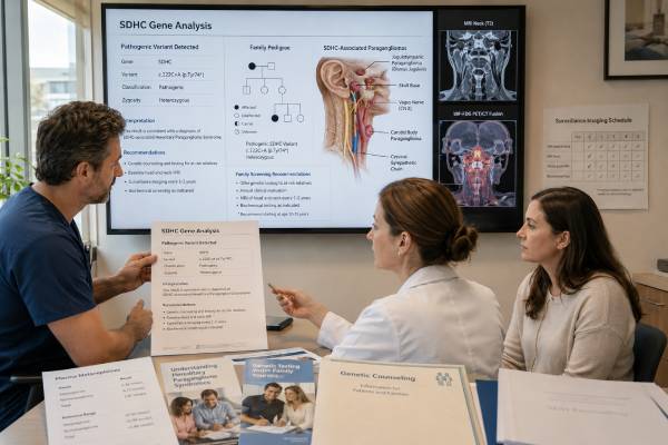
An SDHC genetic test looks for an inherited pathogenic variant that increases susceptibility to paraganglioma and, less often, pheochromocytoma. SDHC is part of the mitochondrial succinate dehydrogenase complex. When the complex loses function, altered cell metabolism can promote tumor growth. SDHC-associated tumors are often found in the head and neck, where they may cause a neck mass, pulsatile tinnitus, hearing change, hoarseness, or swallowing difficulty rather than hormone-related symptoms.
Testing usually uses blood or saliva and should assess both small sequence changes and larger deletions or duplications. A positive germline result supports lifelong surveillance even when the person feels well, because some tumors are silent. It also allows relatives to have focused testing for the exact familial variant. A negative result may be reassuring when it excludes a known family variant, but it does not rule out every hereditary PPGL syndrome in an affected person. A variant of uncertain significance is not a diagnosis and should not direct surgery or predictive testing by itself.
- A pathogenic SDHC result confirms hereditary susceptibility to paraganglioma-pheochromocytoma syndrome.
- SDHC is most often linked to head and neck paragangliomas, although tumors can occur elsewhere.
- Many head and neck tumors do not secrete catecholamines, so normal metanephrines do not replace imaging.
- A negative targeted family test usually means the person did not inherit the familial SDHC risk.
- A VUS should be managed using clinical findings until the laboratory has enough evidence to reclassify it.
Table of Contents
- SDHC and Tumor Development
- Reasons for SDHC Testing
- Testing Methods and Samples
- How to Read SDHC Results
- Associated Tumors and Symptoms
- Surveillance for Carriers
- Inheritance and Family Testing
- Limitations and Care Planning
SDHC and Tumor Development
SDHC encodes a membrane-anchoring subunit of succinate dehydrogenase, also called mitochondrial complex II. This enzyme participates in the citric acid cycle and the electron transport chain. When SDH function is lost, succinate accumulates and disrupts oxygen-sensing and epigenetic enzymes. Cells can behave as though oxygen is scarce even when it is available, a state called pseudohypoxia. That altered signaling can support tumor formation.
A person born with one nonworking SDHC copy still has one functional copy in most cells. A susceptible cell may become a tumor after the working copy is lost or silenced. This explains incomplete penetrance: inheriting a pathogenic variant raises risk, but not every carrier develops a recognized tumor.
SDHC belongs to a family of PPGL susceptibility genes that includes SDHA, SDHB, SDHD, and SDHAF2. All can destabilize the SDH complex, yet their average clinical patterns differ. SDHC variants are uncommon and are most strongly associated with parasympathetic paragangliomas of the head and neck. Thoracic, abdominal, pelvic, or adrenal tumors can occur, so surveillance cannot focus only on the neck.
A paraganglioma develops from paraganglial cells outside the adrenal gland. A pheochromocytoma is the related tumor within the adrenal medulla. Head and neck paragangliomas often arise near the carotid body, jugular bulb, vagus nerve, or middle ear. They are usually highly vascular and may grow slowly, but their location can threaten hearing, swallowing, speech, cranial nerves, or major blood vessels.
Most SDHC findings discussed in hereditary care are germline. A tumor can also show SDH deficiency without an inherited SDHC variant. Tumor immunohistochemistry that shows loss of SDHB protein is a clue that the SDH complex is disrupted, but it cannot identify SDHC specifically. Confirmation requires germline testing and sometimes tumor sequencing.
The genetic result does not classify a known tumor as harmless or metastatic. Metastatic disease is diagnosed when tumor appears in sites that normally lack paraganglial tissue, such as bone, liver, lung, or distant lymph nodes. SDHC-associated metastatic disease appears less common than with SDHB, but it is possible. Tumor size, location, growth, secretion, imaging, and long-term behavior remain important.
Reasons for SDHC Testing
Genetic evaluation is now considered for most people diagnosed with pheochromocytoma or paraganglioma because hereditary causes are common enough that age and family history alone miss carriers. A multigene panel is usually preferred for an affected person because several genes can cause similar tumors. Targeted SDHC testing is most efficient when a relative already has a confirmed SDHC variant.
Findings that may bring SDHC into focus include:
- A head and neck paraganglioma, especially at a younger age.
- Multiple paragangliomas or a tumor on both sides.
- A family history of head and neck masses, glomus tumors, pheochromocytoma, or paraganglioma.
- Loss of SDHB staining in tumor cells with retained SDHA staining.
- An SDH-deficient gastrointestinal stromal tumor.
- A thoracic or other extra-adrenal paraganglioma without another identified cause.
- A previously reported SDHC pathogenic variant in the family.
An apparently isolated head and neck tumor can still be hereditary. Family history may look negative because relatives were never imaged, a tumor was called by a different name, or a carrier remained unaffected. Small kindreds and adoption can also hide inheritance.
Head and neck paragangliomas may be described as carotid body tumors, glomus jugulare tumors, glomus tympanicum tumors, or vagal paragangliomas. These labels are important when collecting records. A relative may remember “ear surgery” or a “vascular neck tumor” rather than the word paraganglioma.
Tumor pathology can guide testing. SDHB immunohistochemistry uses the SDHB protein as a marker of the whole complex. Loss of granular cytoplasmic staining in tumor cells, with intact internal controls, suggests an SDH-deficient tumor. Because SDHC, SDHD, and SDHB defects can all produce this pattern, a panel rather than SDHC-only testing is often appropriate.
Testing healthy relatives is different from testing an affected person. Once the familial variant is known, a focused familial variant test can determine who needs surveillance. It is less expensive and easier to interpret than repeating a broad panel in every relative.
A genetics consultation can also identify whether another syndrome fits better. For example, VHL, RET, NF1, MAX, TMEM127, FH, and other genes can cause PPGL. The tumor’s location, catecholamine pattern, pathology, and associated features help select and interpret the test.
Testing Methods and Samples
Germline SDHC testing usually uses a blood sample or saliva kit. The sample tests DNA present throughout the body, unlike a tumor-only assay. No fasting or medication changes are required for the DNA collection.
A complete assay should include:
- Sequence analysis, which detects most single-letter variants and small insertions or deletions.
- Deletion-duplication analysis, which detects larger missing or extra sections that sequencing may not find.
- Validated splice assessment, when a variant near an exon boundary may alter RNA processing.
- A multigene panel, when the affected person’s presentation could be caused by several PPGL genes.
The report should state the transcript used, the regions analyzed, the method for copy-number changes, and any limitations. Older tests sometimes examined only a small set of variants or omitted deletion analysis. A convincing phenotype may justify updated testing with a modern panel.
Testing often takes two to six weeks. Targeted familial testing may be faster. Results can be delayed by insurance authorization, poor saliva quality, or the need for confirmation and family studies.
Tumor testing can complement germline testing. Useful tumor studies include SDHB and SDHA immunohistochemistry, loss-of-heterozygosity analysis, sequencing, and methylation or metabolite studies in specialized settings. A tumor result can strengthen evidence for an uncertain germline variant, but this work should be coordinated with the laboratory because not every tumor observation proves causality.
A finding on tumor genomic testing must be classified by origin. If the tumor shows an SDHC variant at an allele fraction compatible with germline origin, blood or another normal sample is needed. The same variant can be somatic, germline, or mosaic; a tumor report alone cannot establish family risk.
When blood testing is negative but multiple clinical signs remain, the team may review the raw laboratory coverage, order a broader panel, test stored tumor, or consider mosaicism. Testing another tissue can help when mosaicism is suspected. The next test should address a defined limitation rather than simply repeat the same assay.
How to Read SDHC Results
Laboratories classify variants using several evidence categories. Pathogenic and likely pathogenic findings can guide clinical care. Benign and likely benign findings do not explain the syndrome. A VUS remains unresolved.
Pathogenic or likely pathogenic
A positive germline result confirms SDHC-related hereditary PPGL susceptibility. The person should receive a baseline evaluation even if no symptoms are present. Lifelong surveillance is recommended because tumors can be silent and age of onset varies.
“Likely pathogenic” is generally managed like pathogenic because the probability of disease causation is high. The report should be saved exactly as issued. Relatives need the gene, DNA change, protein change when available, and laboratory classification for targeted testing.
A positive result does not mean cancer is currently present. Many paragangliomas are not malignant, and some carriers never develop a tumor. The result also does not predict the number, location, or growth rate of future tumors.
Negative
A negative result is most definitive when the laboratory looked specifically for a known familial SDHC variant. A relative who does not carry that variant is generally released from SDHC-specific surveillance.
For a person who already has a paraganglioma, a negative panel means no reportable pathogenic variant was found. It does not prove the tumor is nonhereditary. The cause could be another gene, mosaicism, a variant outside the tested regions, an epigenetic mechanism, or a change not yet recognized as disease-causing. Follow-up for the tumor itself remains necessary.
Variant of uncertain significance
A VUS is not a positive diagnosis. It should not be the sole reason for preventive surgery, intensified imaging of healthy relatives, or prenatal testing. Clinical management should follow the personal and family history until the variant is reclassified.
Some SDHC variants are rare because the condition itself is rare, so scarcity alone does not prove pathogenicity. Stronger evidence may include functional loss of SDH activity, segregation with disease, consistent tumor loss of the normal allele, characteristic immunostaining, and multiple unrelated affected carriers. The laboratory, not the patient or clinician alone, should integrate these data under current standards.
A person with a VUS should keep the report and ask how reclassification notices are handled. The concepts in a genetic variant result help explain why uncertain findings can change as new cases and functional evidence appear.
Mosaic or low-level result
A low allele fraction can indicate post-zygotic mosaicism, a technical artifact, or sample issues. Confirmation with a fresh sample or another tissue may be needed. Mosaicism can alter tumor risk and transmission risk, but the extent cannot be inferred from one blood percentage alone.
Associated Tumors and Symptoms
SDHC is most often associated with head and neck paragangliomas. These tumors may grow slowly and remain unnoticed for years. Their behavior is driven as much by anatomy as by growth rate: a modest tumor near the skull base can affect important cranial nerves.
Possible head and neck symptoms include:
- A painless, slowly enlarging neck lump.
- Pulsatile tinnitus, described as hearing the heartbeat in one ear.
- Hearing loss, ear fullness, or recurrent ear symptoms.
- Hoarseness, weak voice, cough with swallowing, or aspiration.
- Difficulty swallowing or tongue weakness.
- Dizziness, facial weakness, shoulder weakness, or other cranial-nerve changes.
Many head and neck tumors are biochemically silent because parasympathetic paraganglia usually do not produce large amounts of catecholamines. Normal plasma or urine metanephrines therefore cannot exclude them. Imaging remains essential for carrier surveillance.
Pheochromocytomas and sympathetic paragangliomas can release catecholamines. Symptoms may include episodic headache, sweating, palpitations, tremor, anxiety-like spells, pallor, high blood pressure, or glucose elevation. Some secretory tumors are discovered before symptoms through screening.
A catecholamine crisis can cause severe hypertension, arrhythmia, heart failure, stroke, or shock. A known carrier with intense headache, chest pain, fainting, new neurologic symptoms, or very high blood pressure needs urgent evaluation. Elective surgery or anesthesia should not proceed when a secretory PPGL is suspected without appropriate endocrine assessment.
SDH-deficient gastrointestinal stromal tumors have been reported in association with SDHx variants, including SDHC. These GISTs can differ from common KIT- or PDGFRA-mutant tumors and may arise in younger patients. Symptoms can include gastrointestinal bleeding, anemia, abdominal pain, early fullness, or an incidental mass. The absolute risk for an SDHC carrier is uncertain, so surveillance is usually driven by specialist protocols and symptoms rather than a single universal endoscopy schedule.
Metastatic PPGL is possible but appears less frequent with SDHC than with SDHB. No histologic feature can guarantee benign behavior. A diagnosed tumor requires long-term follow-up, especially if it is large, extra-adrenal, recurrent, incompletely removed, or shows concerning growth.
Surveillance for Carriers
Surveillance combines symptom review, biochemical screening, and imaging. The aim is to detect secretory tumors before a cardiovascular emergency and nonsecretory tumors before they damage nerves or become difficult to treat. Exact starting ages and intervals vary among expert groups and should be individualized by a hereditary PPGL program.
A baseline evaluation commonly includes:
- Medical history focused on headaches, palpitations, sweating, blood pressure, neck or ear symptoms, voice, swallowing, and family diagnoses.
- Physical examination and blood pressure measurement.
- Plasma free metanephrines or 24-hour urinary fractionated metanephrines.
- MRI of the head and neck, chest, abdomen, and pelvis.
- Functional imaging when a lesion is suspected, multifocal disease is present, or the full extent of disease must be staged.
After a normal baseline, many protocols use annual clinical review, periodic biochemical testing, and whole-body regional MRI about every two to three years. MRI reduces cumulative radiation in a lifelong program. Imaging may be more frequent when there is a prior tumor, a suspicious lesion, a strong family history, or symptoms.
Children in an affected family may begin surveillance before adulthood. The exact timing considers the gene, the youngest family diagnosis, the burden of repeated imaging, and the child’s ability to undergo MRI without sedation. Pediatric endocrinology and genetics teams can create a developmentally appropriate plan.
Metanephrine results require careful preparation and interpretation. Stress, strenuous exercise, illness, posture, caffeine, nicotine, and some medications can cause mild elevations. The clinician may repeat a borderline result under controlled conditions. Strong elevations or a consistent biochemical pattern prompt imaging rather than indefinite repeat testing.
Because head and neck tumors can be nonsecretory, new tinnitus, neck swelling, hoarseness, or swallowing problems should lead to evaluation even when recent metanephrines were normal. Likewise, an imaging abnormality should not be dismissed solely because biochemical tests are negative.
Management of a detected head and neck paraganglioma can include observation with serial imaging, surgery, radiation, or a combination. Choice depends on growth, symptoms, location, age, cranial-nerve function, multiplicity, secretion, and patient preference. Surgery can cure a localized tumor but may cause nerve deficits; observation can avoid immediate harm but requires reliable follow-up. A multidisciplinary head and neck team should review the tradeoffs.
Any planned operation requires biochemical evaluation for catecholamine secretion. A secretory tumor needs specialist preparation, commonly including alpha-adrenergic blockade and volume management, to reduce perioperative risk.
Inheritance and Family Testing
SDHC susceptibility follows autosomal dominant inheritance. A carrier has a 50% chance of passing the variant to each child. Men and women are equally able to inherit and transmit it. Unlike SDHD, SDHC does not have an established parent-of-origin effect that routinely changes clinical risk based on whether the variant came from the mother or father.
Once a familial pathogenic variant is identified, testing usually starts with parents, siblings, and children. The result can clarify which side of the family is at risk. More distant relatives on that branch can then be offered testing.
A positive relative enters surveillance. A negative relative for the exact familial variant generally does not need SDHC-specific imaging or biochemical screening. This targeted approach prevents both missed tumors in carriers and unnecessary lifelong testing in noncarriers.
If neither parent carries the variant in blood, the variant may have arisen de novo. Sibling risk is then low but not zero because parental gonadal mosaicism is possible. The affected person’s children still face a 50% transmission risk.
Testing minors can be ethically appropriate because the result may change childhood surveillance. Counseling should explain the limits clearly: a positive result means increased risk, not that a tumor is inevitable. Families may benefit from a written plan that states when testing, MRI, biochemical screening, and symptom evaluation begin.
Reproductive options include natural conception with or without prenatal diagnosis, in vitro fertilization with preimplantation genetic testing for monogenic disease, donor gametes, or adoption. Chorionic villus sampling and amniocentesis can determine whether a fetus inherited the variant but cannot predict whether disease will occur or how severe it would be.
A family letter should use the exact term SDHC pathogenic variant and list the laboratory result. Descriptions such as “neck tumor gene” are incomplete and may lead clinicians to overlook thoracic, abdominal, adrenal, or gastrointestinal manifestations.
Limitations and Care Planning
SDHC is rare, so penetrance and tumor-risk estimates are less precise than for more common syndromes. Published families may be selected because several members were affected, while population testing can identify carriers with milder histories. Percentages should be treated as group estimates rather than personal predictions.
Laboratory limitations include variants outside routinely analyzed regions, complex structural changes, low-level mosaicism, and incomplete knowledge about rare missense changes. A negative result from an older or narrow test may deserve review. A VUS may change classification as new families and tumor data become available.
Care after a positive result should include:
- Confirmation that the result is germline and classified as pathogenic or likely pathogenic.
- Baseline clinical, biochemical, and MRI assessment.
- A written surveillance calendar with the responsible specialty for each test.
- Clear instructions for new head and neck or catecholamine-related symptoms.
- Preoperative notification and biochemical assessment before anesthesia.
- Targeted testing for relatives.
- Periodic review of the variant and surveillance recommendations.
- Long-term follow-up after any tumor treatment.
Patients should keep copies of the genetic report, pathology, imaging, biochemical trends, and treatment records. A hereditary tumor clinic can coordinate endocrinology, genetics, radiology, head and neck surgery, nuclear medicine, oncology, and primary care.
Surveillance can create anxiety and incidental findings. A structured protocol helps avoid unnecessary tests while ensuring that important regions are imaged. Psychological support is appropriate when repeated scans or family diagnoses become difficult.
The most useful SDHC result is one connected to a clear care pathway. Genetic information alone cannot prevent disease, but timely surveillance can identify tumors at a smaller size, reduce emergency presentations, and give families a rational basis for testing.
References
- Hereditary Paraganglioma-Pheochromocytoma Syndromes 2023 (Review)
- International consensus on initial screening and follow-up of asymptomatic SDHx mutation carriers 2021 (Consensus Statement)
- Multidisciplinary practice guidelines for the diagnosis, genetic counseling and treatment of pheochromocytomas and paragangliomas 2021 (Guideline)
- Pheochromocytoma: an updated scoping review from clinical presentation to treatment 2024 (Review)
- Succinate Dehydrogenase Pathogenic Variants Among Older Patients With Head and Neck Paragangliomas 2025 (Cohort Study)
- Pheochromocytomas and Paragangliomas—Current Management and Future Directions 2025 (Review)
Disclaimer
SDHC results and surveillance plans should be reviewed with genetics and PPGL specialists who know the laboratory method and current guidelines. This article is educational and does not replace individualized diagnosis, treatment, or urgent assessment of severe blood-pressure, cardiac, or neurologic symptoms.