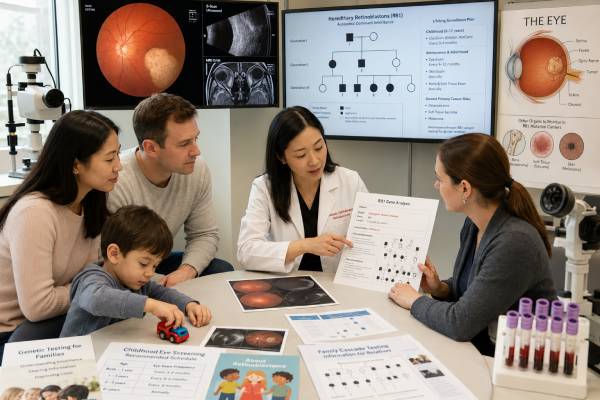
An RB1 genetic test determines whether a child or adult carries a disease-causing change in the RB1 tumor-suppressor gene. The test is central to retinoblastoma care because it can distinguish a heritable cancer predisposition from a tumor change limited to the eye. That distinction affects eye-examination schedules, the chance of tumors in the other eye, screening for relatives, reproductive risks, and long-term awareness of second cancers.
Most children with tumors in both eyes have heritable retinoblastoma, even when no one else in the family has had the disease. Some children with a tumor in only one eye also carry a germline RB1 variant, so appearance alone cannot settle the question. Testing may use blood plus tumor tissue, and sometimes more than one normal tissue, because low-level mosaicism can be missed in blood. A result should be interpreted by a retinoblastoma team and genetics professional who understand the laboratory method, the child’s tumor pattern, and the family history.
- A germline pathogenic RB1 variant confirms heritable retinoblastoma predisposition and creates a 50% transmission risk for each future child.
- Bilateral, multifocal, or familial retinoblastoma is strongly suggestive of an inherited or mosaic RB1 alteration.
- A negative blood result after unilateral disease lowers inherited risk but may not eliminate low-level mosaicism unless testing is sufficiently sensitive.
- Tumor testing can clarify whether both RB1 changes are confined to the tumor and can improve risk estimates for siblings and future children.
- A VUS is not proof of heritable retinoblastoma and should not be used alone to label relatives as affected or unaffected.
Table of Contents
- RB1 and Retinoblastoma
- Who Should Have RB1 Testing
- Blood, Tumor, and Mosaic Testing
- How to Read RB1 Results
- Eye Surveillance and Treatment Planning
- Second-Cancer Risk and Long-Term Care
- Testing Relatives and Future Children
- Limitations and Next Steps
RB1 and Retinoblastoma
RB1 encodes the retinoblastoma protein, which helps control the transition from cell growth to cell division. When both working copies of RB1 are lost in a susceptible retinal cell, that cell can begin the path toward retinoblastoma. This is the classic “two-hit” model of tumor-suppressor genes.
In heritable retinoblastoma, the first nonworking RB1 copy is present in the egg or sperm, develops shortly after conception, or is inherited from a parent. It is therefore present in all or a proportion of the child’s cells. A second change later occurs in one or more retinal cells. Because many retinal cells already carry the first hit, tumors tend to arise earlier, in more than one location, and often in both eyes.
In nonheritable retinoblastoma, both RB1 changes usually occur in a single retinal cell lineage. The tumor is generally unilateral and unifocal, and the change is not present in blood. A small minority of retinoblastomas are driven through another mechanism, such as high-level MYCN amplification without detectable RB1 inactivation. These cases are usually nonheritable but require expert tumor interpretation.
The words germline, constitutional, and heritable overlap but are not always identical in practice. A constitutional variant is present in the body outside the tumor. A germline variant is capable of being passed through egg or sperm. A post-zygotic mosaic variant may be present in only some tissues; it can still be heritable if reproductive cells carry it. The exact tissue distribution cannot always be measured directly.
Retinoblastoma is often the first and most visible sign, but RB1 testing is a cancer-predisposition test rather than a test for eye appearance alone. A positive result may also explain retinoma, a benign retinal lesion that can be a precursor or arrested form of disease, and it identifies a lifelong predisposition to certain second primary cancers.
Who Should Have RB1 Testing
Genetic evaluation is recommended for essentially every child with retinoblastoma because the result can change care for the child and family. The pretest probability differs by presentation, but no family history is required.
Testing is especially important when a person has:
- Retinoblastoma in both eyes.
- More than one tumor focus in one or both eyes.
- Retinoblastoma plus a pineal or other midline intracranial tumor, called trilateral retinoblastoma.
- A close relative with retinoblastoma or retinoma.
- Retinoma without a prior molecular explanation.
- A chromosome 13q deletion that includes RB1.
- Retinoblastoma diagnosed at a very young age, even if only one eye is involved.
- A child or pregnancy at risk for a known familial RB1 variant.
Adults who were treated for retinoblastoma before modern genetic testing may also benefit. Old records may simply say “unilateral” or “bilateral” without defining hereditary status. Establishing the family variant can guide their own long-term care, testing of children, and reproductive decisions.
The most informative person to test first is usually the affected child or adult. Testing an unaffected parent without first finding the child’s variant may produce an unhelpful negative result. When an affected person is unavailable, a genetics team can estimate risk from the number of tumors, eye involvement, age at diagnosis, treatment records, and family history, but uncertainty will remain higher.
A detailed pedigree should include eye tumors, childhood cancers, sarcomas, melanoma, brain tumors, and relatives who had an eye removed in infancy for an unclear reason. Old photographs may reveal leukocoria, the white-pupil reflection associated with retinoblastoma, but photographs cannot replace medical records or testing.
Testing is urgent for newborn relatives at high risk because eye surveillance may need to begin at or shortly after birth. In some families, prenatal diagnosis allows the delivery and first eye examination to be coordinated at a specialist center. A delay of months can matter because small tumors are often easier to treat while preserving vision.
Blood, Tumor, and Mosaic Testing
A complete evaluation may use several complementary methods. The exact pathway depends on whether tumor tissue is available, whether the disease is unilateral or bilateral, and what the initial blood test shows.
Blood or saliva testing
Constitutional testing usually begins with blood. The laboratory performs RB1 sequence analysis and deletion/duplication analysis. Sequence analysis detects single-nucleotide changes and small insertions or deletions. Copy-number testing detects missing or extra exons or a whole-gene deletion. Some assays also assess promoter changes or use RNA analysis for suspected splicing effects.
Saliva can be used in some settings, but blood often provides cleaner DNA and may be preferred for high-sensitivity mosaic analysis. No fasting or medication changes are needed.
Tumor testing
When an eye is removed and tumor tissue is available, paired tumor and blood testing can be particularly informative in unilateral disease. The laboratory can identify the two tumor-driving events, then ask whether either is present outside the tumor. If both are absent from blood with a sensitive assay, the remaining hereditary risk may become very low, although not always zero.
Tumor testing may detect sequence variants, loss of heterozygosity, promoter methylation, copy-number changes, or MYCN amplification. A tumor report is not automatically a germline report. Any potentially inherited finding must be compared with a normal sample, following the same distinction used in tumor genomic testing more broadly.
Mosaicism testing
Mosaicism occurs when an RB1 variant arises after fertilization, so only a fraction of cells carry it. A child can have bilateral or multifocal disease with a low-level variant in blood, or unilateral disease with a variant too rare for a standard assay to detect. High-depth next-generation sequencing, digital methods, or testing a second tissue can improve detection.
A negative blood test does not have the same residual risk in every situation. In a child with bilateral tumors, clinicians generally continue to treat the case as heritable even if initial blood testing is negative, because undetected mosaicism or a difficult-to-detect alteration remains plausible. In a child with one unilateral tumor and two clearly defined tumor-only RB1 changes, residual risk may be much lower.
Chromosome testing
Some children have a larger deletion of chromosome 13q that includes RB1 and neighboring genes. Clues may include developmental delay, growth differences, congenital anomalies, or characteristic facial features. A chromosomal microarray or other copy-number study can define the deletion better than a narrow sequencing test. This broader finding has implications beyond cancer risk and may require developmental, neurologic, cardiac, or other evaluations.
Turnaround time ranges from a few weeks to several months when sequential tumor studies are needed. Clinical eye surveillance should never pause while waiting for a result.
How to Read RB1 Results
The most important questions are where the variant was found, how it was classified, and how sensitive the assay was for mosaicism. The same DNA change can have very different implications when found in blood versus only in tumor.
Pathogenic or likely pathogenic variant in blood
This result confirms constitutional RB1-related predisposition. It explains bilateral, multifocal, familial, or some unilateral retinoblastoma and supports intensive eye surveillance during the years when new retinal tumors can develop.
The result also means:
- Each biological child has up to a 50% chance of inheriting the variant, depending on whether the carrier is mosaic and whether reproductive cells are involved.
- Parents and siblings may need targeted testing.
- The person has increased lifetime risk for second primary malignancies.
- Radiation exposure should be minimized when a clinically suitable nonionizing alternative exists, although medically necessary imaging or treatment should not be withheld without expert discussion.
Some RB1 variants have reduced penetrance. A carrier may have no obvious retinoblastoma, unilateral disease, retinoma, or mild findings. Reduced penetrance explains why a family history can appear negative even when a variant was inherited.
Two pathogenic changes in tumor but not in blood
This pattern supports a tumor-confined, nonheritable event when the blood test is sensitive and the tumor findings fully explain the cancer. It can sharply reduce risk estimates for siblings and future children. The report should state the residual risk of mosaicism and whether parental testing is still advised.
Negative blood test with no tumor result
In unilateral disease, this lowers the chance of a germline variant but does not remove it completely. The residual risk depends on the laboratory’s coverage, mosaic detection limit, and whether deletion/duplication analysis was performed. In bilateral or multifocal disease, a negative blood result does not overturn the clinical evidence for heritable predisposition.
Variant of uncertain significance
A VUS lacks enough evidence to call it disease-causing or benign. It should not be used alone to change eye examinations, advise pregnancy testing, or declare a relative safe. The laboratory may study tumor data, RNA effects, family segregation, and population frequency to clarify it. General principles for a variant of uncertain significance apply, but RB1-specific review by an experienced laboratory is valuable.
Benign or likely benign finding
These variants are not considered the cause of retinoblastoma. They are often listed only in technical data or not reported at all. A benign finding does not make a clinically suspicious case nonheritable; the overall test may still be negative or incomplete.
Eye Surveillance and Treatment Planning
RB1 results help determine who needs repeated retinal examinations and how long surveillance should continue. The schedule is most intensive in infancy and early childhood, when new tumors are most likely to appear. Examinations may require anesthesia so the ophthalmologist can inspect the entire retina, photograph lesions, and treat small tumors during the same session.
A child with a known familial variant or a strong clinical diagnosis usually begins surveillance from birth. Examinations may occur every few weeks or months at first, then become less frequent as the child ages. The precise interval depends on age, prior tumors, treatment response, variant status, and center protocol. Unaffected relatives who test negative for the known family variant can generally stop the special retinoblastoma schedule.
The genetic result does not choose one treatment by itself. Treatment depends on tumor size, location, seeding, eye involvement, visual potential, spread beyond the eye, and prior therapy. Options can include laser therapy, cryotherapy, systemic chemotherapy, intra-arterial chemotherapy, intravitreal chemotherapy, plaque radiotherapy, or removal of the eye. The clinical priorities are survival first, then preservation of the eye and useful vision.
Heritable status does influence how clinicians balance radiation and imaging. External-beam radiation can increase the risk of later cancers in irradiated tissues, especially in RB1 carriers treated at young ages. Modern care therefore favors focal and chemotherapy-based strategies when they can control disease safely. MRI is preferred over CT for many surveillance questions because it avoids ionizing radiation.
Some centers perform brain MRI screening for trilateral retinoblastoma in young children with heritable disease. Protocols differ because the tumor is rare and evidence about the best interval is limited. Families should follow the plan of their specialist center rather than applying a generic schedule.
An urgent eye examination is needed when an infant or child develops leukocoria, new strabismus, an enlarged or painful eye, unexplained redness, reduced vision, or an abnormal red reflex. Genetic testing must not delay evaluation of these signs.
Second-Cancer Risk and Long-Term Care
People with constitutional RB1 variants face a lifelong risk of second primary cancers. The spectrum includes bone and soft-tissue sarcomas, melanoma, and several epithelial cancers. Risk is influenced by age, treatment exposures, smoking, ultraviolet exposure, and the tissues included in prior radiation fields. A second cancer is a new primary malignancy, not the spread or return of the original eye tumor.
There is no single intensive imaging program proven to detect every RB1-associated cancer early. Long-term care therefore combines education, regular clinical review, skin examination, avoidance of tobacco, sun protection, and prompt evaluation of persistent symptoms. Survivorship clinics can integrate genetic risk with late effects of chemotherapy, radiation, eye removal, hearing changes, endocrine problems, and psychosocial needs.
Symptoms that deserve timely assessment include:
- A growing or painful bone or soft-tissue lump.
- Persistent localized bone pain, especially at night or without an injury.
- A changing pigmented skin lesion or a lesion that bleeds.
- Unexplained neurologic symptoms, prolonged headaches, or focal weakness.
- New symptoms within or near a prior radiation field.
- Unexplained weight loss, persistent cough, blood in urine or stool, or abnormal bleeding.
Routine whole-body screening is not universally recommended for all adult RB1 carriers because benefits, false positives, cost, and anxiety must be weighed. Recommendations may change as evidence develops. Survivors should have an individualized plan based on the exact treatment history and current specialist guidance.
Psychological effects also matter. Parents may feel guilt even when a variant arose de novo, while adult survivors may face fear about second cancers and passing the condition to children. Genetic counseling should directly state that inherited variants are not caused by anything a parent did during pregnancy and that a de novo event could not have been predicted or prevented.
Testing Relatives and Future Children
Once the family’s pathogenic RB1 variant is known, relatives can have a targeted known-mutation test. Results are faster and clearer than broad testing because the laboratory knows exactly what to seek.
For a nonmosaic carrier, each child has a 50% chance of inheriting the variant. Penetrance is high for many variants, but the number of tumors, eye involvement, visual outcome, and second-cancer history cannot be predicted with certainty. Reduced-penetrance variants may produce milder or no eye disease in some carriers.
Testing of newborns should be arranged before delivery when possible. If a pregnancy is known to carry the familial variant, the obstetric, neonatal, genetics, anesthesia, and ophthalmology teams can coordinate delivery and early examination. The test result does not usually determine the mode of delivery by itself, although a specialist may recommend specific timing or location.
Reproductive choices include:
- Natural conception followed by newborn testing.
- Prenatal diagnosis through chorionic villus sampling or amniocentesis.
- In vitro fertilization with preimplantation genetic testing for the familial variant.
- Use of donor egg or sperm.
- Adoption or choosing not to have children.
Prenatal testing identifies whether the variant is present but cannot forecast disease severity. Some families use the result to prepare for immediate care rather than to make decisions about continuing a pregnancy. Counseling should be nondirective and respect the family’s values.
If a variant appears de novo in a child and is absent from both parents’ blood, sibling risk is low but not absolutely zero because a parent could have gonadal mosaicism. The affected child’s own future reproductive risk may still approach 50% unless their result is shown to be limited mosaicism.
Limitations and Next Steps
RB1 testing is highly informative, but limitations include low-level mosaicism, variants outside routinely examined regions, complex rearrangements, degraded tumor tissue, and incomplete detection of epigenetic changes. A negative result should be interpreted with the tumor pattern and the laboratory’s technical sensitivity.
Old tests may have examined only selected exons or used methods that were poor at detecting deletions and mosaicism. Adults with an unresolved hereditary status can ask whether updated sequencing, copy-number analysis, or archived tumor testing is possible. Reanalysis may also change a prior VUS classification.
After a report is issued, the family should receive a written summary that states:
- Whether the finding is constitutional, mosaic, tumor-only, or unresolved.
- The exact RB1 variant and classification.
- The estimated risk to siblings, parents, and future children.
- The child’s eye-examination schedule and the clinician responsible for it.
- Whether brain MRI surveillance is recommended.
- The long-term second-cancer and survivorship plan.
- Which relatives should receive targeted testing and how urgently.
Keep the original laboratory report permanently. Treatment summaries and pathology records are also valuable because adult cancer-risk counseling depends partly on radiation fields and chemotherapy exposures. Families who move should transfer records to a center with expertise in ocular oncology, pediatric oncology, and cancer genetics.
The strongest use of RB1 testing is not simply naming hereditary status. It is allowing one child to receive the right surveillance, preventing unnecessary examinations in relatives who did not inherit the variant, and giving carriers a lifelong plan that connects eye care, survivorship, and family planning.
References
- Retinoblastoma 2023 (Review)
- Update on Retinoblastoma Predisposition and Surveillance Recommendations for Children 2025 (Guideline)
- Genetics of Retinoblastoma: An Overview and Significance of Genetic Testing in Clinical Practice 2025 (Review)
- RB1 gene mutations and genetic spectrum in retinoblastoma patients from Turkey 2023 (Cohort Study)
- Retinoblastoma Treatment (PDQ®)–Health Professional Version 2025 (Official Review)
- Recommendations for long-term follow-up of adults with heritable retinoblastoma 2020 (Guideline)
Disclaimer
This article is general educational information and does not replace care from an ocular oncologist, pediatric oncologist, geneticist, or genetic counselor. Eye-surveillance schedules and family risks depend on the exact RB1 finding, tumor pattern, testing method, age, and treatment history. A white pupil, new eye misalignment, painful eye, or other concerning eye change in a child requires urgent medical assessment.