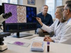
An FLCN genetic test looks for inherited changes that cause Birt-Hogg-Dubé syndrome, a condition associated with kidney tumors, lung cysts, spontaneous pneumothorax, and benign skin growths called fibrofolliculomas. Testing usually uses blood or saliva and should include both sequencing and deletion or duplication analysis. A pathogenic or likely pathogenic result confirms the inherited syndrome and supports lifelong kidney surveillance, counseling about pneumothorax risk, and targeted testing for relatives. A negative result may not fully exclude the diagnosis when clinical features are strong, especially if testing was incomplete or the family’s causal variant is unknown. The condition varies widely: some people first develop collapsed lung, others have facial papules, and some are diagnosed only after a renal tumor or a relative’s result. Because kidney cancers in Birt-Hogg-Dubé syndrome often grow more slowly than FH-deficient renal cancer, management commonly uses imaging surveillance and a size-based surgical threshold under specialist care.
- A pathogenic FLCN variant confirms Birt-Hogg-Dubé syndrome, even when the person has no skin lesions, pneumothorax, or kidney tumor yet.
- Each child of an affected person has a 50% chance of inheriting the variant because the syndrome is autosomal dominant.
- Renal MRI is commonly recommended every one to two years from about age 20, with the exact schedule individualized by an expert team.
- Most lung cysts need no routine treatment, but sudden chest pain or shortness of breath may signal pneumothorax and requires urgent evaluation.
- A negative FLCN test does not always end the evaluation when characteristic lung cysts, fibrofolliculomas, or family history meet clinical criteria.
Table of Contents
- FLCN and Birt-Hogg-Dubé syndrome
- Who should have FLCN testing
- Test methods, samples, and accuracy
- Understanding positive, negative, and uncertain results
- Kidney surveillance and tumor treatment
- Lung, skin, and daily-life care
- Family testing and next steps
FLCN and Birt-Hogg-Dubé syndrome
FLCN provides instructions for folliculin, a protein involved in cellular energy sensing, nutrient signaling, and regulation of the mTOR and AMPK pathways. It acts as a tumor suppressor. A person with Birt-Hogg-Dubé syndrome inherits one altered FLCN copy, and certain cells later lose the remaining working copy. That second event can allow a renal tumor or fibrofolliculoma to develop.
The syndrome is sometimes abbreviated BHD or BHDS. Its three best-established clinical features are:
- Renal tumors. These may be bilateral or multifocal and can include hybrid oncocytic/chromophobe tumors, chromophobe renal cell carcinoma, oncocytoma, clear cell renal cell carcinoma, and other subtypes. The lifetime renal tumor risk is often estimated around 15% to 30%, although figures differ among cohorts.
- Pulmonary cysts and spontaneous pneumothorax. Most affected adults have multiple lung cysts on computed tomography. The cysts often occur in the lower and medial lungs and may have irregular shapes. A cyst can rupture, allowing air to collect around the lung and cause partial or complete collapse.
- Fibrofolliculomas and related skin lesions. Small, smooth, dome-shaped, whitish or skin-colored papules commonly appear on the face, neck, and upper trunk during adulthood. They are benign but can be cosmetically bothersome.
A person does not need all three features. Some families have mostly pneumothorax, while others are recognized through renal tumors. Skin lesions may be subtle, appear later, or be less prominent in some populations. The wide range of expression can delay diagnosis for years.
Birt-Hogg-Dubé syndrome is distinct from more common causes of lung cysts or kidney cancer. Smoking-related emphysema tends to affect different lung regions and has a different cyst pattern. Lymphangioleiomyomatosis, alpha-1 antitrypsin deficiency, connective-tissue disorders, and other inherited pneumothorax syndromes can resemble parts of BHD. Renal tumor pathology and family history help narrow the diagnosis.
The condition is autosomal dominant. One pathogenic FLCN variant is sufficient to create susceptibility, and each child has a 50% chance of inheriting it. Men and women can inherit and transmit the syndrome. Penetrance is incomplete, meaning a carrier may never develop a kidney tumor or pneumothorax.
Colon polyps, colorectal cancer, thyroid nodules, and several other tumors have been reported in affected families, but evidence has not established universal extra screening beyond ordinary population or family-history recommendations. Management should focus on confirmed risks rather than assuming every reported association is part of the syndrome.
Who should have FLCN testing
Testing is appropriate when the combination of lung, kidney, skin, and family findings raises suspicion. A single common feature, such as one spontaneous pneumothorax in a tall young adult, does not always indicate BHD, but characteristic imaging or additional findings can make testing important.
Common reasons for referral include:
- multiple bilateral lung cysts, especially in lower or medial lung regions;
- recurrent or familial spontaneous pneumothorax;
- a first-degree relative with Birt-Hogg-Dubé syndrome or a pathogenic FLCN variant;
- five or more adult-onset fibrofolliculomas or trichodiscomas, with at least one confirmed by skin biopsy;
- bilateral, multifocal, early-onset, or mixed-histology renal tumors;
- chromophobe, oncocytic, or hybrid oncocytic/chromophobe renal neoplasms in a suggestive setting;
- renal cancer plus lung cysts or pneumothorax;
- a tumor-sequencing result showing a potentially germline FLCN alteration; or
- a family pattern that combines kidney tumors, lung collapse, and characteristic facial papules.
Current clinical criteria can support a BHD diagnosis even when no FLCN variant is identified. Major criteria include multiple fibrofolliculomas or a germline pathogenic FLCN variant. Minor criteria include characteristic basal lung cysts, early or multifocal renal cancer, and an affected first-degree relative. The exact diagnostic framework should be applied by a clinician familiar with the syndrome.
A renal cancer patient may receive an inherited kidney cancer panel when several syndromes are possible. FLCN is often analyzed with VHL, FH, MET, BAP1, SDH genes, TSC1, TSC2, and others. This approach can be useful for young-onset, bilateral, multifocal, or unusual renal tumors. It can also produce uncertain findings in genes unrelated to the person’s presentation.
Testing should be considered after a spontaneous pneumothorax when chest CT shows multiple nonapical cysts or when relatives have similar events. A radiologist or pulmonologist may be the first clinician to recognize the pattern. Asking about kidney tumors and small facial papules can reveal the broader syndrome.
A dermatologist may diagnose fibrofolliculomas by biopsy. Several benign lesions look similar, so pathology confirmation can strengthen the indication for testing. Skin lesions alone may be absent in younger adults, and their absence should not dismiss a strong pulmonary or renal pattern.
Before testing, genetic counseling should review the possible results, kidney surveillance, family implications, insurance concerns, and the fact that a positive result may identify risk before symptoms. The family history should include collapsed lung, lung cysts, kidney cancer and histology, age at diagnosis, skin papules, smoking history, and prior chest or abdominal imaging.
Test methods, samples, and accuracy
Germline testing typically uses a blood or saliva sample. The laboratory examines DNA from non-tumor cells. Blood may be preferred when saliva quality is uncertain or when a clinical circumstance could complicate sample interpretation.
A comprehensive FLCN test includes:
- Sequence analysis. This detects single-nucleotide variants and small insertions or deletions. Many pathogenic variants create a premature stop signal that prevents production of functional folliculin.
- Deletion and duplication analysis. This looks for loss or gain of one or more exons or the entire gene. Standard sequencing may not detect these larger changes.
Some laboratories also assess deep intronic or promoter regions, RNA splicing, or mosaicism when routine testing is negative and clinical suspicion remains high. Such analyses are not automatically included in every panel. The report’s methods and limitations matter more than a simple statement that “FLCN was tested.”
A recurrent hotspot in exon 11 contains a run of cytosine bases where small insertions or deletions occur relatively often. Modern assays generally detect these changes, but older methods and low-quality specimens can miss difficult sequence regions.
A multigene hereditary cancer panel may be the best first test when renal tumor histology is mixed or the phenotype overlaps another syndrome. By contrast, targeted FLCN testing is efficient when a relative’s exact variant is already known.
Tumor sequencing may detect FLCN alterations, but it cannot reliably determine inheritance. A renal tumor often has two FLCN hits, while the person’s normal cells may carry only one or neither. A variant found at an allele fraction near 50% can suggest germline origin, but tumor purity, copy-number changes, and loss of heterozygosity can produce similar patterns. Confirmation with blood or saliva is necessary.
A skin biopsy is not a genetic test. It identifies fibrofolliculoma or another lesion under the microscope. Lung CT is also not genetic testing, although the distribution and shape of cysts can support the diagnosis. These clinical findings and DNA results complement each other.
Testing a family member for a known variant has high analytic sensitivity and a clear interpretation. A negative targeted result usually means the person did not inherit the family syndrome. Testing an unaffected person without first testing an affected relative is less informative because a negative result may not explain the family history.
Results often take two to six weeks, depending on the laboratory and whether additional analysis is required. The report should include the transcript, DNA and protein notation, classification, inheritance, assay coverage, and whether deletion or duplication testing was performed.
Understanding positive, negative, and uncertain results
A pathogenic or likely pathogenic result confirms Birt-Hogg-Dubé syndrome. “Likely pathogenic” reflects strong evidence of disease causation and is generally managed the same way as pathogenic. The diagnosis applies even if the person currently has normal kidney imaging and no recognized skin or lung findings.
A positive result does not predict exactly which features will occur. Relatives with the same variant can have different presentations. One may develop recurrent pneumothorax in early adulthood, another may have a renal tumor later in life, and another may remain minimally affected. Surveillance is based on the known syndrome risk, not on an attempt to forecast an individual outcome from the variant alone.
A negative result must be interpreted in context:
- A relative who tests negative for a known familial pathogenic variant has a true negative result and usually does not need BHD-specific renal surveillance.
- A person with characteristic features but no known family variant has an uninformative negative. The syndrome may still be diagnosed clinically, or additional testing may be considered.
- A sequence-only negative is incomplete if deletion and duplication analysis was not included.
- A negative tumor test does not rule out a germline variant because tumor assays may have limited coverage or filtering.
- A negative result on an older panel may warrant review if methods have improved or the phenotype has become clearer.
A variant of uncertain significance, or VUS, is not a confirmed diagnosis. The laboratory has insufficient evidence to determine whether the change disrupts folliculin. Kidney surgery, family predictive testing, and lifelong surveillance should not be based solely on a VUS. Clinical criteria and family history may independently justify surveillance, but the uncertain variant itself should not be treated as pathogenic.
Segregation can sometimes help: a laboratory may examine whether the VUS tracks with characteristic disease in several relatives. Functional studies, population frequency, RNA analysis, and reports from unrelated families may also contribute to future reclassification. Most families should not arrange informal testing without genetics guidance because results can be misleading.
A variant result explanation can clarify why pathogenic, uncertain, and benign findings lead to different actions. Patients should keep the complete report and ask how the laboratory handles reclassification notifications.
Mosaicism is possible when a pathogenic variant arose after conception and is present in only some cells. A low-level result may require confirmation with another tissue and specialist interpretation. Mosaic carriers may have milder or segmental features, but risk cannot be assumed to be negligible.
A result may also identify a benign or likely benign variant. These changes are common genetic differences and do not establish BHD. They should not be used to explain symptoms or test relatives predictively.
Kidney surveillance and tumor treatment
Renal surveillance is the main cancer-prevention strategy. Expert guidelines generally recommend beginning around age 20 because renal tumors are uncommon in childhood. MRI of the kidneys or abdomen is preferred because it avoids radiation and can detect small solid lesions. Scans are commonly repeated every one to two years, although annual imaging may be chosen based on family history, prior findings, local practice, or patient preference.
The imaging order should mention Birt-Hogg-Dubé syndrome so the radiology protocol covers both kidneys carefully. Contrast can improve characterization when medically appropriate. Computed tomography is an alternative when MRI is unavailable or contraindicated, but repeated CT adds cumulative radiation. Ultrasound alone is less sensitive for small or isoechoic tumors and is not preferred as the only surveillance method.
When a lesion is found, the care team reviews size, growth, location, enhancement, and whether tumors are present in both kidneys. Short-interval imaging may clarify an indeterminate lesion. A urologic oncologist experienced in hereditary renal cancer should guide management.
For many BHD-associated solid renal tumors, active surveillance continues until the largest tumor approaches 3 cm. This “3-cm rule” aims to preserve kidney tissue while maintaining a low metastatic risk. It is not a universal rule for every renal mass. Rapid growth, suspicious imaging, symptoms, uncertain pathology, or another tumor syndrome may justify earlier intervention.
Nephron-sparing surgery, usually partial nephrectomy, is preferred when technically safe because new tumors may develop over a lifetime. Removing an entire kidney for each small lesion can create avoidable chronic kidney disease. Complex or centrally located tumors may still require a more extensive operation. Ablation can be considered in selected patients, but surgery often provides clearer pathology and durable local control.
This strategy differs sharply from FH tumor predisposition syndrome, where small renal cancers can behave aggressively and prompt surgery is usually recommended rather than waiting for a 3-cm threshold. Correct genetic diagnosis therefore changes management.
After renal surgery, surveillance continues for the remaining kidney tissue. Kidney function, blood pressure, and medication exposure should be monitored. A renal tumor pathology review may be useful when the original diagnosis was made before BHD was recognized.
There is no proven medicine that prevents BHD renal tumors. Healthy weight, tobacco avoidance, blood-pressure control, and protection from unnecessary kidney injury support general health but do not replace MRI. Blood in the urine, persistent flank pain, or a new abdominal mass should prompt evaluation, although surveillance often finds tumors before symptoms.
Lung, skin, and daily-life care
Most people with BHD lung cysts have normal or near-normal lung function. Routine repeated chest CT is not usually needed in an asymptomatic person after the pattern has been documented because CT exposes the lungs to radiation and cyst burden does not directly determine treatment. Pulmonary follow-up is individualized for symptoms, pneumothorax history, occupation, or another lung disease.
A spontaneous pneumothorax can cause sudden one-sided chest pain, shortness of breath, rapid heart rate, or a dry cough. These symptoms need urgent medical assessment. A small pneumothorax may be observed or treated with oxygen, while a larger or symptomatic event may require drainage. Recurrent events often lead to pleurodesis or surgery intended to reduce recurrence.
Commercial airline cabins are pressurized, and many affected people fly without difficulty. The risk of pneumothorax around air travel is low but not zero, particularly for someone with a recent event or untreated pneumothorax. A clinician should advise when it is safe to fly after treatment. Work in unpressurized aircraft or frequent aviation exposure deserves individualized occupational counseling.
Scuba diving creates pressure changes that can be dangerous when lung cysts are present. Expert guidance generally discourages diving unless a specialist with knowledge of both BHD and diving medicine completes a detailed evaluation. Standard recreational medical forms may not capture the syndrome’s specific risk.
Smoking can add emphysema and other lung injury to an existing cystic condition, so avoidance is strongly advised. Symptoms of sleep apnea, asthma, or chronic obstructive pulmonary disease should be evaluated on their own merits rather than attributed automatically to BHD.
Fibrofolliculomas are benign and do not become kidney cancer. Treatment is optional and mainly cosmetic. Options include carbon dioxide or erbium laser, electrosurgery, shave removal, or other dermatologic procedures. Lesions may recur because treatment removes visible papules but does not correct the inherited predisposition. Sun protection and routine skin care are sensible, though they do not prevent genetically driven lesions.
A baseline dermatology examination can confirm lesion type and document the skin pattern. A rapidly growing, bleeding, ulcerated, or pigmented lesion should be assessed because it may be unrelated to BHD. Not every facial papule in a carrier is a fibrofolliculoma.
There is no established need for extra colon or thyroid screening solely because of FLCN status. Follow general screening guidelines and any enhanced plan justified by personal or family history. This avoids both missed care and unnecessary procedures.
Family testing and next steps
Once a pathogenic FLCN variant is found, first-degree relatives should be offered targeted testing. Parents, siblings, and adult children each may be at risk. Testing is generally offered in late adolescence or early adulthood so renal surveillance can begin around age 20. Earlier testing may be reasonable when family circumstances or unusual early disease warrant it.
A positive relative should receive a coordinated plan for renal MRI, pneumothorax education, and specialist referrals. A negative relative for the known family variant can usually stop BHD-specific screening. They still need ordinary health care and evaluation of any unrelated symptoms.
Reproductive options include natural conception, prenatal diagnosis, or in vitro fertilization with preimplantation genetic testing for the familial variant. Some people use donor gametes or choose not to test pregnancies. A genetic counselor can explain timing, accuracy, cost, and personal considerations without directing the choice.
After receiving a result, ask:
- Is the variant pathogenic, likely pathogenic, or uncertain?
- Did the assay include deletion and duplication analysis?
- Do my clinical features meet diagnostic criteria even if testing was negative?
- When should renal MRI begin, and should it be annual or every two years?
- Which urologic oncology team will evaluate a renal lesion?
- What should I do if sudden chest pain or shortness of breath occurs?
- Are air travel, diving, or my occupation affected by my pneumothorax history?
- Which relatives should receive targeted testing?
Keep copies of the genetic report, renal imaging, chest CT, pathology, and operative notes. Record the exact FLCN variant in standard notation. Make sure primary care, genetics, radiology, urology, pulmonology, and dermatology know who is coordinating care.
If testing is negative but suspicion remains, ask whether the laboratory covered copy-number changes, difficult sequence regions, and mosaicism. A broader multigene test may be useful if another inherited pneumothorax or kidney cancer syndrome is possible. Clinical surveillance may still be appropriate when diagnostic criteria are met.
The most useful result is one connected to a durable follow-up system. The plan should identify the imaging interval, the clinician responsible for reminders, a pathway for urgent pneumothorax care, and a method for relatives to access testing.
References
- Birt-Hogg-Dubé Syndrome 2024 (Clinical Review)
- ERN GENTURIS clinical practice guidelines for the diagnosis, surveillance and management of people with Birt-Hogg-Dubé syndrome 2024 (Guideline)
- Hereditary Renal Cell Carcinoma Syndromes 2023 (Review)
- Recommendations on scuba diving in Birt-Hogg-Dubé syndrome 2023 (Expert Review)
- Diagnosis of renal tumors in Birt-Hogg-Dube syndrome 2024 (Cohort Study)
- Clinical and genetic characteristics of 100 consecutive patients with Birt-Hogg-Dubé syndrome in Eastern Chinese region 2024 (Cohort Study)
Disclaimer
This article is educational and does not replace genetic counseling, renal imaging advice, or emergency assessment. Kidney surveillance and treatment thresholds should be set by clinicians experienced in hereditary renal cancer. Sudden chest pain or shortness of breath may be a pneumothorax and requires urgent medical evaluation.