What is traumatic cataract?
Traumatic cataract is a type of cataract that develops following an eye injury. Unlike age-related cataracts, which typically develop gradually over time as a result of the natural aging process, traumatic cataracts can develop suddenly or over time following an injury. A cataract causes the normally clear crystalline lens of the eye to become opaque or cloudy, resulting in visual impairment. Traumatic cataracts can develop at any age and can be caused by a variety of traumas, such as blunt force, penetrating injuries, radiation, chemical exposure, or electric shock.
Causes of Traumatic Cataracts
The most common cause of traumatic cataracts is physical eye injuries. There are several types of injuries:
- Blunt Trauma: Blunt trauma to the eye, such as a punch, fall, or object striking the eye, can result in the formation of a cataract. The force of the impact may damage the lens fibers or rupture the lens capsule, causing the lens to become cloudy. The cataract can form immediately after the injury or develop gradually over weeks, months, or even years. Sports injuries, car accidents, and workplace accidents are among the most common causes of blunt trauma.
- Penetrating Injuries: Penetrating injuries occur when an object penetrates the eye, causing damage to the lens and possibly other structures inside the eye. Sharp objects, such as knives, glass, and metal fragments, are common causes of penetrating injuries. These injuries can result in immediate lens clouding as well as complications like infection, inflammation, and retinal damage. Penetrating injuries frequently necessitate immediate surgical intervention to prevent additional damage and preserve vision.
- Radiation Exposure: Prolonged exposure to ultraviolet (UV) radiation, such as during radiation therapy for cancer treatment, can cause cataracts to form. Radiation-induced cataracts are typically a delayed response to exposure, appearing months or years after the initial exposure. UV radiation from the sun is a known risk factor for cataract formation, and people who work outside or in UV-rich environments are more likely to develop cataracts.
- Chemical Injuries: Chemical exposure, especially to strong acids or alkalis, can cause serious damage to the eye, including the lens. Chemical burns can cause cataract formation by disrupting the proteins in the lens and making it opaque. The severity of the cataract is determined by the chemical concentration, exposure time, and prompt treatment.
- Electric Shock: Although rare, electric shock injuries can cause cataracts. The mechanism is thought to involve the heat produced by the electric current as it passes through the eye, which can denature the proteins in the lens, resulting in cataract formation. Cataracts caused by electric shock can develop shortly after the injury or later.
Pathology of Traumatic Cataracts
The pathophysiology of traumatic cataract differs according to the type and severity of the injury. The lens is made up of water and proteins that have been precisely arranged to maintain its transparency and refractive properties. Trauma to the eye can disrupt this delicate balance, causing the lens to become opaque.
In blunt trauma, the force of the impact can cause a contusion in the lens, causing changes in the lens fibers and capsule. The lens capsule may rupture, allowing water and other substances into the lens, resulting in swelling (hydropic change) and clouding. Furthermore, the lens fibers themselves may suffer damage, resulting in the formation of a cataract. The force of the impact and the extent of damage to the lens structures determine the severity of the cataract.
Penetrating injuries can cause more severe and immediate cataract formation due to direct damage to the lens. When the lens capsule ruptures, the protective barrier that keeps the lens proteins intact is compromised, resulting in the rapid development of cataracts. Foreign bodies that remain lodged in the lens or other parts of the eye can cause infection, chronic inflammation, and retinal detachment, complicating the clinical picture even more.
Radiation-induced cataracts result from damage to the lens epithelial cells and fibers. The radiation can cause DNA damage and disrupt the normal cell cycle, resulting in the accumulation of damaged cells and proteins inside the lens. This process results in the formation of opacities within the lens, which gradually coalesce into cataracts. The latency period between radiation exposure and cataract formation varies according to radiation dose and individual susceptibility.
Chemical injuries to the eye can cause cataracts by either directly damaging the lens proteins or causing inflammation and scarring within the eye. Strong acids and alkalis can penetrate the eye’s tissues, resulting in necrosis and cataract formation. The nature of the chemical agent, the duration of exposure, and the efficacy of immediate treatment all influence the extent of cataract development. Chemical burns frequently cause complex ocular damage, with cataracts being just one of the possible long-term complications.
Electric shock injuries cause cataract formation due to the thermal effects of the electric current. The heat generated by the current as it passes through the eye can denaturate lens proteins, causing them to aggregate and form opacities. In some cases, the electric shock can cause damage to other ocular structures, such as the retina or optic nerve, exacerbating the vision loss.
Clinical Features of Traumatic Cataract
The clinical presentation of traumatic cataract varies according to the type and severity of the injury. In some cases, the cataract appears immediately following the trauma, while in others, it develops gradually over time. Patients with traumatic cataracts may exhibit a variety of symptoms, including:
- Blurred or Cloudy Vision: The most common sign of a cataract is gradual blurring or clouding of vision. As the lens becomes opaque, it scatters light entering the eye, reducing the clarity of the retinal image.
- Glare and Light Sensitivity: Patients with traumatic cataracts may become more sensitive to bright lights, with glare being a common complaint. This symptom is especially bothersome when driving at night or in areas with bright lights.
- Double Vision (Diplopia): Some patients may experience double vision in their affected eye, especially if the cataract causes irregularities in the lens curvature. Diplopia can be especially disorienting and disrupt daily activities.
- Reduced Color Perception: Traumatic cataracts can cause a reduction in the vibrancy of colors, with patients reporting that they appear faded or dull. This symptom occurs when lens opacities filter out specific wavelengths of light.
- Visible Opacities: In some cases, the cataract may appear to the naked eye as a whitish or cloudy area in the pupil. This is more common in mature cataracts, where the lens has become noticeably opaque.
- History of Ocular Trauma: Diagnosing traumatic cataracts requires a thorough history of ocular trauma, including the type of injury, timing of symptoms, and any previous treatments. Patients may also report other symptoms, such as pain, redness, or blurred vision in the affected eye.
Risk Factors
A traumatic cataract is more likely to develop when:
- Age: While traumatic cataracts can occur at any age, older people may be more prone to developing cataracts following trauma due to pre-existing lens changes associated with aging.
- Occupation: Workers in high-risk occupations, such as construction, manufacturing, or agriculture, are more likely to sustain eye injuries that result in traumatic cataracts. Proper protective eyewear is required in these environments to avoid ocular injuries.
- Sports: Contact sports and projectile-based activities, such as baseball, hockey, and racquet sports, raise the risk of blunt trauma to the eye, which can lead to cataract formation.
- Previous Eye Surgery: People who have had previous eye surgery, such as cataract or refractive surgery, may be at a higher risk of developing traumatic cataracts due to changes in the structural integrity of their eye.
- Radiation Exposure: People who have had radiation therapy or work in environments with high levels of UV radiation are more likely to develop cataracts, including trauma-related cataracts.
Effective Diagnostic Methods for Traumatic Cataract
Traumatic cataract diagnosis entails a combination of clinical examination, imaging studies, and, in some cases, laboratory tests to determine the extent of lens damage and plan an appropriate management strategy. The diagnostic process is critical for distinguishing traumatic cataracts from other types of cataracts and detecting any associated ocular injuries.
Clinical Examination
The clinical examination is the initial step in diagnosing traumatic cataract. An ophthalmologist will start by gathering a detailed medical history, including the nature of the injury, the onset of symptoms, and any prior ocular conditions or surgeries. The physical examination includes a thorough examination of the eye, with emphasis on the lens, cornea, and other ocular structures.
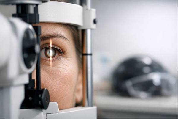
- Slit-Lamp Examination: A slit-lamp microscope is used to examine the eye’s anterior segment, which includes the cornea, lens, and anterior chamber. This enables the ophthalmologist to see the extent of lens opacification, lens capsule rupture, and other anterior segment abnormalities. Slit-lamp examination can also detect other ocular trauma complications, such as corneal scarring, hyphema (blood in the anterior chamber), and iris damage.
- Visual Acuity Test: Visual acuity testing is used to determine the effect of a cataract on a patient’s vision. The Snellen chart is commonly used to assess the ability to see fine detail. The findings establish a baseline for determining the severity of the visual impairment and planning treatment.
- Fundus Examination: A fundus examination, often performed with an ophthalmoscope or through dilated pupils using a slit-lamp, is required to evaluate the posterior segment of the eye. This examination evaluates the retina, optic nerve, and vitreous, which may all be affected by the trauma. Imaging studies, such as ultrasound, may be required in cases where the cataract has advanced and obscures the view of the posterior segment.
Imaging Studies
Imaging studies are frequently used to determine the extent of trauma-related damage, particularly when lens opacities obscure the view of the posterior segment of the eye.
- Ultrasound Biomicroscopy (UBM): UBM is a high-frequency ultrasound technique for visualizing the eye’s anterior segment, which includes the lens, ciliary body, and zonules. It is especially useful when the lens opacity is dense and blocks a clear view of the eye’s internal structures. UBM can help assess lens capsule integrity, detect foreign bodies, and determine anterior chamber depth.
- B-Scan Ultrasound: B-scan ultrasound is a useful tool for evaluating the posterior segment of the eye when a dense cataract obscures the view. This imaging modality can detect retinal detachment, vitreous hemorrhage, and other post-traumatic pathologies in the posterior segment.
- Anterior Segment Optical Coherence Tomography (AS-OCT) generates high-resolution cross-sectional images of the anterior segment, which includes the cornea, iris, and lens. This imaging technique is particularly useful for determining the structural integrity of the lens capsule and detecting subtle changes in the lens fibers.
Additional Diagnostic Tests
In some cases, additional diagnostic tests may be required to assess the overall health of the eye and devise an appropriate management strategy.
- Specular Microscopy: Specular microscopy evaluates the endothelial cell count and morphology of the cornea. This test is important if there has been any trauma to the corneal endothelium, as these cells are essential for maintaining corneal clarity. A low endothelial cell count may influence the decision-making process for cataract surgery.
- Tonometry measures the eye’s intraocular pressure (IOP). Traumatic cataracts can lead to secondary glaucoma, either due to trabecular meshwork damage or inflammatory responses within the eye. Elevated IOP may necessitate additional glaucoma treatment prior to or following cataract surgery.
- Gonioscopy: Gonioscopy is a diagnostic procedure that involves examining the anterior chamber angle, which is where the aqueous humor drains from the eye. It is especially useful in cases of trauma that have resulted in angle recession or other angle abnormalities, which can increase the risk of glaucoma.
- Electrophysiological Testing: If there is a suspicion of optic nerve or retinal damage, electrophysiological tests such as visual evoked potentials (VEP) or electroretinography (ERG) may be performed to evaluate the function of the optic nerve and retina. These tests provide useful information about the integrity of the visual pathway and aid in predicting visual outcomes following cataract surgery.
Traumatic Cataract Management
The severity of the cataract, the extent of other ocular injuries, the patient’s overall health, and the impact on vision all play a role in managing traumatic cataract. The primary goal of management is to restore vision while avoiding further complications like glaucoma, retinal detachment, or chronic inflammation. Traumatic cataract management can include both surgical and non-surgical approaches, with surgical intervention being the most common and effective treatment.
Surgical Management
Surgery is the only definitive treatment for traumatic cataract, especially if the cataract significantly impairs vision or there are other ocular complications. The nature of the cataract and the presence of other ocular injuries determine the type of surgery performed.
- Phacoemulsification is the most commonly used surgical technique for cataract removal. This procedure involves making a small incision in the cornea and using an ultrasonic probe to break up the cloudy lens into tiny fragments that are then aspirated out of the eye. After the cataract is removed, an intraocular lens (IOL) is implanted to replace the natural lens and improve vision. This procedure is usually performed under local anesthesia and requires a short recovery time. However, the success of phacoemulsification in traumatic cataracts is dependent on the lens capsule’s integrity and the absence of any other significant ocular injuries.
- Extracapsular Cataract Extraction (ECCE): If the lens capsule ruptures or if the cataract is too dense for phacoemulsification, ECCE may be necessary. This procedure entails creating a larger incision in the cornea or sclera, removing the cloudy lens in one piece, and implanting an IOL. ECCE is typically reserved for more complex cases where phacoemulsification is not an option. ECCE requires a longer recovery time than phacoemulsification and has a higher risk of complications such as corneal edema or wound dehiscence.
- Secondary IOL Implantation: In some cases, especially when the lens capsule is compromised, the initial surgery may only involve removing the cataract and not immediately implanting an IOL. A secondary IOL implantation can be done after the eye has healed and the ocular anatomy is more stable. Depending on the individual case, this procedure may involve implanting the IOL in the posterior chamber, anterior chamber, or as a scleral-fixated IOL.
- Combined Surgical Procedures: Traumatic cataracts are frequently associated with other ocular injuries, including retinal detachment, vitreous hemorrhage, and glaucoma. In these cases, a combination surgical approach may be required. For example, cataract surgery may be performed concurrently with vitrectomy (removal of the vitreous gel), retinal repair, or glaucoma surgery. The surgical plan is designed to address all ocular issues at the same time, with the ultimate goal of improving visual outcomes.
Non-surgical Management
Non-surgical management of traumatic cataract is typically considered when the cataract does not significantly impair vision or the patient is not a good candidate for surgery due to medical reasons. Non-surgical approaches can also be used as a temporary measure before surgery.
- Observation: In some cases, especially when the cataract is small and not progressing, a period of observation may be advised. The patient’s vision and cataract progression are regularly monitored, and surgery is postponed until the cataract is visually significant or there is a risk of other complications.
- Corrective Lenses: For patients with mild cataracts, prescription glasses or contact lenses can temporarily improve vision. These lenses can help correct cataract-related refractive errors like myopia and astigmatism. However, corrective lenses do not slow the progression of cataracts and are only a temporary solution.
- Medication: Although there is no medication that can reverse or prevent cataracts, medications can be used to treat associated conditions such as inflammation or high intraocular pressure. Topical corticosteroids or nonsteroidal anti-inflammatory drugs (NSAIDs) can be used to reduce inflammation after an ocular injury, potentially delaying cataract progression. Antiglaucoma medications may also be required if the trauma caused an increase in intraocular pressure.
Post-operative Care and Rehabilitation
Postoperative care is critical for achieving a successful outcome following cataract surgery. Patients are usually given a regimen of eye drops that includes antibiotics, corticosteroids, and anti-inflammatory medications to prevent infection, reduce inflammation, and promote healing. Follow-up visits are scheduled to monitor the healing process, check for complications, and determine the surgery’s success.
In some cases, patients may require visual rehabilitation after cataract surgery, especially if they have residual refractive errors or if the surgery did not completely restore vision due to other ocular conditions. Visual rehabilitation may include the use of corrective lenses, low vision aids, or additional surgical procedures.
Overall, managing traumatic cataracts necessitates a tailored approach that takes into account the nature of the injury, the patient’s visual requirements, and the potential risks and benefits of surgery. Early intervention and appropriate management are critical to maintaining vision and avoiding long-term complications.
Trusted Resources and Support
Books
- “Cataract Surgery: Techniques, Complications, and Management” by Roger F. Steinert: This comprehensive book provides an in-depth exploration of cataract surgery, including the management of complex cases such as traumatic cataracts.
- “Trauma and Emergency Care in Ophthalmology” by Urmi V. Shah and Pradeep V. Shah: This book covers various aspects of ocular trauma, including the diagnosis and management of traumatic cataracts.
Organizations
- American Academy of Ophthalmology (AAO): The AAO offers extensive resources and guidelines on cataract management, including the latest updates on surgical techniques and postoperative care.
- National Eye Institute (NEI): The NEI provides information on cataracts, including resources on the causes, diagnosis, and treatment of traumatic cataracts.
- The Royal College of Ophthalmologists: This organization offers professional guidelines and resources for the management of cataracts, with a focus on improving patient care and outcomes.


