Retinitis Pigmentosa (RP) is a group of inherited retinal disorders marked by a slow but relentless degeneration of photoreceptor cells. Over time, this progression erodes peripheral vision, reduces night vision, and eventually compromises central acuity, leaving many patients legally blind. While the onset is often in adolescence or early adulthood, late-stage RP represents one of the greatest therapeutic challenges because much of the photoreceptor layer has already atrophied. Traditional management strategies—such as low-vision aids, vitamin supplements, and environmental adaptations—offer symptomatic relief but do not fundamentally alter the disease course.
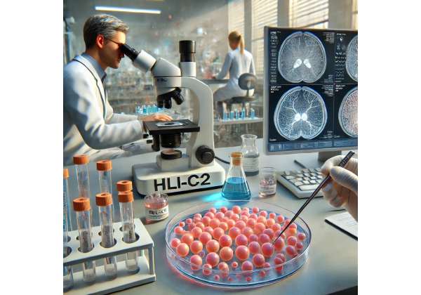
Scientific breakthroughs in regenerative medicine have introduced the possibility of halting or partially reversing the degenerative processes that define RP. One particularly promising avenue centers on the use of retinal progenitor cells. These cells, obtained through specialized procedures, possess the capacity to differentiate into key retinal cell types, potentially supporting or replacing the compromised photoreceptors. HLI-C2, a proprietary line of human retinal progenitor cells, has emerged as a candidate therapy. Early research suggests that transplanted HLI-C2 cells may provide trophic support, reduce inflammation, and perhaps reconstruct lost or atrophied segments of the retinal architecture. Below, we explore this treatment in depth, outlining how the therapy works, the procedure to administer it, recent trial data, its safety record, and the anticipated costs for patients seeking this intervention.
Understanding the Potential of Retinal Progenitor Cell Therapy
Retinal progenitor cells (RPCs) can be thought of as specialized stem-like cells that retain the ability to become various retinal cell types—particularly photoreceptors (rods and cones) and supporting neurons. Over the last two decades, numerous labs have investigated whether transplanting these progenitors could help patients with degenerative diseases of the retina. The impetus behind HLI-C2 arises from a desire to address cases that are no longer amenable to simpler interventions, such as gene therapy for early-stage RP or targeted treatments for mild to moderate disease.
Why Progenitor Cells for Retinitis Pigmentosa?
Retinitis Pigmentosa’s hallmark is the gradual apoptosis of rods and cones. In advanced disease, these cells are severely diminished, leaving behind a thin, nearly functionless retinal layer. Even if a gene fix became possible at this point, the underlying photoreceptor structure is often too compromised to salvage. Progenitor cells, however, bring in a new supply of cells that can integrate (to some degree) and produce essential growth factors. Specifically, RPCs may:
- Differentiate into Photoreceptors: Under the right conditions, transplanted progenitors might form rod or cone cells.
- Release Trophic Factors: Beyond direct replacement, RPCs often secrete a variety of molecules—such as brain-derived neurotrophic factor (BDNF) or ciliary neurotrophic factor (CNTF)—that protect and nourish any remaining native photoreceptors.
- Modulate Local Inflammation: Chronic inflammation can hasten the pace of degeneration. RPCs may calm inflammatory pathways, stabilizing the retinal environment.
By combining these beneficial effects, HLI-C2 attempts to restore a more nurturing ecosystem in the retina. For patients on the cusp of total blindness, the ability to maintain a functional patch of vision can make a profound difference in daily life.
Unique Features of HLI-C2
Retinal progenitor cell lines differ substantially in their derivation, purification steps, and final characteristics. HLI-C2 stands out in its:
- Proprietary Culturing Protocol: Researchers have established a method for expanding these cells in a controlled environment, ensuring purity and consistent phenotype.
- Documented Differentiation Pathways: Preclinical studies highlight HLI-C2’s capacity to generate both rod-like and cone-like cells, which is crucial for broad-spectrum vision.
- Survival and Integration: Data from animal models suggest robust cell survival and some level of structural integration when transplanted, a factor that may influence the longevity of therapeutic effects.
Because late-stage RP typically involves near-total loss of rod photoreceptors first (leading to night blindness), and progressive cone demise after (compromising daytime vision), a product like HLI-C2 must address the complexities of restoring or supporting multiple photoreceptor types. There is an ongoing debate within the scientific community over how many newly formed photoreceptors might actually integrate with the existing retinal circuitry, as opposed to just offering paracrine support. However, even the latter can be beneficial in halting disease progression.
Potential Synergies and Future Directions
While HLI-C2 is being trialed primarily as a stand-alone therapy, the field of regenerative ophthalmology is moving towards combination strategies. For example, gene therapy could address the root genetic defect in earlier disease stages, while progenitor cells might be introduced in advanced cases to rebuild or support degenerating regions. Similarly, bioengineered scaffolds or synthetic retinas might eventually pair with HLI-C2 to form a stable “neo-retina” that offers better phototransduction. Over the next decade, the synergy between cell therapy, gene editing, and advanced implant technology may redefine how RP is managed at various stages.
In essence, HLI-C2 highlights a shift in approach: rather than adapt to progressive vision loss, clinicians and scientists are exploring ways to replace or reinforce the very cells lost to disease. Early results are promising, particularly for patients whose retinas show just enough structural integrity to host transplanted cells. However, turning theory into practice involves carefully orchestrated treatment regimens, which we explore next.
Key Steps for Administering HLI-C2 in Advanced Retinitis Pigmentosa
To maximize the odds of successful cell transplantation, each procedural phase must be optimized—starting with patient selection and culminating in postoperative monitoring. HLI-C2 therapy introduces living cells into the subretinal space, so precision and rigorous aseptic conditions are paramount. The entire process can be broken down into multiple stages that ensure consistent quality and patient safety.
Patient Candidacy and Preoperative Evaluations
Not every individual with late-stage RP qualifies for HLI-C2 therapy. Factors influencing eligibility include:
- Disease Etiology: While RP can stem from myriad genetic mutations, certain backgrounds may respond more favorably to a progenitor approach. Patients generally undergo genetic testing to confirm an RP diagnosis.
- Residual Retinal Structure: Advanced imaging techniques like optical coherence tomography (OCT) or ultra-widefield scanning help ascertain whether any viable retinal layers remain. If the retina is severely thinned without any stable architecture, transplanted cells may lack the substrate needed for integration or contact.
- Overall Ocular Health: Coexisting disorders (like advanced cataracts, severe vitreoretinal traction, or uncontrolled glaucoma) might complicate the procedure. For instance, a chronically inflamed eye is more prone to immunological reactions that sabotage newly introduced cells.
- Systemic Fitness: Because sedation or general anesthesia may be required, patients with significant systemic issues should be cleared before surgery.
Once deemed eligible, patients often receive detailed counseling about realistic expectations. While some improvement in functional vision is possible, particularly if any healthy photoreceptors remain, many participants might see more subtle benefits—like decreased progression, enhanced contrast sensitivity, or improved navigational vision in certain lighting conditions.
Preparation and Storage of HLI-C2 Cells
The foundation of success lies in robust cell preparation:
- Sourcing: HLI-C2 progenitor cells are grown in specialized labs under Good Manufacturing Practice (GMP) conditions. Technicians harvest them at a developmental stage where they maintain the capacity to differentiate into retinal lineages.
- Expansion and Cryopreservation: The culture expands to the desired volume, after which cells are often cryopreserved. They remain in this state until the patient is ready for transplantation, ensuring viability and standardization across batches.
- Quality Assurance: Before shipping or transferring to the surgical center, each lot undergoes rigorous testing for bacterial or fungal contamination, correct cell identity, and stable viability post-thaw.
At the clinical facility, staff thaw the cells (if necessary) and prepare them for injection. This process may involve final washing steps or suspension in a carrier solution that promotes cell survival during transplantation.
Surgical Delivery to the Subretinal Space
The standard technique for delivering HLI-C2 progenitor cells involves a subretinal injection, requiring advanced microsurgical skills:
- Anesthesia: Depending on patient preference and the surgeon’s assessment, local anesthesia with sedation or full general anesthesia may be used.
- Pars Plana Vitrectomy (PPV): A three-port vitrectomy is the norm, providing access to the retina’s surface by removing the vitreous gel. This step ensures a clear path for instruments and better control over fluid dynamics.
- Retinotomy Creation: Using fine-gauge instruments, the surgeon creates a tiny opening (retinotomy) in the retina. A subretinal bleb of fluid might be injected first to separate the photoreceptors from the underlying pigment epithelium, creating a potential space for the cells.
- Cell Infusion: The HLI-C2 suspension is gently introduced via a microcannula, depositing the progenitor cells under the retina. Careful observation ensures that the injection does not cause iatrogenic tears or extensive detachment.
- Closure and Stabilization: Surgeons may use air or gas tamponades to reattach the retina, though the specifics vary by protocol. Postoperative instructions typically advise patients on positioning (e.g., face-down) to keep the transplanted cells in place.
Throughout these steps, preventing trauma to the retina is critical, as mechanical damage can disrupt local tissues needed to support new cell integration. Because advanced RP retinas are often fragile, the surgical approach must be gentle and precisely calibrated.
Postoperative Care and Follow-Up
Following surgery, patients enter a carefully monitored recovery phase. Key components include:
- Inflammation Control: Topical and sometimes systemic corticosteroids or immunosuppressants may be prescribed. Although the eye offers some immune privilege, foreign or newly introduced cells can still trigger inflammation.
- Infection Prevention: Antibiotic drops reduce the risk of endophthalmitis—an infection within the eyeball that can be devastating if unchecked.
- Positioning: If gas or air was used, certain head positioning for a few days may help keep the subretinal bleb stable, maximizing contact between transplanted cells and the host retina.
- Serial Examinations: Multiple visits at intervals (e.g., one week, one month, three months, etc.) let the medical team track integration. Tools such as OCT, fundus autofluorescence, microperimetry, and standard visual acuity tests can detect subtle changes in retinal thickness, cell layering, and functional improvement.
Importantly, because we do not expect instantaneous gains, the success of HLI-C2 therapy may only become apparent over weeks or months. During this period, the transplanted cells must settle, differentiate as needed, and establish beneficial interactions with the existing neuronal network. For some, the outcomes might revolve more around slowed progression than overt improvements in acuity, especially if the disease is extremely advanced.
Breakthrough Data and Ongoing Investigations
Scientific literature on HLI-C2 therapy remains relatively new, yet a variety of preclinical and early-phase clinical trial results are starting to shape our understanding of its promise. While many aspects require further study, the growing body of evidence underscores that retinal progenitor cell transplants can safely coexist in human eyes and, in some cases, yield functional benefits.
Preclinical Cornerstones
Research in animal models of retinitis pigmentosa (e.g., rd1 mice, Royal College of Surgeons [RCS] rats) has paved the way:
- Cell Survival and Differentiation: Investigators have consistently observed that transplanted progenitor cells survive for months post-injection. Some fraction differentiates into rod-like cells, forming rudimentary outer segment structures.
- Anatomical Preservation: In rodent models, the outer nuclear layer (ONL) thickness—a measure of photoreceptor cell bodies—remains more robust in eyes that receive HLI-C2 transplants compared to untreated controls.
- Functional Gains: Electroretinography (ERG) tests show small but significant improvements in scotopic (low-light) response for transplanted rodents. This indicates that at least some transplanted cells or rescued native photoreceptors maintain phototransduction capabilities.
These findings underpin the rationale that, while not every cell becomes a fully mature photoreceptor, the protective or trophic influence alone can sustain part of the retina that would otherwise degenerate.
Early Human Trials
Human clinical trials for advanced RP typically follow a phased approach:
- Phase I Safety Trials: The goal is to confirm that transplanted cells do not provoke severe immunological reactions, lead to tumor formation, or cause undue retinal damage. So far, HLI-C2 trials report mild, transient inflammation but no major adverse events.
- Phase II Efficacy Studies: Investigators expand the participant pool and incorporate objective measures of vision improvement—ranging from best-corrected visual acuity (BCVA) to navigation tests. Early data suggest stabilization or slight improvement in some participants, especially those with patches of viable photoreceptor or RPE layers.
- Open-Label Extensions: Some studies include an option for continuing follow-up beyond one year, observing whether improvements hold or intensify. Investigators also collect valuable data on how immunosuppressant regimens might be tapered over time without jeopardizing the graft.
Promising anecdotal reports from participants describe modest expansions in peripheral vision or the ability to detect low-contrast objects. While these outcomes can be life-changing for severely visually impaired individuals, they also underscore that, at this stage, the therapy’s success often varies from one person to another. Genetic background, residual retina quality, and subtle surgical differences all potentially influence the final outcome.
Research Directions and Next Steps
Given the complexity of advanced RP, many research teams see HLI-C2 therapy as one piece of a larger puzzle. Notable avenues for future exploration include:
- Combination Gene Therapy: If a patient’s genotype is known, gene therapy might reduce ongoing cell damage, while HLI-C2 provides fresh cells or supportive factors.
- Neuroprotective Agents: Administering protective molecules—either systemically or locally—to amplify the transplanted cells’ impact on the diseased retina.
- Structured Scaffolds: Some investigators aim to embed progenitor cells in biodegradable scaffolds that guide them into an organized retinal lamination, possibly boosting integration and yield.
- 3D Imaging and Mapping: Advanced imaging, including adaptive optics, can help track the precise location and maturity of transplanted cells over time, refining our knowledge of how progenitor cells integrate into human retinal circuitry.
Because many forms of RP are rare or “orphan” conditions, collaborating across multiple centers remains paramount. Pooling data from different patient cohorts speeds up the refinement of dosing, injection strategies, and postoperative care. Consequently, the next five years may bring more definitive studies clarifying which subgroups of advanced RP respond best to HLI-C2 transplants.
Evaluating Prospects and Assessing Safety
All regenerative therapies, especially those involving living cells, carry inherent uncertainties. Understanding the potential benefits of HLI-C2 in late-stage RP must be balanced against known and theoretical risks. For patients considering this avenue, transparent discussions with healthcare teams can highlight realistic expectations and potential outcomes.
Confirmed Advantages
- Possibility of Vision Stabilization: The most consistent finding across early reports is a slowing or halting of progressive vision loss. Even minimal gains in visual performance can have significant practical value for patients nearing total blindness.
- Possible Improvements in Residual Vision: Some participants experience modest expansions in visual field or better detection of movement in low-light conditions. While far from a cure, these changes can restore some level of independence.
- No Significant Adverse Effects in Early Data: Large-scale data remain limited, but published studies show minimal safety concerns. Immunological complications and serious infections have been rare, especially when guidelines for sterile technique and post-surgical medication are followed.
Potential Risks and Limitations
- Variable Efficacy: Not all eyes respond equally. Disease stage, underlying gene mutations, or the presence of severe scarring may hinder the ability of transplanted cells to integrate and function.
- Surgical Complexity: As with any subretinal surgery, complications like retinal detachment, hemorrhage, or endophthalmitis can occur, sometimes leading to further vision loss.
- Long-Term Durability: Whether transplanted HLI-C2 cells survive and function for decades remains uncertain. Chronic degenerative processes might eventually overtake any short-term gains, prompting the need for repeated procedures or additional therapies.
- Immunologic Tolerance: Although the retina is partially immune-privileged, the risk of rejection or inflammatory response persists. Most trial protocols incorporate a mild immunosuppressive regimen, introducing side effects that can affect overall health.
For many individuals with advanced RP, these risks may be acceptable in light of the possibility of retaining some usable vision. However, each patient’s decision depends on personal circumstances—like age, lifestyle, support systems, and the pace of their disease. Family members, low-vision specialists, and mental health counselors often play pivotal roles in clarifying goals and coping strategies.
Clinical Best Practices
From the vantage point of emerging consensus, the following strategies enhance safety and outcomes:
- Early Intervention: Even if “late-stage,” some measure of anatomically viable retina is beneficial. Delaying treatment until complete atrophy severely diminishes success rates.
- Sterile Surgical Environment: Thorough staff training and advanced instrumentation reduce infection or mechanical trauma.
- Tailored Immunosuppression: Minimizing steroid or immunosuppressant load is crucial, but abrupt discontinuation may trigger inflammation. Physicians often craft patient-specific taper schedules.
- Detailed Follow-Up: Frequent postoperative imaging and functional testing can catch early problems, such as subtle fluid accumulations or atypical scarring, enabling timely intervention.
Given these guidelines, HLI-C2 therapy remains at the forefront of a broader shift in ocular medicine: harnessing biologic interventions to restore essential vision. Though still an investigational realm, its track record so far provides a hopeful glimpse for those with advanced degenerations once considered irrevocable.
Navigating the Cost of HLI-C2 Treatments
The financial component of a cutting-edge therapy like HLI-C2 can be substantial. In many cases, the overall price may fall between tens of thousands to upwards of a hundred thousand dollars, factoring in surgical facility fees, anesthesia, clinical evaluations, and the proprietary cost of cell production. Participation in clinical trials may reduce or eliminate certain expenses, but coverage often depends on trial design, location, and sponsor. Some private insurance policies consider regenerative therapies experimental and may refuse reimbursement, although advocacy efforts and alternative funding routes—like philanthropic grants—might help alleviate the burden. Patients interested in HLI-C2 should discuss financial options with their care team, clarify what each cost covers, and explore possible payment plans or research protocols.
Disclaimer: This article is for informational purposes only and does not replace professional medical advice. Consult an ophthalmologist or other qualified healthcare provider for personalized guidance regarding Retinitis Pigmentosa and potential treatment options.
We invite you to share this article on Facebook, X (formerly Twitter), or your favorite social media platform. By spreading the word, you can help raise awareness of retinal progenitor cell therapies and offer hope to individuals facing the challenges of advanced Retinitis Pigmentosa.


