Age-related macular degeneration (AMD) and other advanced retinal disorders are leading causes of progressive, sometimes severe vision loss. Millions of people worldwide grapple with the daily realities of decreasing sight—such as difficulty reading, recognizing faces, or navigating spaces that were once familiar. While modern medicine has produced treatments that slow or stabilize certain forms of retinal disease, individuals with advanced stages often face limited options for restoring lost vision.
In recent years, however, retinal prostheses have emerged as a beacon of hope, offering the possibility of renewed light perception or visual function. One of the most exciting developments in this field is the PRIMA system—a miniaturized subretinal implant designed to interface with the damaged retina and transmit visual information to the brain. This revolutionary device aims to improve quality of life for patients who have been told that no effective therapy exists for their advanced macular or retinal conditions. Below, we explore how the PRIMA system operates, who may benefit from it, the procedure to implant it, ongoing research, confirmed advantages, safety considerations, and the financial aspects tied to adopting this life-changing technology.
Demystifying the PRIMA Implant: Key Insights for Hopeful Patients
The PRIMA system is often described as a next-generation retinal implant, building upon earlier devices that sought to bypass damaged photoreceptors and stimulate the remaining retinal cells directly. Its name is shorthand for “Photovoltaic Retinal Implant,” emphasizing its dependence on photovoltaic technology—tiny solar-cell-like units—activated by light signals. Each micro-unit within the implant is engineered to pick up incoming images, convert them to electrical pulses, and then relay these signals to functional retinal cells. From there, the brain interprets these impulses as visual information, thereby partially restoring aspects of sight that were lost.
How the PRIMA Implant Addresses Vision Loss
At the core of many retinal diseases, such as atrophic (dry) AMD or certain hereditary conditions, is the degeneration of photoreceptors—the cone and rod cells that capture light and send signals along the optic nerve to the brain. With these cells compromised, standard treatments like injections or laser therapy may not reverse advanced vision damage. The PRIMA system aims to replace or augment the function of these lost photoreceptors by offering an external means of triggering the surviving inner retinal cells.
In practice, the implant is placed underneath the macula—traditionally the center of detailed vision—to make direct contact with the inner layers of the retina. On top of the eye, or in special glasses, a mini-projector can illuminate the implant’s array of photovoltaic pixels, effectively controlling which micro-units activate and send impulses to the neural tissue. This approach can partially replicate normal photoreceptor activity, albeit in a simplified, lower-resolution manner compared to a healthy macula. While the resulting image typically lacks the sharp detail of natural vision, the improvement from complete central blindness to the ability to see shapes or large letters can be life-changing.
Basic Components of the PRIMA Setup
A typical PRIMA system might encompass:
- Subretinal Implant: A tiny chip, often measuring only a few millimeters on each side, packed with thousands of pixel-like elements.
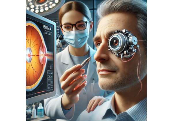
- External Visor or Glasses: This external component projects near-infrared light patterns through the eye’s pupil onto the implant. Some prototypes may integrate a camera that captures real-world images, processes them, and converts them into patterns suitable for the implant to interpret.
- Image Processing Unit: Typically a small, wearable computer that customizes the visual information before sending it to the visor. Through sophisticated algorithms, it can highlight edges or contrast, optimizing the stimulation pattern for clarity and comfort.
- Neural Adaptation Protocol: Because the device effectively rewires how visual signals reach the brain, patients undergo training sessions to help their visual cortex interpret these new inputs meaningfully.
Even though this system is cutting-edge, it doesn’t typically require bulky apparatus around the patient’s head. Manufacturers and research teams prioritize compact, lightweight designs to minimize intrusion into daily life and encourage consistent use.
Who May Benefit from This Technology
The PRIMA system is primarily intended for individuals who have suffered advanced central vision loss—commonly from conditions like dry AMD or certain inherited degenerative diseases. Those who can still process some visual information in peripheral regions may be especially well-suited, as the healthy peripheral retina can continue aiding in orientation while the implant works to restore aspects of central sight. For patients with near-total blindness from retinitis pigmentosa or advanced geographic atrophy in AMD, the technology can potentially provide tangible improvements in object recognition, reading large print, and performing tasks that hinge on central visual detail.
However, not every patient with advanced vision loss is automatically a candidate. Successful adaptation to the PRIMA system relies on:
- Intact Inner Retina: Sufficiently healthy bipolar, amacrine, and ganglion cells to transmit signals to the optic nerve.
- Stable Ocular Health: Limited inflammation, scarring, or other pathologies that could disrupt the subretinal space or compromise implant function.
- Motivation and Cognitive Capability: The ability to engage in post-implant rehabilitation, learning how to interpret artificially generated visual patterns.
When these conditions align, the PRIMA system can truly change a patient’s day-to-day experience, offering an unprecedented chance to reconnect with aspects of the visual world previously lost to disease.
How the PRIMA System Is Placed and Maintained
A high-tech device like the PRIMA implant is only as good as the surgical technique and rehabilitation protocols that accompany it. The journey from preoperative evaluation to gaining functional benefit may be lengthy, but it is carefully structured to maximize safety and improve outcomes. Below is a closer look at the detailed procedures, guidelines, and postoperative strategies that ensure the best possible performance of this advanced retinal prosthesis.
Preoperative Assessment and Candidacy Evaluation
Before scheduling any surgical intervention, prospective recipients go through a comprehensive screening and assessment phase. These evaluations aim to confirm that the individual has:
- Appropriate Retinal Structure: Imaging tests like optical coherence tomography (OCT) help determine if enough of the retinal pathway remains. Surgeons also look for any scarring, thickening, or fluid accumulation in the macular region that could undermine successful implantation.
- Visual History and Stability: A stable condition for at least several months is often crucial to prevent disease flare-ups that might complicate healing or the device’s integration.
- Systemic Health Clearance: Since the procedure involves delicate ocular surgery, patients with uncontrolled hypertension, diabetes, or bleeding disorders may face elevated risks. The surgical team coordinates with primary care providers to mitigate these factors.
- Psychological and Lifestyle Readiness: Installing and using a retinal prosthesis involves a learning curve. Medical professionals will discuss expectations, daily maintenance requirements (if any), and the necessity of vision training post-surgery.
If the patient and medical team determine that the PRIMA system is suitable, they move forward with scheduling the surgery. While many aspects may be covered by specialized research protocols or insurance (depending on region and policy), out-of-pocket expenses can remain substantial (more on that in a later section).
Surgical Procedure for Subretinal Placement
The key step—implantation of the photovoltaic chip—requires precision vitrectomy and subretinal surgery. Under local or general anesthesia, the surgeon typically performs the following:
- Vitrectomy: Removal of the vitreous humor (the gel filling the eye) to access the retina.
- Retinal Incision and Blebs: Creating a small bleb in the subretinal space, the area between the retina’s outer layers and the underlying structures like the choroid. This is where the implant will sit.
- Chip Positioning: Using specialized microinstruments, the surgeon gently slides the PRIMA chip under the retina, ensuring alignment with the macular region. Exact centration is crucial for best functional outcomes.
- Fluid Exchange and Closure: Once the chip is in place, the surgeon may replace any fluid to stabilize the retina, close incisions, and place sutures if needed.
The complexity of this procedure varies depending on the patient’s retinal anatomy, the surgeon’s expertise, and the version of the PRIMA implant used. Some individuals might have coexisting ocular issues (e.g., cataract or epiretinal membranes) that the surgeon addresses simultaneously.
Postoperative Recovery and Initial Assessment
Following surgery, patients usually remain under close observation, especially over the first few days. Eye drops containing antibiotics and steroids help prevent infection and manage inflammation. In some cases, posture guidelines (like keeping the head in a certain position) may be recommended to ensure stable chip integration. Each clinic may have its own protocol, but typical follow-up visits involve:
- Retinal Imaging (OCT): Checking that the implant remains well-positioned without causing significant retinal detachment or fluid accumulation.
- Visual Function Testing: Basic tests to see if the patient can detect light patterns or simple shapes using the new implant. This early stage is crucial for calibrating external components (like the camera or projector) to the patient’s unique retinal response.
- Monitoring for Complications: Includes watching for elevated intraocular pressure, suture leaks, or signs of infection.
Initial visual improvements might be subtle or even imperceptible until the external camera and image processing are fine-tuned and the patient starts a structured rehabilitation program.
Rehabilitation and Ongoing Training
Perhaps the most significant factor determining success with the PRIMA system is the dedication to vision rehabilitation. Even if the surgery is flawless, the user’s brain must adapt to the new mode of seeing. Rehabilitation typically includes:
- Device Calibration: Technicians adjust the external glasses or head-mounted display to deliver optimal brightness, contrast, and resolution. Careful calibration can help minimize glare or flicker that might confuse the user’s visual system.
- Visual Exercises: These training sessions may involve focusing on high-contrast letters or shapes on a screen, learning to interpret the pixelated images that appear in the central field. Over time, the brain can learn to associate these patterns with real-world objects.
- Occupational Therapy: Rehabilitation often extends beyond pure visual tasks. Occupational therapists might help the user practice daily skills—like identifying household objects, reading large-print text, or navigating mild obstacles—while wearing the PRIMA setup.
- Gradual Expansion of Activities: As the patient gains confidence, they often progress to more complex tasks, such as reading short words, distinguishing faces at a close distance, or using the device under varied lighting conditions.
Because the PRIMA system is designed for partial restoration of sight, it’s important to set realistic goals. The best outcomes typically occur when patients integrate the device as a complement to any remaining natural vision, along with other assistive strategies. Over time, many individuals become more comfortable navigating their environment and performing tasks that rely on central detail, a function previously compromised by their retinal disease.
Long-Term Device Maintenance
The subretinal chip is intended as a long-term implant that doesn’t need routine maintenance once placed. However, external components such as the wearable camera or the image processing unit may require occasional software updates or hardware adjustments. If the manufacturer releases improved versions of the external hardware, patients might have the option to upgrade while retaining the implanted subretinal chip.
Follow-up appointments remain essential to watch for any shifting or damage to the implant, as well as changes in the underlying retinal condition that could affect performance. Some conditions—like ongoing atrophic progression in AMD—may alter the retina’s thickness or cause atrophy around the implant, potentially impacting its effectiveness. Early detection of any complications or shifts in ocular anatomy can ensure timely interventions and maintain the best visual outcomes possible.
Recent Discoveries and Clinical Trials Informing PRIMA’s Progress
Like most innovative biomedical technologies, the PRIMA system is under constant scrutiny from scientists, engineers, and clinicians eager to validate and refine its efficacy. Initial feasibility studies and pilot clinical trials pave the way for broader, more rigorous investigations that evaluate safety, performance, user satisfaction, and long-term stability. Below is an overview of some research milestones and the evolving body of knowledge surrounding PRIMA’s capabilities.
Early Feasibility Studies and Prototypes
The underlying concept of subretinal photovoltaics has been around for some time, but the first-generation PRIMA prototypes aimed to prove that microchips powered purely by external infrared light could stimulate the retina meaningfully. Small animal studies, often in models of retinal degeneration, confirmed that an implant with many tiny pixels could elicit distinct local neural responses when properly illuminated. Encouraged by these results, research teams refined the pixel design, improving aspects like:
- Pixel Density: A key factor in resolution, higher pixel counts should yield more detailed vision. However, packing too many micro-photodiodes close together can raise manufacturing complexity and reduce reliability.
- Light Sensitivity: Achieving consistent neural stimulation under typical indoor or outdoor lighting was challenging. That’s why an external projector emitting near-infrared light is often needed, delivering focused, controlled illumination to the implant.
- Biocompatibility: Ensuring the device causes minimal local inflammation or damage to surrounding tissue was paramount. Robust encapsulation materials and special coatings on the microchips emerged to safeguard both the implant and the delicate retinal environment.
International Pilot Trials in Humans
The real test for any visual prosthesis is how well it works in living patients coping with advanced vision loss. Early-phase human trials, often limited to small cohorts, have consistently aimed to answer critical questions:
- Surgical Feasibility: Can ophthalmic surgeons implant these chips with manageable complication rates and minimal damage to the residual retina?
- Functional Improvement: Do patients experience meaningful improvements in visual tasks—like letter recognition or shape discrimination—relative to their baseline performance without the device?
- Reliability and Durability: Will the implant continue functioning steadily over months or years without breakdown, infection, or movement within the subretinal space?
Several high-profile studies in Europe, and more recently in North America, have yielded promising initial data. Participants with profound central vision loss due to advanced AMD reported perceiving flashes, lines, and letters while using the PRIMA device. Over time and with dedicated training, some advanced to reading short words or identifying simple objects on a computer screen. The reported adverse events were generally mild—mostly surgical concerns like minor bleeding or transient inflammation—reinforcing the potential for safe, stable implantation.
Ongoing Research Expanding PRIMA’s Capabilities
As the system’s fundamental feasibility gains traction, multiple lines of research now target enhancements that could broaden PRIMA’s utility or accelerate patient adoption:
- Higher Resolution Arrays: Next-generation prototypes promise to pack more photovoltaic pixels into the same subretinal area, aiming for clearer, more naturalistic images. This might demand advanced microfabrication techniques or even 3D layering of diodes.
- Smart Software for Image Processing: The external imaging system, effectively playing the role of an artificial photoreceptor layer, can incorporate advanced algorithms. Machine learning approaches might highlight edges, enhance contrast, or filter out extraneous background flicker to yield crisper on-chip stimulation patterns.
- Integration with Eye-Tracking Sensors: Future iterations might incorporate eye-tracking to adjust the implant’s focal region on the fly, or possibly to stabilize images during rapid eye movements.
- Cortical-Level Training Techniques: Beyond standard visual rehab, neuroscientists explore brain-centric exercises that reinforce neural plasticity. These might include immersive virtual reality (VR) simulations that help the brain adapt to PRIMA’s unique stimuli.
Experts emphasize that rigorous, large-scale clinical trials remain necessary to confirm improvements in reading speed, facial recognition, or daily independence metrics. Yet the overall direction is undeniably positive: each success story and incremental engineering breakthrough cements the PRIMA system’s standing as a leading candidate in the quest to restore functional vision in advanced retinal disease.
Collaborations and Funding Support
It’s worth noting that no single entity alone propels PRIMA technology forward. Multi-institution collaborations often bring together universities, biotech companies, philanthropic foundations, and government agencies. In some cases, cross-disciplinary synergy fosters breakthroughs at the interface of microelectronics, ophthalmology, and neuroscience. Public-private partnerships help offset the substantial development costs and expedite translation from the lab to the clinic.
Such cooperative energy also benefits patients, as consistent data collection and peer-reviewed publication accelerate acceptance among surgeons, rehabilitation specialists, and insurers. Over time, this momentum can drive broader access, especially if new research underscores robust improvements in functional vision and cost-effectiveness.
Measuring Gains While Managing Risks in the PRIMA Experience
The ultimate value of the PRIMA system rests on its real-world benefits for those facing debilitating vision loss. While preliminary studies have found that many recipients experience meaningful improvements, no intervention exists without potential drawbacks. Below, we dive into the common efficacy markers, documented advantages for daily living, and crucial considerations about device safety.
How Clinicians Evaluate Outcomes
In standard ophthalmic practice, assessing vision typically relies on metrics like best-corrected visual acuity (BCVA) or reading speed. However, such measures were never designed for a subretinal prosthesis scenario where an external camera, specialized glasses, and neural adaptation come into play. Researchers and clinicians, therefore, employ additional or modified parameters:
- Light Perception and Localization: Initially, can the patient detect light? Can they discern where in their visual field light is brightest?
- Pattern Recognition: Over time, does the user manage to differentiate lines, shapes, or simple letters? This is often tested using custom software that displays high-contrast patterns.
- Real-World Functional Tasks: From reading large print to picking up an object from a table, practical tasks measure whether partial vision improvements translate to everyday benefits.
- Patient-Reported Quality of Life (QOL): Surveys and interviews can reveal whether small increments in visual function significantly ease daily living or raise the user’s self-confidence.
These data points guide iterative refinements of the PRIMA system and shape post-implant rehabilitation strategies.
Reported Benefits for Daily Activities
Though every patient’s outcome differs, certain consistent themes arise among early adopters:
- Object Identification and Reading: Many find that even the ability to read large letters—like product labels or short text passages—empowers them to be more self-reliant.
- Spatial Awareness: Those with a complete or near-complete central blind spot can glean essential details about their environment, reducing reliance on others. Being able to detect a piece of furniture or see a door handle offers new autonomy.
- Enhanced Social Interaction: Recognizing faces or at least perceiving the presence of someone in front can lead to stronger social ties, alleviating a major frustration in advanced AMD patients.
- Gradual Vision Expansion: With consistent use and neural training, some recipients report a progressive refinement of perception, going from vague shapes to increasingly distinct outlines or letters.
Overall, such gains tend to come incrementally rather than immediately after surgery. A blend of device calibration, structured rehab, and personal perseverance is often the key to unlocking the full potential of the technology.
Potential Risks and Side Effects
On the surgical side, standard complications of retinal procedures apply—risks of infection, hemorrhage, and retinal detachment. Although these are uncommon with modern techniques, they cannot be dismissed. Patients must also be prepared for:
- Postoperative Discomfort: Mild to moderate eye irritation or visual distortions as the implant settles.
- Device Dislocation or Movement: If the implant shifts from its intended macular location, the prosthetic effect may weaken or cause confusing visual input.
- Possible Chronic Inflammation: Rarely, the subretinal chip may cause ongoing low-grade inflammation, requiring anti-inflammatory medication or device removal in extreme cases.
- Technical Glitches: Malfunctions in the external projection system or power supply may hamper functionality. While the subretinal chip is a passive device, the external electronics are not immune to hardware or software issues.
Proactive risk management includes thorough preoperative screening, well-honed surgical skills, and meticulous postoperative follow-up. In many studies, an overwhelming majority of adverse events were mild or resolved with standard ophthalmic care.
Encouraging Long-Term Stability and Success
Given that AMD or inherited retinal diseases often progress further over time, how well the implant can adapt remains a point of active research. If atrophy spreads beyond the initially affected macular region, the area under the PRIMA chip might eventually suffer from reduced blood supply or other changes. That said, the subretinal placement is designed with the hope of stable integration, and early data suggests that once the device is in position, it remains relatively well tolerated.
Crucially, the patient’s dedication to using the device in everyday life—and to refining their visual skills with therapy—often dictates how far the system’s benefits extend. Although the PRIMA system cannot halt the underlying degenerative disease itself, it can complement other possible treatments (like nutritional supplements, anti-VEGF injections for wet AMD, or future gene therapies) aimed at preserving any remaining natural vision.
Price Points and Funding Options for the PRIMA Prosthesis
Costs for the PRIMA system can vary depending on the clinic, region, insurance policies, and whether the device is still considered investigational. A ballpark figure for the implant procedure and associated hardware might range from around \$30,000 to \$70,000 or more per eye. Ancillary fees for surgical facility use, postoperative visits, and rehabilitation programs can add to the final total. Some patients rely on private insurance or government healthcare coverage to offset these expenses, especially if PRIMA implantation is part of a recognized clinical trial. Certain manufacturers or foundations may provide partial grants or financial assistance, and others offer monthly payment plans to reduce the immediate burden on patients seeking this transformative technology.
Disclaimer: This article is for informational purposes only and does not replace professional medical advice, diagnosis, or treatment. Always consult a qualified healthcare provider regarding any questions you may have about a medical condition or treatment.
We warmly encourage you to share this article with friends, family, or online communities that might benefit from learning about the PRIMA retinal prosthesis. Feel free to use our Facebook and X (formerly Twitter) share buttons—or any other platforms you prefer—to help others discover how this remarkable technology is giving new hope to individuals living with advanced vision loss!


