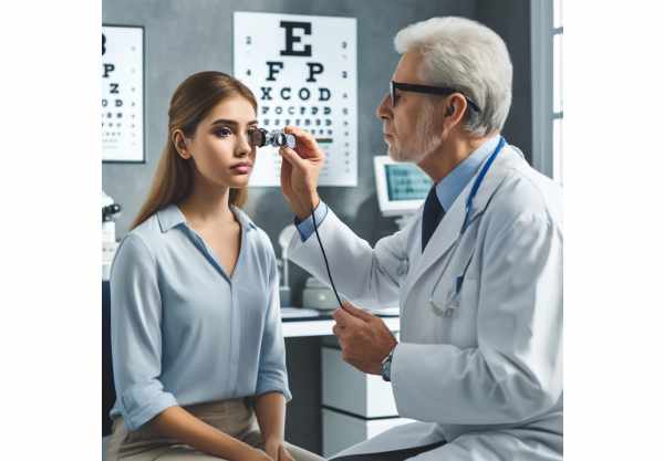
What Is Adie’s pupil?
Adie’s pupil, also known as tonic pupil, is a neurological condition in which the eye’s pupil reacts abnormally to light. This condition usually manifests as a unilateral, enlarged pupil that reacts slowly to direct light but constricts more quickly during near vision tasks, a phenomenon known as light-near dissociation. While Adie’s pupil is typically benign, it can occasionally indicate underlying systemic or neurological conditions. Awareness and early detection are critical for effectively managing symptoms and identifying underlying health issues. A timely diagnosis can help prevent complications and improve the quality of life for those living with this condition.
Insights into Adie’s Pupil
Adie’s pupil is caused by damage to the postganglionic fibers of the eye’s parasympathetic innervation. These nerve fibers control the pupil’s response to light. When these fibers are damaged, the pupil loses its ability to constrict properly in response to light, resulting in the distinctive tonic, or slow-reacting pupil.
Epidemiology and Risk Factors
Adie’s pupil primarily affects young adults, with a higher incidence in women than in men. The exact cause of nerve damage is frequently unknown, but it can be linked to viral or bacterial infections that cause inflammation and damage to the ciliary ganglion, a collection of nerve cells that regulates eye movements and pupil responses. Trauma, surgery, or systemic diseases such as diabetes can all play a role in Adie’s pupil development.
Pathophysiology
The underlying cause of Adie’s pupil is selective damage to the postganglionic parasympathetic fibers that supply the sphincter pupillae muscle. This damage causes muscle denervation, resulting in an abnormally large pupil (mydriasis) that reacts slowly to light. Over time, the remaining functional nerve fibers may undergo a process known as aberrant regeneration. During this process, some fibers intended for other functions, such as accommodation, incorrectly innervate the sphincter pupillae muscle, resulting in light-near dissociation, in which the pupil responds better to near stimuli than to light.
Clinical Manifestations
Adie’s pupil is typically a unilateral condition, but it can occasionally affect both eyes. The main clinical feature is a dilated pupil that reacts poorly to light but may constrict during accommodation (focusing on a nearby object). This can cause several symptoms:
- Blurred Vision: A dilated pupil can make it difficult to focus, resulting in blurred vision, particularly for close-up tasks.
- Photophobia: Increased sensitivity to light caused by an enlarged pupil, which allows more light to enter the eye.
- Difficulty with Night Vision: The impaired response to light can make it difficult to see in low-light situations.
- Anisocoria: A noticeable difference in pupil size between the affected and unaffected eyes, which may be cosmetically concerning for some patients.
Differential Diagnosis
Several other conditions can present with similar symptoms to Adie’s pupil, necessitating an extensive differential diagnosis:
- Third Nerve Palsy: This condition can cause a dilated pupil, but it is usually accompanied by other symptoms such as ptosis (drooping of the eyelid) and restricted eye movement.
- Argyll Robertson Pupil: Characterized by small, irregular pupils that constrict poorly in the presence of light but respond normally to accommodation. This condition is frequently associated with neurosyphilis.
- Pharmacological Dilation: Taking certain medications, such as anticholinergics, can cause pupil dilation. A detailed medication history is required to rule out this cause.
- Iris Sphincter Trauma: Direct injury to the iris can result in a fixed, dilated pupil. This is distinguished by a history of trauma and examination results.
Histopathology
The number of ganglion cells and nerve fibers in Adie’s pupil has decreased, according to histopathology. The remaining fibers may exhibit signs of degeneration and fibrosis. This pathological process emphasizes the importance of early detection and treatment in order to maintain as much nerve function as possible.
Prognosis and Complications
The prognosis for Adie’s pupil is generally favorable, particularly in isolated cases where no underlying systemic disease exists. Most patients adjust to the visual changes over time, and the condition tends to stabilize or improve gradually. However, complications may occur if the condition is part of a larger neurological disorder. Regular follow-up is essential for detecting changes and effectively managing symptoms.
Public Health Implications
Although Adie’s pupil is uncommon and usually harmless, it emphasizes the importance of regular eye exams and neurological assessments, particularly for people with risk factors like a history of infections or systemic diseases. Public health initiatives should prioritize raising awareness of the signs and symptoms of neurological conditions affecting the eyes, encouraging timely medical consultations, and ensuring access to comprehensive eye care services.
Diagnostic methods
Diagnosing Adie’s pupil necessitates a thorough clinical evaluation and the application of various diagnostic techniques to distinguish it from other conditions with similar symptoms.
Clinical Examination
The first step in diagnosing Adie’s pupil is a thorough eye examination by an ophthalmologist. This includes:
- Pupil Assessment: A thorough evaluation of the students’ responses to light and near stimuli. Adie’s pupil is typically dilated and constricts poorly in direct light but better in accommodation (near vision tasks).
- Slit-Lamp Examination: This allows for a more detailed examination of the anterior eye structures and helps rule out other causes of anisocoria (unequal pupil sizes).
Pharmaceutical Testing
Pharmacological tests are commonly used to confirm the diagnosis of Adie’s pupil.
- Pilocarpine Test: Both eyes receive a diluted solution of pilocarpine (0.1% or 0.125%). In Adie’s pupil, the denervated sphincter muscle is extremely sensitive to the drug, causing the tonic pupil to constrict significantly more than the normal pupil.
- Cocaine Test: This test distinguishes Adie’s pupil from Horner’s syndrome. In Horner’s syndrome, the affected pupil does not dilate in response to cocaine, whereas Adie’s pupil usually does not change significantly.
Imaging Techniques
Advanced imaging techniques can provide additional insights into the underlying cause and extent of nerve damage.
- Magnetic Resonance Imaging (MRI): MRI scans can be used to visualize the ciliary ganglion and associated nerve pathways, assisting in the identification of any structural abnormalities or lesions that may be causing the condition.
- Computed Tomography (CT) Scan: CT scans provide detailed images of the brain and orbit, which can aid in determining intracranial causes of pupillary abnormalities.
Electrophysiological Tests
Electrophysiological tests, such as visual evoked potentials (VEP) and electroretinography (ERG), can evaluate the functional integrity of the visual pathways and retina, revealing additional information about the extent of neurological involvement.
Histopathologic Examination
In rare cases where a biopsy is performed, histopathological examination of the ciliary ganglion may reveal decreased ganglion cell count, nerve fiber degeneration, and fibrosis. This can help to confirm the diagnosis and shed light on the pathophysiological mechanisms involved.
Effective Treatments for Adie’s Pupil
Standard Treatments
The management of Adie’s pupil is primarily concerned with symptom relief and improving the patient’s quality of life. While there is no cure for the condition, the following treatments can help manage the symptoms:
- Pilocarpine Eye Drops: These drops constrict the dilated pupil, relieving symptoms like photophobia and blurred vision. Patients are typically given a diluted concentration (0.1% or 0.125%) to avoid excessive constriction and minimize side effects.
- Reading Glasses: For patients who have difficulty with near vision due to accommodative issues, reading glasses or bifocals can be prescribed to improve visual clarity when performing close-up tasks.
- Tinted Lenses: Wearing sunglasses or tinted lenses can help with photophobia by reducing glare and light sensitivity.
Innovative and Emerging Therapies
Research into new and innovative treatments for Adie’s student is ongoing, with several promising approaches being explored.
- Botulinum Toxin Injections: If pilocarpine eye drops are ineffective or poorly tolerated, botulinum toxin injections can be used to cause miosis (pupil constriction). This treatment temporarily paralyzes the muscles that cause pupil dilation, thereby reducing anisocoria and improving vision.
- Neuroprotective Agents: Experimental treatments involving neuroprotective agents seek to preserve remaining nerve function and prevent further degeneration. These therapies are still in the early stages of development, but they show promise for treating nerve-related conditions such as Adie’s pupil.
- Regenerative Medicine: Advances in regenerative medicine, such as stem cell therapy and gene therapy, could provide future treatments for Adie’s pupil. These approaches seek to repair or replace damaged nerve cells, potentially restoring normal pupil function.
- Visual Rehabilitation: Comprehensive visual rehabilitation programs, which include occupational therapy and adaptive devices, can assist patients in dealing with the visual challenges associated with Adie’s pupil, thereby improving their overall quality of life.
- Customized Optical Solutions: Advances in optical technology have resulted in the development of customized contact lenses and intraocular lenses that improve vision for patients suffering from specific refractive errors and accommodative issues related to Adie’s pupil.
Lifestyle and Supportive Measures.
In addition to medical treatments, lifestyle changes and supportive measures can play a critical role in managing Adie’s pupil:
- Avoid Bright Lights: To alleviate photophobia symptoms, patients should avoid bright lights and use dimmer switches or lower-wattage bulbs in their homes.
- Regular Eye Check-Ups: Routine follow-up visits with an ophthalmologist are required to monitor the condition and adjust treatments as necessary.
- Patient Education: Educating patients about their condition and the importance of following prescribed treatments can help them understand and engage in managing Adie’s pupil effectively.
Essential Preventive Measures
- Regular Eye Exams: Seek comprehensive eye exams from an ophthalmologist to detect early signs of pupil abnormalities and other eye conditions.
- Protect Eyes from Infections: Maintain proper hygiene and avoid contact with people who have contagious eye infections. Wear protective eyewear in situations where eye injuries or infections are likely.
- Manage Systemic Health Conditions: Maintain control of chronic health conditions such as diabetes, hypertension, and autoimmune diseases by visiting the doctor on a regular basis and following the prescribed treatment plan.
- Avoid Head and Eye Trauma: Wear appropriate safety gear when participating in activities that could result in head or eye injuries, such as sports or hazardous work environments.
- Educate on Symptom Awareness: Raise patient and healthcare provider awareness of the symptoms of Adie’s pupil and other neuro-ophthalmic conditions in order to promote early diagnosis and intervention.
- Healthy Lifestyle Choices: To support overall neurological and ocular health, maintain a healthy lifestyle that includes a balanced diet, regular exercise, and abstaining from smoking and drinking excessively.
- Use Proper Lighting: When reading or performing tasks, use adequate lighting to reduce eye strain and accommodate Adie’s pupil’s visual difficulties.
- Sunlight Protection: Wear UV-blocking sunglasses to protect your eyes from the potential damage caused by prolonged sun exposure.
Trusted Resources
Books
- “Neuro-Ophthalmology: Diagnosis and Management” by Grant T. Liu, Nicholas J. Volpe, and Steven L. Galetta
- “Clinical Neuro-Ophthalmology: A Practical Guide” by Ambar Chakravarty
- “Neuro-Ophthalmology Illustrated” by Valerie Biousse and Nancy J. Newman
Online Resources
- American Academy of Ophthalmology
- National Eye Institute
- Mayo Clinic – Eye Health
- MedlinePlus – Eye Diseases
- All About Vision
- WebMD – Eye Health


