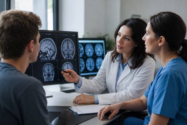
A brain scan can sometimes show medical problems that affect mood, attention, behavior, or thinking, but it usually cannot diagnose depression, anxiety, ADHD, or autism by itself. These conditions are real brain-based conditions, yet they are diagnosed mainly through symptoms, development, functioning, history, clinical interviews, questionnaires, and sometimes psychological or neuropsychological testing.
That distinction can be confusing. Brain imaging is powerful, and research studies often find average brain differences in groups of people with depression, anxiety, ADHD, or autism. But a group-level difference is not the same as a dependable diagnostic test for one person sitting in a clinic. A normal scan does not mean symptoms are “not real,” and an unusual scan does not automatically explain a mental health or neurodevelopmental diagnosis.
Table of Contents
- What Brain Scans Can and Cannot Diagnose
- Why Brain Scans Are Not Diagnostic
- What Research Scans May Show
- When Doctors Order Brain Imaging
- How These Conditions Are Diagnosed
- Scan Types and Commercial Claims
- Questions Before Getting a Scan
- When Symptoms Need Urgent Care
What Brain Scans Can and Cannot Diagnose
A brain scan can show structure, blood flow, metabolism, or electrical patterns, depending on the test, but it cannot usually show a label such as “depression,” “anxiety,” “ADHD,” or “autism” in a direct way. Imaging is most useful when the clinician is asking whether another medical or neurological problem could be contributing to the symptoms.
For example, an MRI may help detect a tumor, stroke, bleeding, inflammation, hydrocephalus, major structural abnormality, some developmental brain differences, or certain patterns of brain atrophy. CT can be useful in urgent situations, especially after head injury or when bleeding is a concern. PET can help in selected dementia, epilepsy, cancer, or movement disorder evaluations. EEG is not a brain scan in the usual imaging sense, but it can detect seizure-related electrical activity.
That is different from diagnosing a psychiatric or neurodevelopmental condition. Depression is diagnosed by patterns such as persistent low mood, loss of interest, sleep or appetite change, guilt, slowed thinking, low energy, and suicidal thoughts. Anxiety disorders are diagnosed by patterns of fear, worry, panic, avoidance, physical arousal, and impairment. ADHD is diagnosed by developmentally inappropriate inattention, impulsivity, or hyperactivity that began in childhood and affects more than one setting. Autism is diagnosed by differences in social communication, restricted or repetitive behaviors, sensory differences, developmental history, and daily functioning.
A scan may still matter in the right context. If a person has new personality change, sudden confusion, seizures, abnormal neurological findings, rapidly worsening memory, severe headaches, or symptoms that began after a head injury, brain imaging may be part of a careful workup. A dedicated brain MRI is often the preferred structural scan when doctors need detailed images of brain anatomy, while a brain CT scan is often used when speed is important.
The practical takeaway is simple: a brain scan may help rule in or rule out certain medical causes, but it does not replace a clinical diagnostic evaluation for depression, anxiety, ADHD, or autism.
Why Brain Scans Are Not Diagnostic
Brain scans are not standard diagnostic tests for these conditions because the findings are usually too variable, overlapping, and nonspecific for individual diagnosis. The brain is involved in every thought, mood, sensation, and behavior, but that does not mean one scan can reliably separate one mental health condition from another.
One problem is overlap. Depression, anxiety, ADHD, autism, sleep deprivation, trauma, chronic stress, substance use, medication effects, learning differences, and medical illness can all affect attention, mood, motivation, sleep, and emotional regulation. They can also influence brain networks involved in arousal, reward, threat detection, executive function, and sensory processing. A brain pattern that appears more often in one group may also appear in people with other conditions or in people without a diagnosis.
Another issue is individual variation. Brains differ naturally by age, sex, development, handedness, sleep quality, hormone status, medication exposure, substance use, education, stress, and many other factors. Even scanner type, image-processing method, and study design can affect results. That makes it hard to create a single reliable cutoff that says, “This scan proves ADHD,” or “This scan rules out anxiety.”
This is why research findings need to meet a high bar before becoming clinical tests. A useful diagnostic biomarker must work well across different clinics, scanners, populations, ages, and coexisting conditions. It also needs to change medical decisions in a way that improves outcomes. Many promising brain-based measures are being studied, but most are not ready to guide routine diagnosis for an individual patient. For a broader explanation of this standard, see brain and mental health biomarkers.
There is also a difference between correlation and diagnosis. Suppose a study finds that, on average, people with major depression have differences in a mood-related network. That does not mean every person with depression has that pattern, that every person with that pattern is depressed, or that the scan can identify the cause of symptoms. It may reveal a clue about biology, not a complete diagnostic answer.
For the same reason, a normal scan should not be used to dismiss symptoms. Many people with severe depression, disabling anxiety, ADHD, or autism have structurally normal MRI results. Their symptoms can still be clinically significant and deserving of care.
What Research Scans May Show
Research scans can show meaningful patterns across groups, even when those patterns are not precise enough for routine individual diagnosis. This is where much of the confusion comes from: brain imaging can tell scientists a great deal about mental health and neurodevelopment, but that information does not always translate into a clinical “yes or no” test.
In depression research, MRI and functional MRI studies have examined networks involved in mood regulation, reward, self-focused thought, sleep, stress response, and cognitive control. Some studies report differences in areas such as the prefrontal cortex, hippocampus, amygdala, anterior cingulate cortex, and default mode network. These findings help researchers study why depression can affect memory, motivation, threat sensitivity, rumination, and emotional recovery. They do not, on their own, diagnose a depressive disorder.
In anxiety research, studies often focus on threat processing, fear learning, bodily arousal, avoidance, and regulation of worry. Brain regions and networks involving the amygdala, insula, prefrontal cortex, and anterior cingulate are often discussed. Yet anxiety disorders are diverse. Panic disorder, generalized anxiety disorder, social anxiety disorder, OCD, PTSD, phobias, and illness anxiety have different symptom patterns and can overlap with depression, ADHD, autism, trauma, and medical conditions.
In ADHD research, imaging studies often examine attention networks, executive control, reward processing, motor regulation, and timing. Some group studies show differences in frontostriatal circuits, cerebellar regions, cortical maturation, white matter pathways, or resting-state connectivity. Machine-learning approaches have been tested to classify ADHD from imaging data, but results can vary by dataset, site, age group, preprocessing method, and whether the model generalizes to new populations.
In autism research, imaging studies have explored brain growth patterns, social communication networks, sensory processing, connectivity, white matter organization, and developmental differences. Some MRI-based classification studies have reported moderate performance in research settings, but autism is highly heterogeneous. Two autistic people can have very different language profiles, sensory needs, cognitive strengths, support needs, coexisting ADHD or anxiety, and developmental histories.
The most useful way to read this research is not “the scan can diagnose the condition.” A better interpretation is: “Brain imaging is helping scientists understand possible biological pathways, subgroups, and treatment targets.” That work may eventually lead to more personalized care, but it is not the same as a routine diagnostic scan available for everyday clinical decision-making.
When Doctors Order Brain Imaging
Doctors usually order brain imaging for mental health or developmental symptoms when something about the history, exam, age of onset, or symptom pattern suggests a possible neurological or medical cause. Imaging is not ordered simply because someone feels depressed, anxious, inattentive, socially different, or overwhelmed.
Brain imaging may be considered when symptoms are new, sudden, severe, unusual for the person, or accompanied by neurological signs. Examples include new seizures, one-sided weakness, vision loss, trouble speaking, severe balance problems, fainting with concerning features, severe or unusual headaches, head injury, rapidly worsening memory, sudden confusion, or major personality change later in life.
Imaging may also be used when clinicians are evaluating conditions that can mimic or complicate psychiatric symptoms. These can include stroke, brain tumor, traumatic brain injury, epilepsy, multiple sclerosis, dementia, autoimmune or inflammatory brain disease, hydrocephalus, infection, or complications from substance use. In these situations, the scan is not being used to “find depression.” It is being used to check whether another brain condition might be present.
A simplified comparison can help clarify the usual roles of common tests:
| Test | What it can show | Common role | Key limitation |
|---|---|---|---|
| MRI | Detailed brain structure, some tissue changes | Evaluating neurological causes, memory loss, tumors, inflammation, stroke patterns | Does not diagnose depression, anxiety, ADHD, or autism by itself |
| CT | Bleeding, fracture, major structural problems | Urgent assessment after head injury, stroke concern, acute neurological symptoms | Less detailed than MRI for many non-urgent brain questions |
| PET | Metabolism or specific molecular targets | Selected dementia, epilepsy, cancer, or movement disorder evaluations | Not a routine psychiatric diagnostic test |
| EEG | Electrical activity patterns | Seizure evaluation, certain episodes of altered awareness | Not a general test for mood, anxiety, ADHD, or autism |
If a scan is abnormal, the next step depends on the finding. Some abnormalities are incidental and unrelated to symptoms. Others need follow-up with a neurologist, neurosurgeon, psychiatrist, developmental specialist, or primary care clinician. For many people, an imaging result is only one piece of a larger workup, not the final answer.
How These Conditions Are Diagnosed
Depression, anxiety, ADHD, and autism are diagnosed by matching symptoms and life history to established clinical criteria, while also checking for other explanations. A good evaluation looks at what the person experiences, when symptoms began, how long they have lasted, how much impairment they cause, and what else might be contributing.
For depression, clinicians typically ask about mood, interest, sleep, appetite, energy, concentration, guilt, movement changes, thoughts of death or suicide, medical history, medications, substance use, grief, bipolar symptoms, trauma, and physical health. Screening tools can help organize symptoms, but the diagnosis is not just a questionnaire score. A fuller discussion of depression screening and diagnosis explains how clinicians use scores alongside interviews and follow-up assessment.
For anxiety, the evaluation focuses on worry, panic, avoidance, physical symptoms, triggers, duration, impairment, trauma exposure, obsessive thoughts, compulsions, social fear, phobias, sleep, substances, and medical conditions that can mimic anxiety. Thyroid disease, heart rhythm problems, medication side effects, caffeine, stimulant use, withdrawal, and respiratory problems can sometimes look like anxiety. Screening can be useful, but clinical context is essential. For more detail, see anxiety screening.
For ADHD, diagnosis requires more than trouble focusing. Clinicians look for a long-standing pattern of inattention, hyperactivity, or impulsivity that began in childhood, appears in more than one setting, and causes functional problems. They also consider sleep deprivation, anxiety, depression, trauma, learning disabilities, substance use, thyroid disease, hearing or vision issues, and high-stress environments. Adults may need a careful developmental history because childhood symptoms are not always obvious in hindsight.
For autism, diagnosis is based on social communication differences, restricted or repetitive behaviors, sensory patterns, developmental history, support needs, and how traits appear across settings. The evaluation may include caregiver history, school records, direct observation, standardized tools, speech-language assessment, cognitive testing, adaptive functioning assessment, or occupational therapy input. Autism cannot be ruled in or ruled out by a normal MRI.
Testing is often most useful when it answers a specific practical question: What condition best explains the symptoms? What else needs to be ruled out? What supports are needed at school, work, or home? What treatment options fit the person’s needs? Brain imaging sometimes contributes to that process, but the core diagnosis is still clinical.
Scan Types and Commercial Claims
Commercial brain scan claims should be approached carefully, especially when they promise to diagnose depression, anxiety, ADHD, autism, trauma, addiction, or personality traits from a scan alone. Some clinics market colorful brain images as if they offer a definitive answer, but impressive images are not the same as validated diagnosis.
SPECT scans, for example, show patterns related to blood flow, and some commercial services claim to use SPECT to identify psychiatric conditions or guide medication choices. SPECT has legitimate medical uses, but it is not a standard stand-alone diagnostic test for depression, anxiety, ADHD, or autism. A person considering this route should ask whether the test is recommended by mainstream clinical guidelines for their specific question, how the result will change treatment, and whether the claim has been validated in independent clinical populations. A deeper review of SPECT scans for brain disorders can help separate established uses from broader marketing claims.
qEEG, sometimes called “brain mapping,” measures electrical activity from the scalp and compares patterns with reference databases. It may have research or selected clinical uses, and EEG can be very important for seizure evaluation. But qEEG is not a routine diagnostic test for ADHD, depression, anxiety, or autism. It can generate complex-looking reports that may be overinterpreted if they are not tied to a careful clinical evaluation. For more context, see qEEG brain mapping claims.
Functional MRI is another source of confusion. fMRI can show changes in blood oxygen signals while a person rests or performs a task. It is a major research tool and is sometimes used clinically for presurgical brain mapping. But standard clinical fMRI is not used to diagnose most psychiatric or neurodevelopmental conditions. Research fMRI can reveal patterns across groups, but individual-level classification remains limited.
PET scans are also sometimes discussed in mental health. PET is valuable in certain neurological and medical contexts, such as selected dementia evaluations, epilepsy surgery planning, and cancer care. In depression, anxiety, ADHD, and autism, PET findings are mostly research tools rather than routine diagnostic tests.
The safest rule is to be skeptical of any service that claims a scan can replace a detailed clinical evaluation. Imaging can be useful when ordered for the right reason, interpreted by qualified specialists, and integrated with history and examination. It becomes less useful when it is sold as a shortcut to a complex diagnosis.
Questions Before Getting a Scan
Before paying for or agreeing to brain imaging for mood, attention, or developmental concerns, ask what specific clinical question the scan is meant to answer. A good reason for imaging is usually concrete, medically relevant, and tied to next steps.
Useful questions include:
- What condition are we trying to rule out or confirm?
- Is this scan recommended by clinical guidelines for my symptoms?
- What finding would change my diagnosis, treatment, referral, or safety plan?
- What are the possible false positives or incidental findings?
- What are the risks, costs, radiation exposure, or contrast-related concerns?
- Who will interpret the scan, and what are their qualifications?
- Will the result be reviewed alongside my history, exam, and symptoms?
These questions are especially important when the scan is not covered by insurance or is being offered by a clinic that also sells a specific treatment package. A test can look scientific and still have limited clinical value for your situation.
Also consider what evaluation has already been done. For many people, the more useful first steps are a primary care visit, mental health evaluation, sleep assessment, medication review, substance use review, lab testing when appropriate, school or workplace history, and standardized symptom measures. For attention concerns, clinicians may consider ADHD, anxiety, sleep loss, depression, trauma, learning disability, hearing or vision issues, and medical causes. For social communication or sensory concerns, a developmental evaluation may be more informative than imaging.
Cost is another practical issue. MRI, PET, SPECT, and private “brain mapping” services can be expensive. If the result is unlikely to change care, the money may be better spent on a thorough diagnostic assessment, therapy, coaching, sleep evaluation, occupational therapy, school supports, or evidence-based treatment.
A second opinion can be helpful when a scan is recommended for unclear reasons or when a commercial scan report seems to make broad claims. The best interpretation comes from a clinician who can connect the imaging result to the person’s actual symptoms, exam findings, medical history, and goals.
When Symptoms Need Urgent Care
Some mental health and neurological symptoms should not wait for an outpatient scan or routine appointment. Seek urgent medical care if symptoms suggest immediate danger, possible stroke, seizure, serious head injury, sudden confusion, or risk of self-harm or harm to others.
Emergency evaluation is important for sudden weakness or numbness on one side of the body, trouble speaking, new facial droop, sudden vision loss, a first seizure, repeated seizures, severe head injury, fainting with neurological symptoms, a sudden worst-ever headache, fever with confusion or stiff neck, rapidly worsening confusion, or a major personality change with disorientation. These symptoms may need urgent imaging, lab tests, neurological assessment, or hospital-level care.
Urgent mental health evaluation is also needed for suicidal thoughts with intent or plan, recent suicide attempt, command hallucinations, violent impulses that feel hard to control, severe agitation, mania with dangerous behavior, psychosis with inability to stay safe, or inability to care for basic needs. In those situations, the immediate priority is safety, stabilization, and professional assessment. For a more detailed safety-oriented discussion, see when to go to the ER for mental health or neurological symptoms.
For non-urgent but concerning symptoms, start with a clinician who can evaluate the whole picture. That may be a primary care doctor, psychiatrist, psychologist, neurologist, developmental pediatrician, neuropsychologist, or another qualified specialist, depending on the symptoms and age. The right professional can decide whether imaging is useful or whether another assessment should come first.
The central point is not that brain scans are unimportant. They can be essential in the right medical context. The point is that depression, anxiety, ADHD, and autism are not diagnosed by looking for one visible mark on a scan. A careful diagnosis comes from understanding the person, the timeline, the symptoms, the impairments, the medical context, and the possible alternatives.
References
- Human Brain Magnetic Resonance Imaging Studies for Psychiatric Disorders: The Current Progress and Future Directions 2024 (Review)
- Machine Learning and Brain Imaging for Psychiatric Disorders: New Perspectives 2023 (Review)
- The diagnosis of ASD with MRI: a systematic review and meta-analysis 2024 (Systematic Review and Meta-analysis)
- A systematic literature review of neuroimaging coupled with machine learning approaches for diagnosis of attention deficit hyperactivity disorder 2024 (Systematic Review)
- Depression in adults: treatment and management 2022 (Guideline)
- Attention deficit hyperactivity disorder: diagnosis and management 2018 (Guideline)
Disclaimer
This article is for general educational purposes only and is not a substitute for professional medical advice, diagnosis, or treatment. If you have new neurological symptoms, severe mood symptoms, suicidal thoughts, or concerns about ADHD, autism, depression, or anxiety, seek care from a qualified clinician who can evaluate your individual situation.
If you found this article helpful, consider sharing it on Facebook, X, or your preferred platform so others can better understand what brain scans can and cannot show.