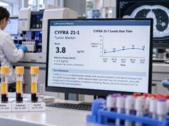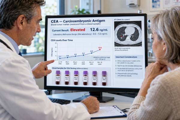
The carcinoembryonic antigen (CEA) blood test can provide supportive information when a person already has lung cancer, especially when the marker is elevated before treatment and can be followed over time. It is not accurate enough to diagnose lung cancer by itself, rule lung cancer out, or screen healthy people. CEA is a glycoprotein involved in cell adhesion that can be released into blood by several cancers, including some lung cancers. A single high result can have many explanations, so the most useful interpretation compares the value with the laboratory’s reference range, the patient’s own baseline, the timing of treatment, imaging findings, symptoms, and other medical conditions. Many laboratories use an upper reference limit near 3 ng/mL for nonsmokers and near 5 ng/mL for smokers, but the exact cutoff is assay- and laboratory-specific. When the marker falls during effective treatment, that trend may support the imaging picture; when it rises persistently, it can prompt closer assessment.
- What it measures: blood concentration of carcinoembryonic antigen (CEA).
- Reference range: Many laboratories use an upper reference limit near 3 ng/mL for nonsmokers and near 5 ng/mL for smokers, but the exact cutoff is assay- and laboratory-specific.
- High does not equal cancer progression: benign disease, smoking or organ dysfunction can raise some tumor markers, and the exact causes depend on the marker.
- Best use: serial measurements are most informative when the marker was elevated at baseline and is measured with the same assay during treatment.
- Preparation: Fasting is usually not required, but some assays have supplement instructions; for example, Mayo Clinic Laboratories advises avoiding biotin-containing supplements for 12 hours before its CEA specimen collection.
Table of Contents
- What the carcinoembryonic antigen (CEA) test measures
- Reference ranges and what a high level means
- How the marker is used in lung cancer
- Why trends over time matter more than one result
- Non-cancer causes of abnormal results
- How the blood test is done and how to prepare
- Limitations and common interpretation mistakes
- What to do with an abnormal or changing result
What the carcinoembryonic antigen (CEA) test measures
CEA is measured in serum or plasma with an immunoassay. The number is a concentration, usually reported in ng/mL. CEA is a glycoprotein involved in cell adhesion that can be released into blood by several cancers, including some lung cancers. The assay detects the marker in blood, not in a CT image and not directly inside every tumor cell.
That distinction matters. Tumors vary in how much marker they produce and release. Two people with tumors of similar size can have very different marker levels. A small tumor may release a measurable amount, while a larger tumor may produce little. For that reason, clinicians do not convert a result such as 6 ng/mL or 60 ng/mL into a tumor diameter, stage, or exact cancer-cell count.
CEA is used most often in lung adenocarcinoma and other non-squamous NSCLC rather than as a highly specific marker for one histologic subtype. The marker can also be used together with other serum markers in research or selected clinical workflows, but combining several nonspecific blood markers does not make them equivalent to tissue diagnosis or molecular profiling.
Blood tumor markers answer a different question from genomic biomarkers. An EGFR, ALK, BRAF, or KRAS result identifies a tumor driver that may select targeted therapy. CEA is a protein-level marker that may reflect tumor biology or burden in some patients, but it generally does not select a gene-targeted drug. When molecular treatment decisions are needed, a dedicated tumor or ctDNA biomarker panel is still required.
Reference ranges and what a high level means
Many laboratories use an upper reference limit near 3 ng/mL for nonsmokers and near 5 ng/mL for smokers, but the exact cutoff is assay- and laboratory-specific. A result should therefore be interpreted against the reference interval on that specific laboratory report, not a cutoff found on the internet or on an old report from another laboratory. Assays from different manufacturers can produce systematically different values.
A high result means the blood concentration exceeds the reference range; it does not automatically mean that lung cancer is growing. Possible explanations include lung cancer activity, other cancers, cigarette smoking, chronic obstructive pulmonary disease, pancreatitis, inflammatory bowel disease, liver disease, and other inflammatory or benign conditions. The probability of each cause depends on the person’s diagnosis, smoking status, kidney and liver function, recent illness, treatment, and prior marker history.
A high CEA is associated with worse prognosis in many NSCLC cohorts, but prognosis for one person cannot be calculated from CEA alone. Stage, imaging, performance status, pathology, molecular findings, treatment response, and many other factors remain more important for individual decisions.
Very high values can raise concern when they are consistent with known cancer, but there is no universal level above which progression is guaranteed. Likewise, a normal result cannot prove that the cancer is absent. Some lung cancers simply do not release enough carcinoembryonic antigen into the bloodstream to become a useful marker.
The most useful baseline is often the patient’s own pretreatment value. If the marker starts normal, serial testing may provide little additional value. If it starts clearly elevated and then tracks closely with imaging, it may become a helpful adjunct for later monitoring.
How the marker is used in lung cancer
The strongest practical role is monitoring, not screening. Clinicians may obtain a baseline result near diagnosis or before a new systemic treatment and then repeat it at selected follow-up visits. The timing should be coordinated with the treatment plan and imaging schedule rather than ordered randomly.
A falling value after treatment can support a clinical impression of response, especially when symptoms and imaging also improve. A stable value may be reassuring if the cancer is otherwise stable. A persistently rising value may justify reviewing the trend, checking for benign explanations, and considering whether imaging should be performed or interpreted more closely. It should not trigger a major treatment change by itself without corroborating evidence.
Serum markers can be attractive because blood draws are simple and inexpensive compared with repeated imaging or biopsies, but ease of testing can lead to overuse. More frequent testing is not automatically better. Small short-term fluctuations can create anxiety without changing management.
If the clinical goal is to understand the tumor’s genomic drivers rather than its serum-marker trend, CEA testing in other cancer settings is not a replacement for comprehensive tumor profiling. For patients who have both CEA and CYFRA 21-1 measured, CYFRA 21-1 monitoring can provide complementary context, but each marker has its own reference interval and confounders.
A marker may also be used in research to build prognostic models or predict response to chemotherapy, immunotherapy, or targeted therapy. These group-level associations are scientifically useful, but most are not validated as stand-alone rules for choosing or stopping treatment in an individual patient.
Why trends over time matter more than one result
Serial interpretation asks whether the marker is moving in a consistent direction and whether that movement agrees with the rest of the clinical picture. A single isolated rise can reflect biologic variation, assay variation, temporary inflammation, organ-function changes, or timing around treatment. Repeating the test under comparable conditions often clarifies whether the change is real.
Percent change can be more informative than a tiny absolute change, but there is no single universal percentage that defines response or progression for every assay and treatment. Studies have used different thresholds and sampling schedules. The oncology team should use a trend rule that fits the laboratory method and clinical protocol rather than borrowing a cutoff from an unrelated study.
Treatment can also create complex patterns. Tumor-cell death may temporarily release proteins, while effective therapy may produce a delayed fall. Conversely, a slow rise may appear before clear radiologic progression in some patients, but it may also remain nonspecific for weeks. This is why marker kinetics should be synchronized with scans and symptoms.
Whenever possible, serial samples should be run by the same laboratory and method. If a patient changes hospitals, a sudden “jump” could reflect a platform difference rather than a biologic change. Keeping the old reports makes it easier to identify whether the reference range or assay manufacturer changed.
A useful monitoring plan therefore focuses on three questions: Was the marker elevated before treatment? Is the trend reproducible over more than one sample? Does the trend match imaging, symptoms, and other clinical data? When the answers line up, the marker has more value than when it is interpreted alone.
Non-cancer causes of abnormal results
Tumor markers are not tumor-exclusive proteins. Lung cancer activity, other cancers, cigarette smoking, chronic obstructive pulmonary disease, pancreatitis, inflammatory bowel disease, liver disease, and other inflammatory or benign conditions can contribute to an elevated value. The degree of elevation and the pattern over time may help, but no simple cutoff perfectly separates benign from malignant causes.
Smoking history deserves special attention for CEA, while kidney function is particularly relevant for several protein markers, including CYFRA 21-1. Liver dysfunction can affect clearance or production of some circulating proteins. Acute infections and inflammatory conditions can also create transient changes. These factors should be reviewed before interpreting a modest rise as cancer progression.
Laboratory interference is less common but possible. Immunoassays can be affected by heterophile antibodies or other analytical problems. If a result changes dramatically without any clinical explanation, repeating the measurement or using a different method may be appropriate.
The timing of other medical events matters too. Surgery, radiation, infection, hospitalization, or a major inflammatory episode can change blood markers around the same time that cancer is being monitored. A careful timeline prevents a laboratory number from being disconnected from what was happening physiologically.
Because benign causes are common, tumor-marker testing performs poorly as a general lung-cancer screening strategy. People at high risk for lung cancer should follow evidence-based screening recommendations, which rely on low-dose CT for eligible populations, not on serum CEA alone.
How the blood test is done and how to prepare
The test requires a routine blood draw from a vein. A laboratory separates serum or plasma and measures the marker with an automated immunoassay. The blood collection itself usually takes only a few minutes.
Fasting is generally not required for CEA. Some laboratory methods have assay-specific preparation. Mayo Clinic Laboratories, for example, advises avoiding multivitamins or supplements containing biotin for 12 hours before specimen collection for its CEA assay. Follow the instructions from the laboratory performing your test. Medications should not be stopped unless the treating team specifically advises it.
The most useful “preparation” is consistency. If the marker is being used for serial monitoring, try to have blood drawn at similar points in the treatment cycle and, when possible, at the same laboratory. Record major events such as infections, hospitalizations, kidney-function changes, or smoking-status changes that could affect interpretation.
Results may be available the same day or within a few days depending on the laboratory. The number should be reviewed in context rather than acted on as soon as it appears in a patient portal. A value flagged high by the computer may be only slightly above the cutoff and may have a very different meaning from a large persistent rise in a person whose marker previously tracked with disease.
There is no radiation exposure and no cancer-specific procedural risk from the marker test itself beyond ordinary blood-draw risks such as brief pain, bruising, dizziness, or, rarely, infection at the needle site.
Limitations and common interpretation mistakes
The first mistake is treating carcinoembryonic antigen (CEA) as a diagnostic test. An elevated result cannot confirm lung cancer, and a normal result cannot rule it out. Diagnosis requires imaging and usually pathologic evaluation of tissue or cytology.
The second mistake is treating a single rise as proof of progression. Cancer monitoring is a pattern-recognition task. Imaging, symptoms, examination, treatment timing, and laboratory confounders all matter. When the marker and scan disagree, clinicians usually investigate the reason rather than automatically trusting one source.
The third mistake is comparing values across laboratories without checking the assay. Different analytical platforms can have different calibration and reference limits. A serial marker loses interpretive power if the method changes repeatedly.
The fourth mistake is assuming that more frequent testing creates earlier useful knowledge. Very frequent measurements can amplify random variability. The interval should be chosen because a result could change a decision, not simply because the test is available.
The fifth mistake is interpreting population-level prognostic studies as an individual prediction. A high CEA is associated with worse prognosis in many NSCLC cohorts, but prognosis for one person cannot be calculated from CEA alone. A biomarker can be statistically associated with survival in a cohort while still being too imprecise to forecast one person’s outcome.
Finally, serum tumor markers should not displace molecular testing. Protein markers and genomic drivers answer different questions. A high or low CEA does not tell whether a tumor has EGFR, ALK, ROS1, BRAF, KRAS G12C, MET, RET, NTRK, or HER2 alterations, and it does not provide a PD-L1 TPS score.
What to do with an abnormal or changing result
If the result is mildly high for the first time, the next step is usually to review the laboratory range, prior values, smoking status, kidney and liver function, recent infection or inflammation, and the reason the test was ordered. If there is no prior baseline, one number has limited interpretive power.
If the value is rising on repeated measurements in a person with known lung cancer, the oncology team may compare the slope with symptoms and the timing of the next CT, PET/CT, MRI, or other planned imaging. A marker change can justify closer assessment, but imaging and clinical evaluation usually determine whether the cancer is actually progressing.
If the value falls substantially during therapy and imaging also shows response, the marker may be useful for future trend monitoring. If it never tracks with the cancer, continuing to order it may add little. A test is only valuable when it helps answer a clinical question.
Ask the care team:
- What reference range does this laboratory use, and has the assay changed?
- Was the marker elevated before treatment?
- How large is the change compared with prior values?
- Could smoking, kidney function, liver disease, infection, or another condition explain the result?
- Does the marker trend agree with imaging and symptoms?
- Will repeating the test or moving imaging earlier change management?
- Is this marker actually useful for this individual tumor?
These questions keep the marker in its proper role: a convenient adjunct to lung-cancer monitoring, not a stand-alone verdict on whether treatment is working.
References
- Serum tumor markers for response prediction and monitoring of advanced lung cancer: A review focusing on immunotherapy and targeted therapies. 2023 (Review)
- Prognostic value of blood-based protein biomarkers in non-small cell lung cancer: A critical review and 2008-2022 update 2023 (Review)
- Relevance of tumor markers for prognosis and predicting therapy response in non-small cell lung cancer patients: A CEPAC-TDM biomarker substudy. 2024 (Clinical Study)
- Prognostic Value of Pretreatment Serum Carcinoembryonic Antigen Level in 1130 Patients With Non-small Cell Lung Cancer: A Propensity Score Matching Cohort Study and Cumulative Meta-analysis. 2023 (Meta-Analysis)
- CEA – Overview: Carcinoembryonic Antigen (CEA), Serum 2026 (Official Laboratory Reference)
Disclaimer
This article is for general education about carcinoembryonic antigen (CEA) testing in lung cancer. Reference ranges, assay methods, and clinical uses vary, and an abnormal result should be interpreted by the treating team together with imaging, pathology, symptoms, organ function, and the full treatment history.