
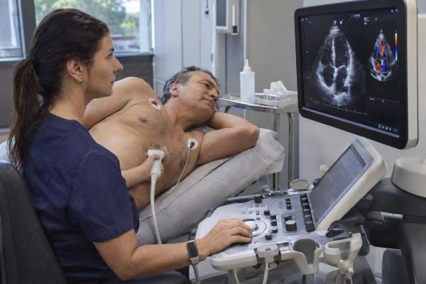
An echocardiogram is an ultrasound of the heart that shows how the chambers, valves, heart muscle, and major blood-flow patterns are working. The most common type, a transthoracic echocardiogram, is performed by placing an ultrasound probe on the chest. The report may include dozens of measurements, but a few usually matter most: left ventricular ejection fraction, chamber size, wall thickness, diastolic function, valve narrowing or leakage, right-heart function, estimated pulmonary pressure, and whether fluid is present around the heart. One abnormal number rarely tells the whole story. Measurements depend on body size, image quality, blood pressure, heart rhythm, and the method used by the laboratory. Terms such as “mild regurgitation,” “grade 1 diastolic dysfunction,” or “borderline reduced EF” can sound alarming even when they do not require urgent treatment. The key is to interpret the final impression with your symptoms, medical history, and prior studies, especially when deciding whether a finding is new, progressive, or clinically important.
Table of Contents
- What an Echocardiogram Shows
- Ejection Fraction and Pumping Strength
- Chamber Size, Wall Thickness, and Diastolic Function
- Valve Results: Stenosis and Regurgitation
- Right Heart, Pulmonary Pressure, and Pericardium
- Common Echo Phrases and What They Usually Mean
- What Happens After an Abnormal Echocardiogram
What an Echocardiogram Shows
A standard transthoracic echocardiogram, or TTE, uses sound waves to create moving images of the heart. Unlike an EKG, which records electrical activity, an echo shows anatomy and motion. Doppler techniques also measure the direction and speed of blood flow.
A comprehensive echo can evaluate:
- Left and right ventricular size and pumping function
- Left and right atrial size
- Heart-muscle thickness
- Relaxation and filling of the left ventricle
- Aortic, mitral, tricuspid, and pulmonic valve structure and function
- Blood-flow velocities and pressure gradients
- Estimated pulmonary artery pressure
- The aortic root and portions of the ascending aorta
- The pericardium, including fluid around the heart
- Congenital defects that are visible on ultrasound
- Some masses, clots, or abnormal communications between chambers
The report usually contains a measurement section followed by an interpretation or “impression.” For most readers, the impression is the best starting point because the cardiologist integrates multiple measurements rather than judging each number independently.
A value can be slightly outside a reference range for technical reasons or because reference limits differ by sex, age, and body size. A chamber that looks “mildly enlarged” in raw centimeters may be normal when indexed to body surface area. Blood pressure at the time of the study can also affect valve leakage and filling pressures.
Echo quality matters. Obesity, lung disease, chest-wall anatomy, prior surgery, and limited acoustic windows can make images difficult. The report may say “technically difficult study” or “limited visualization.” That does not mean the test failed, but it lowers confidence in some measurements. Contrast-enhancing agents, transesophageal echocardiography, cardiac MRI, or CT may be used when a key question remains unresolved.
The type of echo also matters. A transthoracic echocardiogram is the routine chest-wall study. A transesophageal echocardiogram (TEE) places a specialized probe in the esophagus to obtain closer images of structures such as the mitral valve, aortic valve, left atrial appendage, prosthetic valves, and suspected infections. A stress echocardiogram compares images at rest and after exercise or medication-induced stress to look for inducible wall-motion changes or dynamic valve and pressure abnormalities.
Some studies use agitated saline, often called a “bubble study,” to look for abnormal right-to-left passage of blood such as a patent foramen ovale. Others use an ultrasound-enhancing contrast agent when the left-ventricular border is difficult to see. These additions answer specific questions; their presence does not mean the original study was dangerous or that a serious abnormality was already found.
Understanding which type of echo you had prevents misreading the report. A routine TTE may be excellent for chamber size and EF yet still be unable to answer a question that requires TEE, cardiac CT, or MRI.
Ejection Fraction and Pumping Strength
Left ventricular ejection fraction, or LVEF, is one of the most recognized echo numbers. It estimates the percentage of blood in the left ventricle that is pumped out with each contraction.
A typical normal LVEF is roughly in the mid-50s to around 70%, although the exact reference range varies by laboratory, sex, and measurement method. An EF of 60% does not mean the heart pumps only 60% of the body’s blood or that 40% is “stuck.” The ventricle normally retains some blood after each beat.
A practical interpretation is:
| LVEF description | General meaning | Important context |
|---|---|---|
| Normal | Overall left-ventricular squeezing function is preserved | Does not rule out valve disease, diastolic dysfunction, or coronary disease |
| Low-normal or borderline | Pumping function is near the lower end of normal or mildly reduced | Comparison with prior studies and symptoms is important |
| Mildly reduced | Some loss of systolic function | May reflect cardiomyopathy, prior injury, valve disease, or other causes |
| Moderately or severely reduced | Meaningful systolic dysfunction | Usually prompts evaluation for cause and guideline-based treatment |
EF is not the only measure of contraction. The report may describe regional wall-motion abnormalities, meaning one area of the ventricle moves less than others. This can be seen after a prior heart attack, with active ischemia, in certain cardiomyopathies, or because image quality makes a segment hard to judge.
Some echo labs also report global longitudinal strain, or GLS. Strain measures how much heart-muscle fibers deform during contraction and can detect subtle dysfunction before EF becomes clearly abnormal in some conditions. It is especially useful in selected cardiomyopathies and cardio-oncology, but the number is vendor- and method-dependent and should be interpreted by the lab.
A normal EF does not exclude heart failure. Heart failure with preserved ejection fraction, or HFpEF, occurs when the heart squeezes reasonably well but fills at abnormally high pressures or has impaired relaxation and stiffness. That is why diastolic function and left atrial pressure estimates matter.
If your report says reduced EF, symptoms such as shortness of breath, swelling, fatigue, or exercise intolerance help determine urgency and treatment. A single EF estimate can also vary by several percentage points between readers or studies, so small changes should not be overinterpreted.
Chamber Size, Wall Thickness, and Diastolic Function
An echo measures all four chambers and the thickness of the heart muscle. These findings often reveal the long-term effects of blood pressure, valve disease, rhythm disorders, lung disease, or cardiomyopathy.
Left ventricular size and wall thickness
The left ventricle can enlarge when it handles excess volume, as in significant valve regurgitation, or when the muscle weakens. Thickened ventricular walls are commonly associated with long-standing high blood pressure but can also occur in hypertrophic cardiomyopathy, athletic remodeling, infiltrative disease, and other conditions.
“Left ventricular hypertrophy” on an echo means the wall thickness or calculated mass is increased. The significance depends on the pattern and degree. Concentric hypertrophy, for example, often reflects chronic pressure load such as hypertension or aortic stenosis.
If hypertension is part of the picture, accurate home blood-pressure measurement helps determine whether ongoing pressure load may be contributing.
Left atrial enlargement
The left atrium can enlarge when it is exposed to chronically elevated filling pressure, mitral valve disease, or atrial fibrillation. A larger left atrium does not identify the cause by itself, but it can serve as a marker of long-standing pressure or volume stress.
Diastolic function
Diastole is the phase when the left ventricle relaxes and fills. The 2025 American Society of Echocardiography update emphasizes a multiparameter approach rather than relying on one number. Measurements may include mitral inflow velocities, tissue Doppler velocities, left atrial size or strain, tricuspid regurgitation velocity, and other clues to filling pressure.
Reports may describe diastolic function as normal, indeterminate, or abnormal, sometimes with grades. Mild impaired relaxation is common with aging and hypertension. Higher-grade dysfunction generally indicates higher filling pressures and deserves more attention, particularly when a person has shortness of breath or exercise intolerance.
“Diastolic dysfunction” does not automatically equal heart failure. HFpEF is a clinical diagnosis that combines symptoms and signs of heart failure with evidence of elevated filling pressure or structural/functional abnormalities. A person can have mild diastolic dysfunction on an echo and no heart-failure syndrome.
Valve Results: Stenosis and Regurgitation
An echocardiogram is the primary test for evaluating heart valves. It shows whether a valve opens fully, closes properly, and creates abnormal pressure gradients or backward flow.
Stenosis means a valve is narrowed and obstructs forward flow. Regurgitation means a valve leaks backward when it should be closed.
Mild regurgitation is common. Trace or mild leakage of the mitral, tricuspid, or pulmonic valve can occur in otherwise healthy hearts and often needs no treatment. The importance increases when regurgitation is moderate or severe, causes chamber enlargement, raises pulmonary pressure, or produces symptoms.
Aortic stenosis is commonly assessed using peak velocity, mean pressure gradient, and calculated aortic valve area. The 2025 ESC/EACTS valvular guideline stresses an integrated interpretation because no single measurement should be used blindly when values disagree.
Mitral regurgitation is also graded using multiple features, including color Doppler appearance, vena contracta, regurgitant volume or fraction, effective regurgitant orifice area, pulmonary vein flow, and effects on the left atrium and ventricle. Technical limitations and blood pressure at the time of the study can affect apparent severity.
A report may use terms such as:
- Sclerosis: thickening or calcification without enough obstruction to qualify as significant stenosis.
- Prolapse: a valve leaflet bows backward, most often the mitral valve.
- Flail leaflet: part of a leaflet has lost normal support and moves abnormally, often causing significant regurgitation.
- Bicuspid aortic valve: the aortic valve has two functional leaflets rather than the usual three; it can be associated with stenosis, regurgitation, and enlargement of the aorta.
If valve disease is more than mild, follow-up frequency depends on the valve, severity, symptoms, ventricular response, and rate of change. Serial echoes are often more informative than one isolated result because they show whether the condition is stable.
Right Heart, Pulmonary Pressure, and Pericardium
The right ventricle pumps blood through the lungs, and the echo can assess its size, motion, and response to pressure or volume stress.
The 2025 ASE right-heart guideline emphasizes a multiparametric approach because the right ventricle has a complex shape and cannot be judged well by one measurement. Common measures include TAPSE, tissue Doppler S’ velocity, fractional area change, and sometimes right-ventricular strain or 3D volumes.
A report may list an estimated right ventricular systolic pressure, or RVSP. This is often calculated from the speed of tricuspid regurgitation plus an estimate of right atrial pressure. RVSP can be a clue to elevated pulmonary artery pressure, but it is an estimate, not a direct catheter measurement.
A mildly elevated RVSP can be caused by left-heart disease, lung disease, sleep apnea, chronic blood clots, pulmonary vascular disease, or measurement uncertainty. Pulmonary hypertension is not diagnosed from one borderline echo value alone. When the clinical suspicion is important, additional testing and sometimes right-heart catheterization are needed.
The echo also evaluates the inferior vena cava, which can help estimate right atrial pressure, although hydration status and breathing mechanics affect it.
Pericardial effusion
A pericardial effusion is fluid between the heart and the surrounding pericardial sac. Small effusions can occur with viral illness, inflammation, kidney disease, autoimmune conditions, cancer, after surgery, or without an obvious cause.
The key issue is not only the amount of fluid but whether it is impairing heart filling. Cardiac tamponade is the emergency state in which pressure from the fluid compromises circulation. Echo signs are interpreted together with blood pressure, heart rate, jugular venous pressure, breathing changes, and symptoms.
Common Echo Phrases and What They Usually Mean
Echo reports often use technical shorthand. The following phrases are common:
“Normal LV size and systolic function.” The left ventricle is not enlarged and its overall squeezing function is preserved.
“No regional wall-motion abnormalities.” No segment is clearly moving less than expected on the images obtained. This does not completely rule out coronary artery disease.
“Grade I diastolic dysfunction” or “impaired relaxation.” The ventricle relaxes more slowly than ideal. This is often seen with age or hypertension and may not cause symptoms by itself.
“Mild concentric LVH.” The left ventricular walls are mildly thickened in a relatively symmetric pattern, commonly from chronic high blood pressure.
“Mild mitral/tricuspid regurgitation.” A small amount of backward flow is present. Mild leakage is often monitored rather than treated.
“Moderately dilated left atrium.” The left atrium is enlarged, often reflecting chronic filling pressure, valve disease, or atrial fibrillation. In people with AFib, echo assessment of chamber size and valve function is part of the standard evaluation. Atrial fibrillation symptoms can help connect rhythm findings with the structural information from echo.
“RVSP mildly elevated.” The estimated pressure on the right side is above the lab’s usual range. It is a clue, not a stand-alone diagnosis of pulmonary hypertension.
“Technically difficult study.” Some structures could not be seen clearly. The conclusion may still be adequate, but specific measurements may be less certain.
“No significant valvular disease.” Minor or physiologic leakage may still be present, but no valve abnormality is considered hemodynamically important.
“Compared with prior study, no significant change.” This is often one of the most reassuring statements because stability over time can be more important than a mildly abnormal value.
What Happens After an Abnormal Echocardiogram
The next step depends on the abnormality, symptoms, and whether the finding changes treatment.
Mild findings often lead to no immediate testing. A clinician may focus on controlling blood pressure, treating sleep apnea, managing cholesterol, or monitoring symptoms. Moderate or severe valve disease may require a repeat echo on a defined schedule or referral to a valve specialist.
Reduced EF usually prompts evaluation for cause. Depending on the situation, that can include an EKG, blood tests, coronary evaluation, cardiac MRI, rhythm monitoring, or medication review. Treatment for heart failure with reduced EF is based on the clinical syndrome and not on the echo number alone.
An enlarged or thickened heart may lead to closer blood-pressure assessment, evaluation for hypertrophic cardiomyopathy, or additional imaging. The 2024 hypertrophic cardiomyopathy guideline recommends echocardiography as a core tool for diagnosis and follow-up, with cardiac MRI when echo is inconclusive or more tissue detail is needed.
If the echo suggests pulmonary hypertension or significant right-heart dysfunction, clinicians may look for lung disease, sleep apnea, chronic clots, left-heart disease, or pulmonary vascular disease. Right-heart catheterization is sometimes required to confirm pulmonary hypertension and measure pressures directly.
If images are inadequate, a transesophageal echocardiogram may provide a closer view of valves, the left atrial appendage, prosthetic valves, or suspected infection. Because the probe is placed in the esophagus, TEE requires more preparation than a standard chest ultrasound.
When reading your own report, focus on five questions:
- Is the ejection fraction normal or reduced?
- Are any chambers enlarged or walls thickened?
- Is valve disease mild, moderate, or severe?
- Is right-heart function or pulmonary pressure abnormal?
- Did the interpreting cardiologist recommend comparison, follow-up, or another test?
If the report is from a follow-up study, the comparison line may be more useful than any isolated measurement. A stable mild abnormality over years can have a very different meaning from rapid progression.
An echocardiogram is powerful because it combines anatomy, motion, and blood-flow information in one test. Its weakness is that it produces many numbers that can be misread outside context. Use the final interpretation as the starting point, then discuss how the findings relate to your symptoms and cardiovascular risk rather than treating every highlighted value as a separate diagnosis.
References
- Guidelines for the Standardization of Adult Echocardiography Reporting: Recommendations From the American Society of Echocardiography 2025 (Guideline)
- Recommendations for the Evaluation of Left Ventricular Diastolic Function by Echocardiography and for Heart Failure With Preserved Ejection Fraction Diagnosis: An Update From the American Society of Echocardiography 2025 (Guideline)
- Guidelines for the Echocardiographic Assessment of the Right Heart in Adults and Special Considerations in Pulmonary Hypertension: Recommendations from the American Society of Echocardiography 2025 (Guideline)
- 2025 ESC/EACTS Guidelines for the management of valvular heart disease 2025 (Guideline)
- 2023 ACC/AHA/ACCP/HRS Guideline for the Diagnosis and Management of Atrial Fibrillation: A Report of the American College of Cardiology/American Heart Association Joint Committee on Clinical Practice Guidelines 2023 (Guideline)
- 2024 AHA/ACC/AMSSM/HRS/PACES/SCMR Guideline for the Management of Hypertrophic Cardiomyopathy: A Report of the American Heart Association/American College of Cardiology Joint Committee on Clinical Practice Guidelines 2024 (Guideline)
Disclaimer
This article provides general education and cannot interpret an individual echocardiogram without the complete report, images, symptoms, and medical history. Seek urgent medical care for severe shortness of breath, chest pain, fainting, signs of shock, or rapidly worsening heart-failure symptoms.