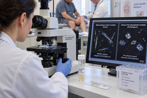
Synovial fluid crystal analysis is a microscopic examination of fluid removed from a swollen joint. Its main purpose is to identify the crystals responsible for gout or calcium pyrophosphate deposition disease, often called pseudogout. In gout, the laboratory looks for monosodium urate crystals—not free “uric acid crystals”—inside or around white blood cells. In calcium pyrophosphate disease, it looks for calcium pyrophosphate crystals. Because the symptoms of crystal arthritis can closely resemble infection, rheumatoid arthritis, injury, or another inflammatory condition, joint aspiration can provide information that a blood uric acid level cannot. A positive crystal result may establish the cause of an acute attack, but it does not automatically exclude septic arthritis. A negative result also requires caution because crystals may be sparse, sampling may be limited, and identification depends on specimen handling and microscopy skill. The best interpretation combines the crystal findings with the joint fluid cell count, Gram stain and culture when indicated, examination findings, medical history, imaging, and the pattern of previous attacks.
- Monosodium urate crystals support gout; calcium pyrophosphate crystals support calcium pyrophosphate deposition disease.
- Crystal analysis is performed on aspirated joint fluid with polarized light microscopy.
- A serum uric acid result cannot confirm which crystal, if any, is causing a current swollen joint.
- Crystals and bacterial infection can occur together, so culture may still be necessary.
- A negative microscopy report lowers confidence but does not completely exclude crystal arthritis.
Table of Contents
- Why Crystal Analysis Is Different
- How Joint Fluid Is Obtained and Examined
- Monosodium Urate Crystals and Gout
- Calcium Pyrophosphate Crystals and Pseudogout
- What Positive, Negative, and Mixed Results Mean
- Crystal Analysis Versus Blood Tests and Imaging
- What Happens After the Result
Why Crystal Analysis Is Different
Crystal arthritis occurs when microscopic mineral crystals accumulate in or around a joint and trigger inflammation. The two major forms are gout, caused by monosodium urate crystals, and calcium pyrophosphate deposition disease, caused by calcium pyrophosphate crystals. Both can produce abrupt pain, warmth, redness, swelling, and severe loss of motion. Either can look like septic arthritis, and chronic crystal disease can resemble osteoarthritis or autoimmune inflammatory arthritis.
Crystal analysis differs from most arthritis testing because it seeks direct evidence of the material causing inflammation. A serum urate test measures a risk factor for gout. An X-ray may show deposits or damage. Ultrasound may display patterns associated with crystals. By contrast, polarized microscopy can reveal crystals within the fluid from the symptomatic joint.
The test is especially useful in situations such as:
- A first episode of sudden monoarthritis
- A hot, swollen joint without a confirmed diagnosis
- An attack in an unusual location
- Suspected gout despite a normal blood urate level
- Suspected calcium pyrophosphate disease in a knee, wrist, shoulder, ankle, or elbow
- A flare that is more severe or different from prior attacks
- A swollen joint in someone who could also have infection
- Chronic inflammatory symptoms that might be caused by more than one disorder
A crystal result should answer a defined question: Are monosodium urate or calcium pyrophosphate crystals present in this sample? It does not by itself determine why the patient formed crystals, how long they have been present, whether joint damage has occurred, or whether another condition is present at the same time.
The term “pseudogout” is still widely recognized, but calcium pyrophosphate deposition disease is more accurate because the condition has several forms. It may cause sudden inflammatory attacks, chronic arthritis, cartilage calcification without symptoms, or a pattern that overlaps with osteoarthritis. Similarly, gout is not simply an isolated attack. Persistent urate crystal deposition can continue between flares and may eventually produce tophi and structural damage.
How Joint Fluid Is Obtained and Examined
The sample is collected by arthrocentesis, also called joint aspiration. After the skin is cleaned and usually numbed, a clinician inserts a sterile needle into the joint and withdraws fluid. Ultrasound guidance may be used for a small effusion, a deep joint, or a difficult anatomical location. The procedure can also reduce pressure and pain when a large effusion is present.
The laboratory places a drop of fluid on a glass slide and examines it with compensated polarized light microscopy. Ordinary light microscopy can reveal cells and larger structures, while polarized light helps distinguish crystals according to their shape and optical behavior. The examiner may look both outside cells and inside neutrophils that have engulfed crystals during an acute attack.
The key observations include:
| Feature | Monosodium urate | Calcium pyrophosphate |
|---|---|---|
| Disease association | Gout | Calcium pyrophosphate deposition disease |
| Typical shape | Needle-like | Rhomboid, rod-like, or rectangular |
| Birefringence | Strong negative | Weak positive |
| Common shorthand under a compensator | Yellow when parallel, blue when perpendicular | Blue when parallel, yellow when perpendicular |
Color descriptions are teaching aids, not substitutes for trained interpretation. Crystal orientation matters, and calcium pyrophosphate crystals may be faint or difficult to see. Other particles—including corticosteroid crystals after an injection, cholesterol crystals, basic calcium phosphate, and debris—can complicate the field.
Timing and handling influence sensitivity. Fresh fluid is preferred, especially when cells, organisms, and crystal location are being assessed together. Crystals may remain detectable after refrigeration or delayed review, but prolonged delay and poor storage can alter cell integrity and make interpretation harder. Small sample volume can force prioritization among microscopy, cell count, Gram stain, and culture.
Observer training is important. Studies show that compensated polarized microscopy can perform well, but agreement is not perfect, particularly for weakly birefringent calcium pyrophosphate crystals or samples with few crystals. A report should therefore be interpreted in light of the laboratory’s expertise and the strength of the clinical suspicion.
Monosodium Urate Crystals and Gout
A positive result for monosodium urate crystals strongly supports gout. These crystals form when urate in body fluids reaches conditions that permit crystallization. Hyperuricemia is the central biological risk, but the relationship between blood urate and an individual attack is not one-to-one. Many people with high serum urate never develop gout, and some people with proven gout have a normal serum level during an acute flare.
The phrase “uric acid crystals” is common in everyday language, but joint crystals in gout are primarily monosodium urate monohydrate. Uric acid stones in the urinary tract are chemically different from monosodium urate crystals in joints. This distinction matters because a uric acid blood test estimates systemic urate burden, while crystal microscopy demonstrates what is present in the joint fluid.
Gout often begins in the first metatarsophalangeal joint at the base of the big toe, but it may affect the ankle, knee, midfoot, wrist, fingers, elbow, or multiple joints. An attack often reaches peak intensity quickly and may be triggered by illness, surgery, dehydration, rapid changes in urate, alcohol exposure, or medication changes. None of these features is exclusive to gout.
Monosodium urate crystals are classically slender needles with strong negative birefringence. During active inflammation, they may be visible inside neutrophils. Crystals can also remain in joints between attacks, so a positive aspirate does not necessarily prove that every current symptom is caused by gout. The clinician still considers whether the degree and pattern of inflammation fit the presentation.
When crystals are found for the first time, additional assessment commonly includes serum urate, kidney function, medication review, prior flare frequency, tophi, cardiovascular and metabolic conditions, and kidney stone history. Treatment of the acute attack is distinct from long-term urate lowering. Anti-inflammatory therapy controls the flare; urate-lowering treatment aims to dissolve the body’s crystal burden over time by maintaining serum urate below a target.
A positive crystal analysis is therefore both diagnostic and directional. It confirms the crystal type but does not replace long-term risk assessment or individualized treatment planning.
Calcium Pyrophosphate Crystals and Pseudogout
Calcium pyrophosphate crystals support calcium pyrophosphate deposition disease, or CPPD. An acute inflammatory episode is sometimes called acute CPP crystal arthritis or pseudogout. The knee and wrist are frequent sites, although shoulders, elbows, ankles, and other joints may be affected. Attacks can be sudden and intense, but they may last longer than typical gout flares.
Under compensated polarized microscopy, calcium pyrophosphate crystals are commonly rhomboid or rod-shaped and weakly positively birefringent. They are often less conspicuous than monosodium urate crystals. Some are small, lie within cells, or show minimal birefringence, which can make detection difficult. A careful search may be required.
CPPD becomes more common with age. It can occur without an identifiable underlying disorder, but earlier-onset or extensive disease may prompt evaluation for associated metabolic conditions. Depending on age and presentation, clinicians may consider:
- Hemochromatosis
- Hyperparathyroidism
- Hypomagnesemia
- Hypophosphatasia
- Thyroid disease in selected contexts
- A familial pattern of CPPD
Calcium pyrophosphate crystals differ from the basic calcium phosphate crystals associated with some calcific conditions. Basic calcium phosphate particles are usually too small to be identified by routine polarized microscopy and are not birefringent. A standard “no crystals seen” report therefore does not exclude every type of mineral deposition.
Imaging may show chondrocalcinosis, a linear or punctate calcification within cartilage or fibrocartilage. Chondrocalcinosis supports CPP deposition but is neither perfectly sensitive nor perfectly specific for symptomatic disease. Some people have radiographic deposits without inflammatory attacks, while others with crystal-proven CPPD have limited findings on a particular X-ray.
The 2023 ACR/EULAR classification criteria recognize synovial fluid calcium pyrophosphate crystals in a symptomatic joint as highly important evidence. Classification criteria are designed mainly for research consistency, however. Clinical diagnosis remains an individualized process that incorporates symptoms, examination, microscopy, imaging, and exclusion of competing causes.
Unlike gout, CPPD currently has no established treatment that reliably lowers a circulating target and removes all deposits. Management focuses on controlling attacks, reducing recurrence in selected patients, addressing associated metabolic abnormalities, and treating chronic joint symptoms.
What Positive, Negative, and Mixed Results Mean
A laboratory report may state that monosodium urate crystals, calcium pyrophosphate crystals, both, other crystals, or no crystals were identified. Each result has a different meaning.
Monosodium urate crystals present: This strongly supports gout. The result is most persuasive when the crystals have the expected morphology and birefringence and the sample comes from a clinically affected joint. The report does not indicate how high the serum urate is or whether infection is absent.
Calcium pyrophosphate crystals present: This supports CPPD in the appropriate clinical setting. The result can explain an acute inflammatory attack, but CPP crystals may also be found in joints with osteoarthritis or between symptomatic episodes.
Both crystal types present: Mixed crystal deposition can occur, particularly in older adults or people with longstanding gout. The finding is not a laboratory contradiction. The clinician decides which process is most likely driving the current episode and whether both require attention.
No crystals seen: This result does not fully exclude gout or CPPD. Possible explanations include a very small sample, low crystal concentration, sampling from a joint that is not actively involved, delayed analysis, limited observer experience, or crystals that are difficult to visualize. If suspicion remains high, options may include expert slide review, repeat aspiration during a flare, ultrasound, dual-energy CT for urate in selected cases, or radiographs for chondrocalcinosis.
Atypical or unidentified crystals: Corticosteroid crystals may appear after an intra-articular injection and can be strongly birefringent. Cholesterol crystals can occur in chronic effusions. Basic calcium phosphate generally requires specialized methods. The laboratory may recommend correlation or additional testing rather than assigning a definitive disease label.
Most importantly, crystals do not exclude infection. A joint can contain monosodium urate or calcium pyrophosphate crystals and bacteria simultaneously. The concern is greater with fever, systemic illness, immune suppression, a prosthetic joint, recent surgery or injection, skin infection, bacteremia risk, or an unusually severe course. In these circumstances, the sample should generally undergo a complete synovial fluid analysis, including microbiologic testing.
Crystal Analysis Versus Blood Tests and Imaging
No single diagnostic method is best in every situation. Crystal microscopy, blood tests, and imaging answer different questions.
Serum urate measures the concentration of urate in blood. A persistently elevated result increases gout risk and is essential for monitoring urate-lowering therapy. It does not prove that a current painful joint contains urate crystals. During an acute flare, serum urate may transiently fall, so a normal result should not be used to dismiss gout. If the level is measured during an attack and is unexpectedly normal, repeating it after the flare may be useful.
C-reactive protein, erythrocyte sedimentation rate, and peripheral white blood cell count can rise in gout, CPPD, infection, and autoimmune inflammation. They measure inflammatory response rather than crystal identity. Very high values may increase concern about severe disease, but they cannot reliably distinguish crystal arthritis from septic arthritis.
Plain radiographs may show chronic gout erosions, tophi-related changes, osteoarthritis, or chondrocalcinosis. Early gout can have normal X-rays. Ultrasound can detect urate-associated features such as the double-contour sign and can identify deposits or effusions in CPPD. Dual-energy CT can map urate deposits and may help when aspiration is not possible or microscopy is negative despite strong suspicion. Imaging can also guide needle placement.
Imaging has limitations. Artifacts, operator skill, equipment, disease stage, and overlapping abnormalities can affect interpretation. A structural deposit does not always establish the cause of acute inflammation. When an accessible joint contains fluid and infection is in the differential diagnosis, aspiration often remains the most direct route because it permits crystals, cell count, and culture to be assessed from the same sample.
The tests are therefore complementary. Microscopy identifies visible crystals, blood tests assess systemic inflammation and metabolic context, and imaging evaluates deposits and structural consequences.
What Happens After the Result
After monosodium urate crystals are confirmed, immediate care focuses on controlling pain and inflammation. Common options include colchicine, a nonsteroidal anti-inflammatory drug, or a glucocorticoid, selected according to kidney function, cardiovascular risk, anticoagulation, gastrointestinal history, drug interactions, and attack severity. Aspiration itself and an intra-articular glucocorticoid may be considered after infection has been reasonably excluded.
Long-term gout care is based on the pattern of disease rather than the microscopy result alone. Recurrent flares, tophi, radiographic damage, chronic kidney disease, very high serum urate, or certain stone histories may support urate-lowering therapy. Serum urate is then followed to a treatment target. Crystal disappearance takes time, so flares can continue during the early months of urate lowering even when treatment is working.
After calcium pyrophosphate crystals are identified, an acute attack may be treated with similar anti-inflammatory options. Recurrent or chronic disease may require a different strategy, and younger patients or those with an atypical pattern may undergo metabolic testing. There is no direct equivalent of urate-lowering therapy that reliably dissolves calcium pyrophosphate deposits.
If infection remains possible, treatment should not wait for the crystal diagnosis to “win” the differential. The synovial fluid culture, blood cultures, cell count, clinical stability, and specialist assessment guide decisions about antibiotics and drainage.
Patients should seek urgent care for a rapidly worsening hot joint, fever or chills, inability to bear weight, confusion, significant immune suppression, or new swelling after joint surgery or injection. These features require prompt evaluation even in someone with a history of proven gout or CPPD.
Useful questions for the clinician include: Which crystal was identified? Was it seen inside white blood cells? Were Gram stain and culture also performed? Could both crystal disease and infection be present? Is repeat serum urate needed after the flare? Does the result change long-term treatment? The answers turn a microscopic finding into a practical care plan.
References
- Agreement Among Multiple Observers on Crystal Identification by Synovial Fluid Microscopy. 2023. Research article.
- The 2023 ACR/EULAR Classification Criteria for Calcium Pyrophosphate Deposition Disease. 2023. Classification criteria.
- Application of the 2023 ACR/EULAR classification criteria for calcium pyrophosphate deposition disease in a cohort of patients with acute arthritis. 2024. Validation study.
- Synovial fluid analysis with compensated polarization light microscopy: results of an online survey among rheumatologists. 2024. Research article.
- 2020 American College of Rheumatology Guideline for the Management of Gout. 2020. Clinical guideline.
Disclaimer
This article provides general educational information and is not a diagnosis or treatment plan. A hot, swollen joint can be caused by infection even when crystals are present and may require urgent medical care. Crystal results should be interpreted by a qualified clinician together with the full fluid analysis, symptoms, examination, and medical history.