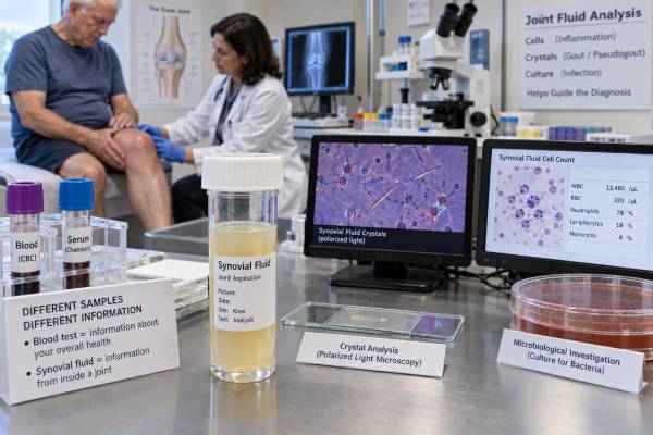
A synovial fluid analysis examines the liquid inside a joint to help explain pain, swelling, warmth, stiffness, or reduced movement. The sample is obtained by arthrocentesis, also called joint aspiration, and may be assessed for appearance, white and red blood cells, crystals, bacteria, and selected chemical features. Unlike a blood test, this analysis looks directly at the affected joint environment. That makes it especially valuable when clinicians must distinguish among infection, gout or calcium pyrophosphate deposition, autoimmune inflammation, bleeding, injury, and degenerative disease. No single fluid finding answers every question, however. A high white blood cell count can occur in both infection and severe crystal arthritis, crystals can coexist with infection, and prior antibiotics can reduce culture yield. Results therefore need to be interpreted alongside symptoms, examination findings, imaging, blood tests, medical history, medications, and the speed at which the problem developed. In a suddenly hot, swollen joint, timely aspiration may be central to preventing permanent joint damage or systemic complications.
- Synovial fluid analysis evaluates the joint itself rather than only measuring inflammation in the bloodstream.
- The most common components are visual inspection, cell count with differential, crystal examination, Gram stain, and culture.
- A very inflammatory sample raises concern for infection but does not prove it; lower counts do not safely exclude infection.
- Finding urate or calcium pyrophosphate crystals can establish crystal arthritis, yet culture may still be necessary.
- Results are most useful when the sample is collected promptly and interpreted as a pattern rather than as isolated numbers.
Table of Contents
- What Synovial Fluid Analysis Is
- When Joint Aspiration Is Needed
- How the Sample Is Collected
- Appearance and Basic Fluid Features
- White Blood Cells, Crystals, and Culture
- Interpreting Common Result Patterns
- Limitations, Next Steps, and Urgent Signs
What Synovial Fluid Analysis Is
Synovial fluid is the lubricating liquid within freely moving joints such as the knee, ankle, wrist, elbow, shoulder, and hip. In a healthy joint, only a small amount is present. It is usually clear to pale yellow and has a slippery, string-like quality created largely by hyaluronic acid. The fluid reduces friction, supports cartilage nutrition, and helps movement remain smooth.
When a joint becomes irritated, injured, inflamed, infected, or filled with blood, the volume and composition of the fluid can change. Synovial fluid analysis is not one test but a group of laboratory assessments chosen according to the clinical question. A standard evaluation often includes:
- Gross appearance, including color, clarity, and viscosity
- Total nucleated or white blood cell count
- White blood cell differential, particularly the percentage of neutrophils
- Red blood cell count when bleeding or a traumatic tap is possible
- Polarized light microscopy for crystals
- Gram stain and bacterial culture when infection is a concern
- Additional cultures or molecular tests in selected circumstances
Some laboratories also report glucose, protein, lactate, pH, or other biochemical measurements. These may provide supportive information, but they generally do not replace the core combination of cell count, differential, crystal analysis, and microbiologic testing.
The main advantage of joint fluid testing is anatomical specificity. C-reactive protein and erythrocyte sedimentation rate can show that inflammation exists somewhere in the body, but they cannot identify why one particular joint is swollen. Rheumatoid factor or anti-CCP antibodies may support rheumatoid arthritis in the right clinical setting, but they cannot reliably distinguish an acute rheumatoid flare from septic arthritis in a single joint. Fluid findings can narrow that uncertainty more directly.
Synovial fluid analysis may classify an effusion as broadly noninflammatory, inflammatory, hemorrhagic, or septic-appearing. These categories are guides, not final diagnoses. Considerable overlap exists, and the clinician must integrate the complete pattern.
When Joint Aspiration Is Needed
Arthrocentesis is commonly considered when a joint has a new or unexplained effusion, especially if it is painful, warm, red, or difficult to move. It is particularly important for acute monoarthritis, meaning a sudden problem affecting one joint. In that setting, the immediate priorities often include excluding infection and identifying crystal disease.
Possible reasons for aspiration include:
- A hot, swollen joint with concern for septic arthritis
- Sudden severe pain that could represent gout or calcium pyrophosphate arthritis
- A first episode of unexplained joint swelling
- An unusual flare in a person with known inflammatory arthritis
- Suspected bleeding into the joint after trauma or in someone using anticoagulants
- Persistent or recurrent effusion without a clear cause
- Joint swelling after surgery, injection, or implantation of prosthetic material
- A need to relieve pressure from a large, painful effusion
Septic arthritis deserves special urgency. Bacteria in a joint can damage cartilage rapidly, and infection may spread into the bloodstream. Fever increases concern but is not always present, especially in older adults, immunosuppressed people, and those who have already taken antibiotics. A person can also have infection despite a normal peripheral white blood cell count or only modest elevation of blood inflammatory markers.
Risk rises with factors such as advanced age, diabetes, immune suppression, recent joint surgery, an artificial joint, skin infection, injection drug use, bacteremia, or a recent procedure that crossed the skin. Yet septic arthritis can occur without an obvious risk factor. Clinical suspicion should therefore be driven by the presentation, not only by a checklist.
Aspiration is also useful when an established diagnosis does not fully explain the current episode. A person with gout can develop septic arthritis, and someone with rheumatoid arthritis can have an infected joint that resembles a flare. In these situations, assuming that every swollen joint is “more of the same” can delay necessary treatment.
Not every painful joint requires aspiration. Pain without an effusion, a clearly mechanical injury, or a typical mild flare with no concern for infection may be evaluated in other ways. Ultrasound can confirm whether fluid is present and can guide aspiration of small or deep joints. The decision depends on the balance between diagnostic value, procedural feasibility, and urgency.
How the Sample Is Collected
During arthrocentesis, the clinician positions the joint, cleans the skin with antiseptic, and often uses local anesthetic. A sterile needle is advanced into the joint space, and fluid is withdrawn into a syringe. Superficial joints such as the knee can often be aspirated using anatomical landmarks. Ultrasound guidance may improve accuracy when the effusion is small, the joint is deep, or nearby structures make access more difficult.
The amount and character of fluid vary widely. A large knee effusion may produce many milliliters, while a small finger or wrist joint may yield only a few drops. Even a limited sample can be valuable, so the laboratory priorities should match the clinical concern. If infection is possible and sample volume is scarce, culture and cell count are often prioritized. Crystal microscopy is also important when gout or calcium pyrophosphate deposition is suspected.
Specimen handling affects the usefulness of results. Fluid intended for culture should be placed in sterile containers and transported promptly. In some settings, part of the sample is inoculated into blood culture bottles to improve microbial recovery. The clinician should tell the laboratory about suspected organisms, antibiotic exposure, prosthetic material, immune suppression, or unusual exposures because these factors can change the requested culture methods and incubation time.
When feasible, aspiration should occur before antibiotics are started because antibiotics may make cultures negative. Treatment should not be dangerously delayed, however, in a person who is unstable or appears septic. Blood cultures may be obtained when fever, systemic illness, or bloodstream infection is suspected.
Preparation is usually minimal. Patients should report anticoagulants, antiplatelet medicines, bleeding disorders, allergies, and prior joint procedures. Anticoagulation does not automatically prevent aspiration, but the clinician must assess individual bleeding risk. Afterward, mild soreness can occur. A small dressing, brief rest, ice, and activity guidance may be recommended.
Complications are uncommon when sterile technique is used. Potential risks include bleeding, infection introduced by the procedure, temporary pain, injury to nearby structures, and a dry tap in which no fluid is obtained. Increasing redness, drainage, fever, or worsening pain after aspiration should be reported promptly.
Appearance and Basic Fluid Features
The clinician may learn something before the sample reaches the laboratory. Normal fluid is usually clear, pale yellow, and viscous. Inflammation often makes it cloudy and less stringy because inflammatory enzymes break down hyaluronic acid. Markedly turbid or purulent fluid raises concern for intense inflammation or infection, but visual appearance alone cannot determine the cause.
Color and clarity can suggest several broad patterns:
- Clear or nearly clear, straw-colored fluid: often seen in noninflammatory conditions, although early or mild inflammation remains possible.
- Cloudy yellow fluid: commonly reflects an increased number of white blood cells and may occur in rheumatoid arthritis, gout, calcium pyrophosphate arthritis, reactive arthritis, or infection.
- Opaque or pus-like fluid: strongly concerning for septic arthritis, though severe sterile inflammation can sometimes appear similar.
- Bloody fluid: may result from trauma, a bleeding disorder, anticoagulation, tumor, synovial injury, or accidental vessel puncture during the procedure.
- Xanthochromic or yellow-orange fluid: may occur after prior bleeding as red blood cells break down.
A bloody specimen requires context. If sequential collection tubes become progressively clearer, the blood may have been introduced by the needle rather than being present throughout the joint. Persistent blood in all tubes can support true hemarthrosis, but this distinction is not always reliable.
Viscosity is sometimes assessed with a string test. Healthy fluid forms a relatively long strand; inflammatory fluid often forms a shorter strand. The mucin clot test is an older measure of hyaluronic acid quality. These observations may help categorize fluid, but modern diagnosis relies more heavily on cell counts, microscopy, and cultures.
Chemical measurements have limited stand-alone value. Low synovial glucose compared with blood glucose can occur in bacterial infection or inflammatory arthritis. High lactate or altered pH may support infection in some studies, but thresholds and laboratory methods vary. Such tests should be considered adjuncts rather than replacements for microbiologic evaluation.
White Blood Cells, Crystals, and Culture
The synovial white blood cell count and differential estimate how strongly immune cells have entered the joint. Results are usually reported as cells per microliter, with the percentage of polymorphonuclear neutrophils. Broadly, noninflammatory fluid often contains fewer than about 2,000 white blood cells per microliter, while inflammatory fluid commonly falls between roughly 2,000 and 50,000. Counts above 50,000, especially with a high neutrophil percentage, increase concern for bacterial infection.
These ranges overlap substantially. Gout and calcium pyrophosphate arthritis can produce counts above 50,000 and occasionally far higher. Conversely, septic arthritis can present below 50,000, particularly early in infection, after antibiotics, in prosthetic joints, or in people whose immune systems cannot mount a strong response. A count should therefore modify the probability of infection, not function as a pass-or-fail cutoff. The separate synovial fluid white blood cell count result is most useful when interpreted with the differential, Gram stain, culture, and clinical presentation.
Crystal analysis uses compensated polarized light microscopy. Needle-shaped monosodium urate crystals with strong negative birefringence support gout. Rhomboid or rod-shaped calcium pyrophosphate crystals with weak positive birefringence support calcium pyrophosphate deposition disease, often called pseudogout. Crystal identification depends on specimen quality, observer experience, crystal concentration, and timely microscopy. A negative examination does not completely exclude crystal disease if suspicion remains high.
Finding crystals is diagnostically powerful, but it does not sterilize the joint. Gout or calcium pyrophosphate arthritis can coexist with bacterial infection. If the joint is unusually severe, systemic symptoms are present, risk factors exist, or the clinical course is atypical, culture remains important even when crystals are seen. More detail is available in the synovial fluid crystal analysis guide.
Gram stain looks for bacteria directly under the microscope. A positive result can guide early therapy, but sensitivity is limited, so a negative Gram stain cannot rule out septic arthritis. Culture is more definitive because it attempts to grow and identify the organism and may permit antibiotic susceptibility testing. The synovial fluid culture test usually requires time, and yield may be reduced by prior antibiotics, small sample volume, fastidious organisms, or inadequate handling.
Additional tests may be selected for mycobacteria, fungi, gonorrhea, Lyme disease, or other organisms based on exposure, geography, immune status, age, and symptom duration. Molecular assays can be useful in selected cases but are not universal replacements for culture.
Interpreting Common Result Patterns
A synovial fluid report is best read as a multidimensional pattern. The diagnosis rarely depends on one column.
Noninflammatory pattern: Fluid may be clear or slightly cloudy, with relatively low white blood cell counts and a low neutrophil proportion. Osteoarthritis, internal mechanical injury, and some chronic degenerative effusions commonly fit this pattern. A noninflammatory count does not identify the exact structural cause, so examination and imaging may still be needed.
Inflammatory, crystal-positive pattern: The fluid is often cloudy, the white blood cell count is elevated, neutrophils predominate, and urate or calcium pyrophosphate crystals are identified. This can establish gout or calcium pyrophosphate crystal arthritis in the appropriate setting. A serum uric acid blood test may support long-term assessment, but blood urate can be normal during an acute gout attack and does not replace crystal confirmation.
Inflammatory, crystal-negative pattern: Possible causes include rheumatoid arthritis, psoriatic arthritis, reactive arthritis, other autoimmune disease, partially treated infection, or crystal disease missed on microscopy. The distribution of affected joints, symptom duration, skin or eye findings, antibodies, inflammatory markers, and imaging become especially important.
Septic-appearing pattern: Very cloudy or purulent fluid, a high white blood cell count, marked neutrophil predominance, and a positive Gram stain or culture strongly support infection. Yet culture-confirmed infection can occur without every classic feature. Clinical urgency may justify drainage and empiric antibiotics before final culture results return.
Hemorrhagic pattern: A high red blood cell count or visibly bloody sample can follow trauma, ligament or cartilage injury, fracture, anticoagulation, hemophilia, or less commonly a proliferative synovial disorder or tumor. The cause is investigated with history, coagulation studies, and imaging.
Mixed or conflicting pattern: Real-world results are often messy. Crystals may be present with a positive culture. A high cell count may accompany a negative Gram stain. A low count may occur in an immunosuppressed patient with infection. A traumatic aspiration may distort the red and white cell counts. In such cases, the safest interpretation gives greater weight to serious diagnoses that have not yet been adequately excluded.
Repeat aspiration may be considered when symptoms worsen, the first sample was inadequate, cultures are pending but suspicion remains high, or drainage is part of treatment. Serial fluid counts alone are not always necessary; the decision depends on the condition and response to therapy.
Limitations, Next Steps, and Urgent Signs
Synovial fluid analysis has several limitations. Reference ranges vary by laboratory, joint type, native versus prosthetic joint, age, and clinical context. Cell counts can change after antibiotics or anti-inflammatory treatment. Crystals may be missed if they are sparse or microscopy is delayed. Gram stain has limited sensitivity, and cultures can be falsely negative. No single result reliably excludes infection in every patient.
The next step depends on the working diagnosis:
- Suspected septic arthritis generally requires urgent specialist assessment, joint drainage, cultures, and antimicrobial treatment.
- Crystal arthritis may be treated with anti-inflammatory therapy, followed by evaluation of recurrence risk and long-term prevention when appropriate.
- Autoimmune inflammatory arthritis may prompt rheumatology evaluation, targeted blood tests, ultrasound or MRI, and disease-modifying treatment planning.
- Hemarthrosis may require investigation for trauma, coagulation problems, medication effects, or structural lesions.
- Noninflammatory effusion may lead to imaging, physical therapy, activity modification, or orthopedic assessment depending on the cause.
Seek urgent care for a rapidly worsening hot or swollen joint, inability to bear weight or move the joint, fever or chills, confusion, low blood pressure symptoms, a new joint problem after surgery or injection, or acute swelling in an immunocompromised person. These features can signal infection even when blood tests are not dramatic.
For patients reviewing a report, useful questions include: Were crystals specifically examined under polarized light? Was a culture sent before antibiotics? What was the neutrophil percentage? Could medication or immune suppression have lowered the cell count? Does the result fully explain the symptoms, or is repeat testing needed? These questions help place the laboratory data into a clinical decision rather than treating the report as self-interpreting.
References
- Synovial Fluid Analysis. 2024. MedlinePlus Medical Test.
- Synovial fluid analysis. 2025. MedlinePlus Medical Encyclopedia.
- Guideline for management of septic arthritis in native joints (SANJO). 2023. Clinical guideline.
- Native Joint Septic Arthritis. 2024. Review article.
- Synovial pH, lactate, and glucose as adjunct biomarkers to differentiate septic arthritis from noninfectious causes of acute arthritis. 2025. Research article.
Disclaimer
This article is for general educational purposes and does not diagnose or treat any condition. A hot, swollen, or rapidly worsening joint may require urgent medical assessment because septic arthritis can cause permanent damage. Laboratory results should be interpreted by a qualified clinician who knows the symptoms, examination findings, medications, and full medical history.