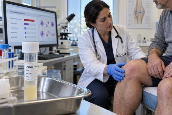
A synovial fluid white blood cell count measures the number of immune cells in fluid aspirated from a joint. It helps clinicians judge whether an effusion is noninflammatory, inflammatory, or strongly suspicious for septic arthritis. The result is usually reported as cells per microliter together with a differential showing the percentage of neutrophils. Higher counts and greater neutrophil predominance generally increase concern for bacterial infection, but there is no universal number that proves or excludes septic arthritis. Gout, calcium pyrophosphate arthritis, rheumatoid arthritis, and other sterile inflammatory conditions can produce very high counts. Infection may produce a lower count early in its course, after antibiotics, or in a person with impaired immunity. Native joints and prosthetic joints also use different interpretive frameworks. For these reasons, the count must be combined with the fluid’s appearance, crystal examination, Gram stain, culture, blood tests, symptoms, risk factors, and clinical stability. In a hot, swollen joint, a reassuring-looking number should never override a concerning presentation.
- The total white blood cell count reflects the intensity of joint inflammation, not its exact cause.
- The neutrophil percentage adds context but also overlaps between infection and crystal arthritis.
- More than 50,000 cells per microliter raises concern for septic arthritis but is not diagnostic by itself.
- Fewer than 50,000 cells per microliter does not safely rule out infection.
- Antibiotics before aspiration can lower the count, neutrophil percentage, and culture positivity.
Table of Contents
- What Is Being Counted
- How Results Are Reported
- Interpreting Low, Intermediate, and High Counts
- The Role of Neutrophil Percentage
- Why the Count Can Mislead
- Native Joints, Prosthetic Joints, and Special Cases
- Using the Count in a Clinical Decision
What Is Being Counted
Synovial fluid normally contains relatively few cells. When the synovial lining is irritated or infected, chemical signals recruit leukocytes—white blood cells—from the bloodstream into the joint. The total nucleated cell count estimates how many of these cells are present in each microliter of fluid. In most acute arthritis evaluations, the reported value is effectively treated as the synovial white blood cell count.
The laboratory may count cells manually with a chamber or use an automated analyzer validated for body fluids. A stained slide or analyzer also separates the cells into categories. Neutrophils, sometimes labeled polymorphonuclear cells or PMNs, are particularly important because they often dominate in acute bacterial infection and crystal-induced inflammation. Lymphocytes, monocytes, macrophages, eosinophils, and other cells may provide additional clues in selected conditions.
The count is obtained as part of a broader synovial fluid analysis. The same aspiration may provide information about:
- Color, clarity, and viscosity
- Total white blood cell count
- Neutrophil percentage
- Red blood cell count
- Monosodium urate or calcium pyrophosphate crystals
- Gram stain
- Aerobic and anaerobic culture
- Specialized microbiology or chemistry when indicated
The count is attractive because it returns faster than culture and gives a numerical measure of inflammation. Its limitation is biological overlap. Neutrophils respond vigorously to both microbes and sterile crystals, and not every infected patient generates the same response. The number is therefore a probability modifier rather than a microbial identification test.
Units may appear as cells/µL, cells/mm³, or ×10⁶/L. A microliter and a cubic millimeter are equivalent, so 50,000 cells/µL is the same concentration as 50,000 cells/mm³. Understanding the unit prevents a result from appearing larger or smaller merely because the laboratory uses a different format.
How Results Are Reported
A typical report includes a total white blood cell count and a differential. For example, it might state “WBC 38,000 cells/µL; 91% neutrophils.” The total count describes the magnitude of cellular inflammation. The differential describes which cells account for that total.
Some reports use “total nucleated cells” rather than WBC. In synovial fluid, this may include cells from the joint lining as well as leukocytes, but the value is generally interpreted within the laboratory’s body-fluid reference system. The report may also flag the sample as clotted, diluted, excessively bloody, or insufficient. These comments matter because they can affect accuracy.
Broad educational categories are often used for native adult joints:
| General pattern | Approximate synovial WBC count | Typical interpretation |
|---|---|---|
| Normal or minimally cellular | Often below 200 cells/µL | Expected in healthy fluid |
| Noninflammatory | Usually below about 2,000 cells/µL | Osteoarthritis or mechanical conditions are more likely |
| Inflammatory | Roughly 2,000–50,000 cells/µL | Crystal or immune-mediated arthritis is common; infection remains possible |
| Strongly inflammatory or septic-appearing | Often above 50,000 cells/µL | Infection risk rises, especially with neutrophil predominance, but overlap is substantial |
These ranges are not rigid reference intervals. Different sources and laboratories use slightly different boundaries. The type of joint, patient population, duration of illness, treatment exposure, and whether a prosthesis is present can change the meaning.
A count should also be interpreted on a continuum. The probability of septic arthritis generally rises as the number increases, especially above 50,000 and again at very high levels such as 100,000 or more. Yet the increase is gradual, not a switch that flips at one threshold. A count of 49,000 is not biologically reassuring while 51,000 is automatically infected.
The clinician also checks whether the sample was collected before antibiotics, whether crystals were seen, and whether culture is pending. A preliminary count often informs immediate decisions, but it does not replace the final microbiology.
Interpreting Low, Intermediate, and High Counts
Low counts are more consistent with noninflammatory fluid, particularly when the sample is clear, neutrophils are not predominant, and the clinical problem is chronic or mechanical. Osteoarthritis, meniscal injury, and some degenerative effusions commonly fall in this range. However, a low count cannot be used as a universal exclusion test for infection. Early infection, partially treated infection, immunosuppression, and certain organisms can produce unexpectedly modest inflammation.
Intermediate counts create the greatest diagnostic uncertainty. Results from about 2,000 to 50,000 cells/µL can occur in rheumatoid arthritis, psoriatic arthritis, reactive arthritis, gout, calcium pyrophosphate arthritis, Lyme arthritis, and bacterial infection. The count narrows the fluid to an inflammatory pattern but usually cannot identify the cause. The symptom timeline, joint distribution, crystals, cultures, and systemic features become decisive.
Counts above 50,000 cells/µL are traditionally considered suggestive of septic arthritis in a native joint. Current guidance emphasizes that the threshold increases suspicion but is not sufficient for diagnosis. Severe gout or calcium pyrophosphate arthritis can exceed 50,000 and sometimes 100,000. Conversely, many culture-proven infections do not reach 50,000.
Very high counts make intense inflammation undeniable. When the sample is purulent, neutrophils are dominant, and the patient is systemically unwell, urgent treatment for infection is commonly justified while cultures are pending. Even then, crystal analysis should be performed because crystal disease can coexist or provide an alternative explanation.
The 25,000-cell threshold is sometimes discussed as a lower point. Counts below approximately 25,000 may reduce the probability of septic arthritis in a native joint, but they do not exclude it. A low post-test probability is not the same as zero risk, especially when the consequences of missing infection are severe.
No interpretation is complete without asking what the pretest probability was before aspiration. A count of 20,000 in a healthy person with a classic gout presentation and visible urate crystals carries a different meaning from the same count in an immunosuppressed patient with bacteremia and a newly swollen knee.
The Role of Neutrophil Percentage
The differential count usually highlights the percentage of polymorphonuclear neutrophils. Bacterial infection commonly produces neutrophil predominance, and percentages above 75% to 90% may increase concern depending on the total count and context. The highest suspicion occurs when a markedly elevated total count is paired with a very high neutrophil percentage.
The neutrophil percentage cannot independently distinguish infection from sterile inflammation. Monosodium urate and calcium pyrophosphate crystals activate a powerful neutrophilic response. An acute gout aspirate can therefore look “septic” by both total count and differential. Rheumatoid or reactive arthritis may also be neutrophil-predominant during an active flare.
Absolute numbers can be informative. A sample containing 10,000 cells/µL with 90% neutrophils has about 9,000 neutrophils/µL. A sample containing 100,000 cells/µL with 80% neutrophils has about 80,000 neutrophils/µL. The percentages alone conceal a large difference in inflammatory burden. Most clinical frameworks still use the total count and percentage together rather than relying on a calculated absolute synovial neutrophil count.
Other differential patterns may suggest alternatives but are rarely definitive. Lymphocyte predominance can occur in some chronic infections or inflammatory states. Eosinophils are uncommon and may appear with air introduced during aspiration, allergy-related phenomena, parasites, or other unusual conditions. Macrophages containing blood products may support prior bleeding. These findings require specialist laboratory and clinical interpretation.
A differential can be less reliable when the sample is old, clotted, or damaged during transport. Neutrophils deteriorate over time. Prompt laboratory processing is therefore important, particularly when the specimen volume is small and the diagnosis urgent.
Why the Count Can Mislead
Several factors can shift the number or distort its meaning.
Antibiotics before aspiration: Research in native-joint septic arthritis shows that preaspiration antibiotics can lower the synovial WBC count, reduce the neutrophil percentage, and decrease culture positivity. In a stable patient, obtaining fluid before antibiotics is preferable. In sepsis or shock, treatment should not be dangerously delayed.
Immune suppression: Glucocorticoids, biologic therapies, chemotherapy, advanced illness, or severe immune deficiency may blunt leukocyte recruitment. A modest count can coexist with serious infection.
Early infection: The inflammatory response evolves over time. Aspiration soon after symptoms begin may occur before the count reaches a typical range.
Crystal arthritis: Gout and CPPD can drive the count into a range commonly associated with infection. A separate synovial fluid crystal analysis is therefore essential when crystals are possible.
Bloody or traumatic aspiration: A needle may introduce peripheral blood into the specimen, adding circulating leukocytes and red blood cells. Correction formulas exist, especially in prosthetic-joint workups, but they are imperfect and should not replace clinical judgment.
Clotting: Synovial fluid can clot when inflammation increases fibrinogen. Cells trapped in a clot may make automated or manual counts inaccurate.
Dilution: Saline introduced during a procedure or a very small sample mixed with other fluid may lower the measured concentration.
Delayed processing: Cell breakdown can reduce count accuracy and alter the differential.
Different disease settings: Native-joint infection, prosthetic-joint infection, postoperative inflammation, and infection after ligament reconstruction do not share one universal threshold.
The most dangerous error is false reassurance. A patient with fever, bacteremia, rapid functional loss, or a purulent aspirate should not be declared infection-free because the count is “only” 30,000. The opposite error also matters: treating every count above 50,000 as bacterial can lead to unnecessary surgery and antibiotics in severe crystal disease. Culture and the broader pattern are indispensable.
Native Joints, Prosthetic Joints, and Special Cases
The commonly cited 50,000-cell threshold applies mainly to suspected acute infection in a native joint. A prosthetic joint requires a separate diagnostic approach. Chronic prosthetic joint infection can occur with much lower cell counts because biofilm-associated organisms may produce a less dramatic inflammatory response. Thresholds also differ by joint, time since surgery, and whether the concern is acute postoperative infection, acute hematogenous infection, or chronic implant infection.
Recent surgery itself raises synovial cells. Applying a chronic prosthetic-joint cutoff immediately after an operation can create false-positive results. Orthopedic and infectious disease teams use timing-specific criteria, cultures, tissue samples, serum markers, and sometimes alpha-defensin or other tests.
Children also require age- and joint-specific assessment. Hip infection can be difficult to distinguish from transient synovitis, and clinical prediction tools may support—but not replace—aspiration and microbiology. Pediatric management should follow pediatric guidelines rather than adult thresholds alone.
Gonococcal arthritis may produce lower counts than classic nongonococcal bacterial arthritis, and joint-fluid culture can be negative. Nucleic acid testing from genital, pharyngeal, or rectal sites may be important based on exposure. Lyme arthritis can produce an inflammatory count with substantial neutrophil predominance and often requires serologic testing rather than routine bacterial culture alone.
Mycobacterial and fungal arthritis may be indolent and less cellular. Specialized cultures, prolonged incubation, biopsy, and exposure history become more important. A chronic low-to-intermediate count is therefore not automatically noninfectious.
Small joints and bursae also generate smaller sample volumes, and laboratory reference assumptions developed for the knee may be less directly transferable. The number must be interpreted in the anatomy from which it came.
Using the Count in a Clinical Decision
The count is most useful when it changes probability and guides action. Clinicians typically combine several questions:
- How likely was infection before aspiration based on symptoms, risk factors, and examination?
- Does the total count make infection more or less likely?
- Is there strong neutrophil predominance?
- Are monosodium urate or calcium pyrophosphate crystals present?
- What did the Gram stain show?
- Was a synovial fluid culture obtained before antibiotics?
- Is the patient stable enough to await further data, or is immediate drainage and empiric treatment required?
A high-risk pattern might include abrupt severe pain, fever, an artificial or recently operated joint, purulent fluid, a high count with neutrophil predominance, and bacteremia. A lower-risk pattern might include recurrent classic gout, urate crystals, no systemic illness, and a rapid response to anti-inflammatory treatment. Many cases fall between these extremes.
Blood CRP and ESR can support the assessment but do not replace aspiration. Imaging can locate fluid and identify adjacent osteomyelitis or abscess. Repeat aspiration or surgical sampling may be needed when the first sample is inadequate, culture is negative after antibiotics, or the patient fails to improve.
Patients reviewing a report should avoid interpreting the number in isolation. Useful questions include: Was this a native or prosthetic joint? Had antibiotics already been given? What was the neutrophil percentage? Were crystals and culture checked? Was the sample bloody or clotted? What diagnosis best explains the complete pattern?
Changes over time may also matter, but repeat counts are not routinely required for every patient. A second aspiration is most useful when fluid reaccumulates, symptoms worsen, the initial sample was unreliable, or drainage is being used therapeutically. Clinical improvement, falling systemic inflammatory markers, restored movement, and culture information may be more meaningful than forcing the synovial count to reach a particular number.
Urgent evaluation is warranted for a rapidly worsening hot joint, fever or chills, inability to bear weight, severe pain with movement, confusion, new symptoms after joint surgery or injection, or acute swelling during immune-suppressing treatment. The white blood cell count is an important clue, but safe care depends on treating the patient rather than a cutoff.
References
- Guideline for management of septic arthritis in native joints (SANJO). 2023. Clinical guideline.
- Effect of Antibiotic Administration Before Joint Aspiration on Synovial Fluid White Blood Cell Count in Native Joint Septic Arthritis. 2024. Research article.
- Effect of Antibiotic Therapy Before Arthrocentesis on Synovial Fluid Cell Count and Differential in Native Joint Septic Arthritis. 2024. Research article.
- Native Joint Septic Arthritis. 2024. Review article.
- Enhanced Diagnostic Accuracy for Septic Arthritis Through Multivariable Analysis of Serum and Synovial Biomarkers. 2025. Research article.
Disclaimer
This article is educational and does not provide a diagnosis or individual medical advice. Septic arthritis can cause rapid, permanent joint damage and systemic illness, so a hot, swollen, or rapidly worsening joint requires prompt professional assessment. Synovial white blood cell counts must be interpreted with cultures, crystals, prior treatment, examination findings, and the type of joint involved.