A sleep-deprived EEG is a brain-wave test performed after you have had less sleep than usual. Doctors most often order it when they are looking for signs of seizure activity that may not appear during a standard awake EEG.
The test is painless and noninvasive, but the preparation can be tiring. You may be asked to stay awake most or all of the night before the appointment, avoid caffeine, arrive with clean hair, and arrange a ride home. The goal is to make it easier for you to become drowsy or fall asleep during the recording, because sleep and sleepiness can reveal electrical patterns that are sometimes missed when you are fully awake.
Table of Contents
- What a Sleep-Deprived EEG Is
- Why Doctors Order It
- How to Prepare
- What Happens During the Test
- Risks and Safety
- What Results Can Show
- Other Tests and Next Steps
What a Sleep-Deprived EEG Is
A sleep-deprived EEG is an electroencephalogram done after a planned period of limited sleep. It records the brain’s natural electrical activity through small electrodes placed on the scalp.
An EEG does not send electricity into the brain. The electrodes only detect electrical signals already being produced by brain cells. These signals appear as wave patterns that a trained specialist reviews for normal rhythms, sleep changes, slowing, spikes, sharp waves, or other patterns that may help explain symptoms.
The “sleep-deprived” part matters because the brain behaves differently when it is tired, drowsy, or asleep. In some people with epilepsy, abnormal electrical discharges are more likely to appear during drowsiness or certain stages of sleep than during a brief awake recording. Sleep deprivation can also make it easier for a person to fall asleep in the lab without sedation.
A sleep-deprived EEG is usually similar to a routine EEG in how it is performed. The main difference is the preparation beforehand and the effort to capture drowsiness or sleep. During the recording, you may still be asked to open and close your eyes, breathe deeply for a short period if safe for you, or look at flashing lights. These activation procedures are used because certain EEG patterns may become more visible under specific conditions.
A sleep-deprived EEG is not the same as a sleep study. A sleep study, or polysomnography, is designed to evaluate sleep disorders such as obstructive sleep apnea, periodic limb movements, or abnormal behaviors during sleep. A sleep-deprived EEG is mainly used to evaluate brain electrical activity, especially when seizures or epilepsy are part of the concern. When sleep apnea, severe daytime sleepiness, or chronic insomnia is the main issue, doctors may use different testing, such as polysomnography or home sleep apnea testing.
A sleep-deprived EEG also differs from brain imaging. MRI and CT scans show brain structure; EEG shows electrical function over time. A person can have a normal MRI and still have abnormal EEG activity, or have an abnormal scan with an EEG that does not show epileptiform activity during the recording. These tests answer different questions, which is why clinicians often interpret them together with the symptom history.
Why Doctors Order It
Doctors usually order a sleep-deprived EEG when they need a better chance of seeing seizure-related electrical patterns. It is especially common after a routine EEG is normal or unclear but the person’s symptoms still raise concern for seizures.
The most common reason is evaluation of possible epilepsy. A person may have had an episode of loss of awareness, shaking, staring, confusion, unusual sensations, unexplained falls, nighttime events, or repeated “spells” that could represent seizures. A routine EEG may be the first test, but because EEG records only a sample of brain activity, it can miss intermittent abnormalities. Sleep deprivation and sleep recording can increase the chance of capturing relevant patterns.
A clinician may also order a sleep-deprived EEG when symptoms happen around sleep or waking. Examples include episodes shortly after falling asleep, unusual movements during the night, morning jerks, unexplained tongue biting, waking with confusion, or events that occur after a night of poor sleep. Some seizure types are closely linked with sleep-wake transitions, and the EEG may be more informative when the recording includes drowsiness or sleep.
The test can also help classify seizure type. This matters because focal seizures, generalized seizures, absence seizures, myoclonic seizures, and other seizure types can have different EEG patterns and may require different treatment choices. EEG findings are not the only factor, but they can help the neurologist decide whether the pattern fits a particular epilepsy syndrome.
A sleep-deprived EEG may be used in children as well as adults. In children, sleep can be especially helpful because some epileptiform patterns become more visible during sleep. Instructions for sleep restriction are usually age-specific, because a toddler, school-age child, teenager, and adult cannot safely or realistically follow the same sleep-deprivation plan.
The test is not ordered only to “prove” epilepsy. Sometimes it helps doctors consider other possibilities. Fainting, migraine, panic attacks, sleep disorders, movement disorders, medication effects, and psychogenic nonepileptic seizures can sometimes resemble epileptic seizures. A sleep-deprived EEG cannot sort out every diagnosis by itself, but it can add useful evidence to the broader evaluation. When events are frequent or unclear, video EEG monitoring may be more useful because it can match symptoms on video with EEG activity at the same time.
It is also important to know what the test is not designed to do. A sleep-deprived EEG does not measure intelligence, personality, memory performance, or mental health symptoms directly. It cannot diagnose depression, anxiety, ADHD, or autism on its own. For a broader explanation of when EEG is used and what it measures, see EEG testing.
How to Prepare
Preparation instructions vary by clinic, so follow the exact directions you were given. The general goal is to arrive tired enough to become drowsy or fall asleep during the EEG while still staying safe.
Many adult instructions involve sleeping only a few hours the night before, staying awake all night, or waking much earlier than usual. Some clinics ask adults to sleep for about three to five hours; others ask for no sleep at all. Children usually receive different instructions based on age. Parents should not improvise a stricter sleep-deprivation plan, because too little sleep can make the day harder and may increase seizure risk in susceptible children.
Do not stop anti-seizure medication, psychiatric medication, sleep medication, stimulants, or other prescribed drugs unless the ordering clinician specifically tells you to. Medication changes can affect both safety and test interpretation. If you are unsure whether to take a medicine the morning of the test, contact the clinic before the appointment rather than guessing.
Common preparation steps include:
- Wash your hair the night before or morning of the test.
- Avoid gels, oils, sprays, heavy conditioners, dry shampoo, or styling products.
- Remove hair extensions, wigs, or tightly braided styles if the clinic instructs you to.
- Eat a normal meal unless told otherwise.
- Avoid caffeine if your instructions say to, because caffeine may make it harder to sleep.
- Bring a medication list and relevant medical history.
- Wear comfortable clothing that allows you to rest.
- Bring a hairbrush, comb, or hat for after the electrodes are removed.
Transportation is a major safety issue. Because you will be sleep deprived, you should not drive yourself to or from the test unless your clinic has clearly said it is safe. Even if you feel alert, sleep loss can slow reaction time and judgment. Arrange for a responsible adult to take you home, use a taxi or rideshare with support if appropriate, or ask the clinic what they recommend.
The night before the test can be difficult. Choose quiet, low-risk activities to stay awake, such as reading, folding laundry, watching calm television, or doing light tasks at home. Avoid alcohol, recreational drugs, intense exercise, swimming, bathing alone, climbing ladders, cooking when extremely drowsy, or caring for small children without another alert adult present. If you have known seizures triggered by sleep loss, ask your clinician whether you need extra precautions.
For children, planning matters. A tired child may fall asleep in the car on the way to the appointment, which can reduce the usefulness of the test. Some families bring another adult to keep the child awake during travel or schedule the appointment at a time that fits the child’s sleep-deprivation instructions. Bring comfort items if allowed, such as a blanket, stuffed toy, bottle, pacifier, or quiet music, because a relaxed child may fall asleep more easily once the recording begins.
What Happens During the Test
The test usually takes about 60 to 120 minutes, depending on the lab protocol and how long setup takes. The recording itself is painless, and most of the appointment involves placing electrodes, recording brain activity, and trying to capture drowsiness or sleep.
A technologist will usually begin by reviewing your name, symptoms, medication list, and the reason for the test. They may ask when you last slept, whether you had caffeine, whether you had any recent seizures or episodes, and whether you followed the sleep-deprivation instructions. This information helps the interpreting clinician understand the recording.
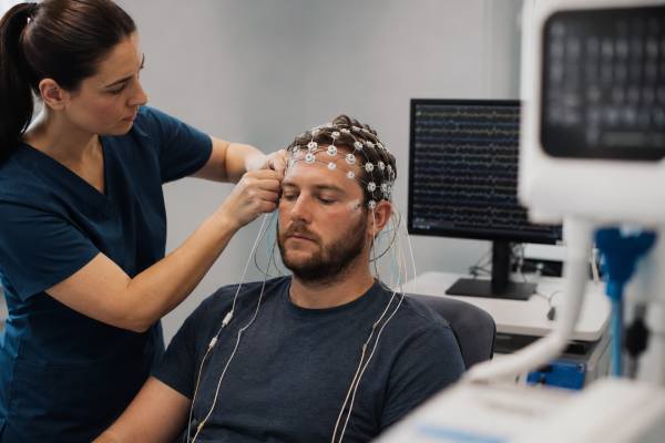
Next, the technologist measures your head and marks electrode locations with a washable pencil. Small areas of the scalp may be gently rubbed with a mild abrasive gel to improve signal quality. Electrodes are then attached with paste, adhesive, or a cap. Many labs use about 20 or more scalp electrodes, and some add electrodes near the eyes, on the chest, or on the shoulders to help distinguish brain activity from eye movement, muscle activity, or heart rhythm artifact.
You will usually sit in a reclining chair or lie on a bed. The room may be dimmed, and you may be asked to relax with your eyes closed. The technologist may give simple instructions at different points, such as opening and closing your eyes. You may also be asked to breathe deeply for a few minutes, called hyperventilation, if it is appropriate for your age and medical history. Some people feel lightheaded or notice tingling around the mouth or fingers during this part; it usually passes after normal breathing resumes.
Photic stimulation may also be used. This involves looking toward a flashing light at different speeds, usually with eyes open and closed at certain points. It is used because some people have EEG changes triggered by flashing light. If you know that flashing lights trigger seizures or severe symptoms, tell the technologist before the test begins.
After these parts, you may be allowed to fall asleep naturally. The technologist continues monitoring the EEG and may record video as well. Video is useful because movements, facial changes, breathing patterns, or symptoms during the test can be compared with the EEG. If you have an event during the recording, staff can document what happened and respond according to the lab’s safety procedures.
You will not feel your brain waves being recorded. The machine does not read thoughts, emotions, memories, dreams, or intentions. Muscle tension, blinking, jaw clenching, scalp movement, and drowsiness can all affect the tracing, which is one reason the technologist may ask you to relax, stay still, or repeat a brief instruction.
When the recording is finished, the electrodes are removed and the paste is cleaned from your hair as much as possible. Your hair may still feel sticky or gritty until you wash it. Most people can return home soon afterward, but because you are sleep deprived, it is wise to keep the rest of the day low-risk and avoid driving, alcohol, important decisions, or hazardous activities until you have recovered.
Risks and Safety
A sleep-deprived EEG is generally safe, painless, and noninvasive. The main added risk is that sleep deprivation, hyperventilation, or flashing lights may provoke a seizure in someone who is susceptible.
For many people, this risk is small, and the test is performed in a setting where staff know how to respond. In some cases, provoking abnormal activity is part of why the test is useful. Still, the goal is controlled observation, not unnecessary danger. Tell the clinic before the test if you have frequent seizures, recent seizure clusters, a history of prolonged seizures, pregnancy, significant heart or lung disease, severe migraine triggered by flashing lights, or any medical condition that could make sleep deprivation unsafe.
Possible minor side effects include fatigue, headache, scalp irritation, dizziness during deep breathing, or feeling emotionally worn down from lack of sleep. Some people feel anxious because they worry about having a seizure during the recording. Let the technologist know if you feel unwell. They can stop or adjust parts of the test when needed.
Safety planning matters most before and after the appointment. Do not drive while severely sleep deprived. Avoid operating machinery, cycling in traffic, swimming, bathing alone, climbing heights, or cooking over high heat when very tired. If you have a known seizure disorder, follow your usual seizure safety plan and ask whether someone should stay with you after the test until you have slept.
Seek urgent medical care if a seizure lasts longer than your emergency plan allows, if repeated seizures occur without full recovery, if breathing or injury is a concern, if a first-ever seizure occurs, or if new neurological symptoms appear, such as one-sided weakness, severe sudden headache, persistent confusion, trouble speaking, or loss of consciousness that does not resolve normally. For broader warning signs, a resource on ER care for neurological symptoms may help clarify when symptoms should not wait.
For children, safety includes the whole household. A sleep-deprived child may be irritable, impulsive, unsteady, or more likely to nap unexpectedly. Plan a quiet day after the test when possible. Schools, sports, swimming, biking, and climbing activities may need to wait until the child has rested and is back to baseline.
Some people worry that the test itself can cause epilepsy. It cannot. EEG recording does not create a seizure disorder. The concern is that sleep loss or activation procedures may trigger a seizure in someone already prone to seizures. If that happens, the event may provide important diagnostic information, but it should still be handled with appropriate precautions and follow-up.
What Results Can Show
A sleep-deprived EEG can show normal brain rhythms, sleep stages, slowing, epileptiform discharges, or seizures during the recording. The meaning depends on the pattern, the person’s age, symptoms, medications, sleep state, and medical history.
One possible result is a normal EEG. This can be reassuring, but it does not completely rule out epilepsy or seizures. EEG is a sample in time. If abnormal activity does not occur during the recording, the EEG may look normal even in someone who has seizures. This is one reason doctors interpret EEG results alongside the event description, witness accounts, video if available, neurological exam, imaging, and other tests.
Another possible result is epileptiform activity. This term usually refers to spikes, sharp waves, spike-and-wave patterns, or other discharges that suggest an increased tendency toward seizures. The location and pattern can help distinguish focal from generalized seizure tendencies. For example, activity from one temporal region may point toward focal epilepsy, while generalized spike-and-wave activity may suggest a generalized epilepsy syndrome. These distinctions can affect medication choices and further testing.
The EEG may also show slowing. Slowing is not the same as epileptiform activity. It can reflect drowsiness, medication effects, recent seizure activity, metabolic problems, structural brain changes, inflammation, injury, or other causes depending on the pattern. Focal slowing may lead doctors to consider whether brain imaging, such as brain MRI, is needed or should be reviewed.
Occasionally, the test captures a typical event. This can be very useful. If a person has their usual spell and the EEG shows a seizure pattern at the same time, that supports an epileptic seizure diagnosis. If the typical event occurs without seizure activity on EEG, the clinician may consider other explanations, though interpretation can be complex. Some focal seizures may be hard to see on scalp EEG, especially if they arise deep in the brain or involve limited regions.
Results are usually not given immediately by the technologist. The recording must be reviewed by a specialist, often a neurologist or clinical neurophysiologist. The report may describe the background rhythm, sleep captured, activation procedures, abnormalities, and clinical impression. The referring clinician then explains what the result means for your situation.
It is reasonable to ask specific questions when results come back:
- Was sleep actually recorded?
- Were epileptiform discharges seen?
- Did the EEG show focal or generalized findings?
- Were any findings nonspecific?
- Does the result change the diagnosis?
- Does it change treatment or activity precautions?
- Is more testing needed?
EEG language can sound alarming, but not every abnormal phrase means the same thing. “Nonspecific abnormality,” “mild slowing,” “artifact,” and “epileptiform discharge” have different implications. If the report is unclear, ask the clinician to translate the findings into practical next steps.
Other Tests and Next Steps
A sleep-deprived EEG is one tool in a broader diagnostic process. What happens next depends on whether the test is normal, abnormal, or incomplete, and whether your symptoms continue.
If the sleep-deprived EEG shows a clear seizure tendency that matches the clinical history, the doctor may diagnose epilepsy or refine the seizure type. Treatment may include anti-seizure medication, seizure safety counseling, driving restrictions based on local rules, and follow-up with neurology. Some people also need MRI, blood tests, medication-level monitoring, or review of possible triggers such as sleep loss, alcohol, missed medication, or illness.
If the EEG is normal but episodes continue, the next step may be longer monitoring. Routine and sleep-deprived EEGs are limited by time. If symptoms happen only once every few days or weeks, a brief recording may miss them. Ambulatory EEG can record at home for a longer period, while inpatient video EEG monitoring can capture events under close supervision. These approaches may be especially useful when the main question is whether repeated events are epileptic seizures or something else.
If symptoms occur mainly during sleep, a sleep study may be considered. Some nighttime events are seizures, but others are parasomnias, sleep apnea-related arousals, REM sleep behavior disorder, panic episodes, or movement disorders. The right test depends on the details: timing in the night, movements, breathing changes, awareness, confusion after the event, injuries, and whether the person remembers anything.
The table below shows how common tests differ.
| Test | Main purpose | Typical use |
|---|---|---|
| Routine EEG | Records a short sample of brain electrical activity | Often the first EEG for possible seizures or unexplained episodes |
| Sleep-deprived EEG | Records brain activity after limited sleep, often during drowsiness or sleep | Used when seizure-related patterns may be easier to see with sleep or after a normal routine EEG |
| Ambulatory EEG | Records EEG over many hours or days during usual activities | Used when events are intermittent and may not occur during a short lab visit |
| Video EEG monitoring | Records EEG and behavior together for a longer period | Used to classify seizures, capture unclear events, or evaluate difficult-to-diagnose episodes |
| Polysomnography | Measures sleep, breathing, oxygen levels, movement, and related signals | Used for sleep apnea, unusual sleep behaviors, and other sleep disorders |
| Brain MRI or CT | Shows brain structure rather than moment-to-moment electrical activity | Used when doctors need to look for structural causes, injury, stroke, tumor, inflammation, or other brain changes |
Sometimes the next step is not another test but a better event history. A phone video of a typical episode, a written timeline, details from a witness, or notes about sleep, missed medications, alcohol, illness, menstrual cycle, stress, and timing can be extremely helpful. For children, school observations may also matter.
A normal sleep-deprived EEG should not be used to dismiss serious or recurring symptoms without follow-up. Likewise, an abnormal EEG should not be interpreted in isolation. Some EEG patterns increase the likelihood of epilepsy, but the diagnosis still depends on whether the clinical events fit. The most useful result is one that is interpreted in context by the clinician who knows why the test was ordered.
References
- Routine and sleep EEG: Minimum recording standards of the International Federation of Clinical Neurophysiology and the International League Against Epilepsy 2023 (Guideline)
- The diagnostic value of sleep-deprived EEG in epilepsy 2024 (Meta-analysis)
- Influence of sleep on seizures and interictal epileptiform discharges in epilepsy 2025 (Review)
- EEG (electroencephalogram) 2026 (Patient guidance)
- Sleep Deprived Electroencephalography (SDEEG) 2025 (Patient guidance)
- Epilepsy and Seizures 2026 (Government medical overview)
Disclaimer
This article is for general educational purposes only and is not a substitute for medical advice, diagnosis, or treatment. Follow the preparation instructions from your EEG clinic, and contact your clinician if sleep deprivation, medication changes, seizures, pregnancy, or another medical condition raises safety concerns.
Share this article on Facebook, X, or your preferred platform to help others understand what a sleep-deprived EEG involves.