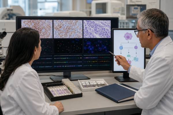
HER2, also called ERBB2, can be tested as a protein, a gene copy-number change, or an activating gene mutation. These are related but distinct biomarkers. In breast and gastric cancer, laboratories usually begin with HER2 immunohistochemistry to measure protein on tumor-cell membranes and may use in situ hybridization to assess ERBB2 gene amplification. In non-small cell lung cancer, next-generation sequencing is used to detect activating ERBB2 mutations, especially insertions in exon 20. The result can determine eligibility for a HER2-directed antibody, antibody-drug conjugate, tyrosine kinase inhibitor, or clinical trial. Interpretation must follow the scoring system validated for the tumor type; a breast-cancer IHC score should not automatically be applied to gastric, colorectal, endometrial, or other tumors. HER2 can also differ between the primary tumor and a metastasis or change after treatment, so repeat testing may be useful when disease recurs or progresses.
- HER2-positive breast cancer usually means IHC 3+ or ERBB2 amplification by in situ hybridization, while IHC 2+ generally requires reflex amplification testing.
- HER2-low breast cancer means IHC 1+ or IHC 2+/ISH-negative, and HER2-ultralow refers to faint membrane staining within an IHC 0 category under a treatment-specific definition.
- HER2-positive gastric or gastroesophageal junction cancer is commonly IHC 3+ or IHC 2+/ISH-positive, using a gastric-specific scoring pattern.
- HER2-mutant lung cancer is identified by DNA sequencing, not by assuming that protein overexpression or amplification represents the same biomarker.
- A negative or borderline result may need repeat or alternative testing when tissue is limited, fixation is poor, staining is heterogeneous, or a new metastasis is available.
Table of Contents
- What HER2 and ERBB2 testing measures
- HER2 results in breast cancer
- Gastric and other solid-tumor results
- ERBB2 mutations in lung cancer
- Samples and testing methods
- Targeted treatment and safety
- Discordant results and next steps
What HER2 and ERBB2 testing measures
ERBB2 is the official gene symbol, while HER2 is the common name for the receptor protein it encodes. HER2 is a receptor tyrosine kinase in the epidermal growth factor receptor family. When too much HER2 is produced or the receptor is activated by a mutation, growth signals can drive cancer-cell survival and proliferation.
A “HER2 test” can refer to three different biological findings:
- Protein overexpression. Immunohistochemistry, or IHC, uses antibodies to show how much HER2 protein is present on tumor-cell membranes and how complete and intense the staining appears.
- Gene amplification. In situ hybridization, or ISH, counts ERBB2 gene signals, often in relation to the chromosome 17 centromere. Amplification can lead to excess protein production.
- Activating mutation. DNA sequencing detects a sequence change that turns on the receptor. Mutations are especially important in lung cancer and may occur without amplification or strong protein expression.
These categories cannot be substituted for one another. A tumor can be amplified and strongly overexpress HER2 without having an activating ERBB2 mutation. Another tumor can carry a kinase-domain mutation with little or inconsistent IHC staining. The treatment evidence and companion diagnostic may require one specific biomarker.
The term HER2-positive is also context dependent. In traditional breast and gastric testing, it usually refers to strong overexpression or amplification. In a tumor-agnostic treatment setting, it may refer to IHC 3+ across selected solid tumors. In lung cancer, “HER2-positive” can be ambiguous unless the report specifies mutation, amplification, or overexpression.
HER2 is usually a somatic tumor biomarker. An ERBB2 finding in a cancer specimen does not ordinarily mean the person has an inherited cancer syndrome, and healthy relatives do not need predictive HER2 testing. Rare germline ERBB2 variants have been studied, but they are not a routine explanation for hereditary breast, gastric, or lung cancer.
This distinction matters when a commercial report lists both inherited and tumor findings. A genetics referral should be driven by the family history or a separate germline result, not by HER2 positivity alone.
A result should always identify the specimen, tumor type, method, score or exact variant, and interpretation. A portal message saying “HER2 negative” may hide clinically important detail such as IHC 1+, IHC 0 with faint membrane staining, or a technically limited sample.
HER2 results in breast cancer
All newly diagnosed invasive breast cancers should have HER2 testing because the result affects systemic treatment. Testing is also commonly repeated on recurrent or metastatic disease when a new biopsy is available, especially if the prior result was negative, borderline, old, or from a small specimen.
Immunohistochemistry scores
Breast-cancer IHC is scored from 0 to 3+ according to membrane-staining intensity, completeness, and the proportion of invasive tumor cells involved.
- IHC 3+ is HER2-positive. It generally shows complete, intense circumferential membrane staining in more than 10% of invasive tumor cells.
- IHC 2+ is equivocal for conventional HER2-positive classification. Reflex ISH is usually required.
- IHC 1+ is HER2-negative for amplification-based classification but can meet a HER2-low treatment definition in an appropriate metastatic setting.
- IHC 0 is negative by the traditional classification. Some IHC 0 tumors have faint, incomplete membrane staining in a small proportion of cells and can meet a HER2-ultralow definition for a specific drug indication.
Distinguishing 0 from 1+ and recognizing ultralow staining can be difficult. The test was originally optimized to separate strongly positive tumors from nonamplified tumors, not to measure very low expression. Preanalytic handling, antibody platform, tissue age, interpretation, and tumor heterogeneity can change the category near the boundary.
“HER2-low” is not a separate genetic disease subtype in the same sense as HER2-amplified cancer. It is a treatment-selection category that identifies some tumors with enough surface protein for an antibody-drug conjugate to deliver its payload. HER2-low status can change between samples and over the disease course.
In situ hybridization results
ISH includes fluorescence, chromogenic, silver-enhanced, and dual-probe methods. The laboratory counts ERBB2 signals and may calculate an ERBB2-to-chromosome-17 ratio and average copy number per cell. Current breast guidelines define several ISH groups and require correlation with IHC for uncommon patterns.
A simple “amplified” or “not amplified” interpretation is usually provided, but the underlying values can be useful in borderline or discordant cases. IHC 2+/ISH-amplified tumors are HER2-positive. IHC 2+/ISH-negative tumors are conventionally HER2-negative and may be considered HER2-low.
Testing must focus on the invasive cancer rather than an in situ component. Ductal carcinoma in situ may show strong HER2 staining even when the adjacent invasive tumor does not. Pathologists select the correct area and may repeat testing on a resection when a small core biopsy appears discordant with tumor grade or morphology.
Treatment meaning
HER2-positive early breast cancer can be treated with HER2-directed regimens in neoadjuvant, adjuvant, or both settings. In metastatic disease, treatment options include combinations built around trastuzumab, pertuzumab, antibody-drug conjugates, kinase inhibitors, chemotherapy, and endocrine therapy when hormone receptors are present. Exact sequencing changes as clinical evidence and approvals evolve.
HER2-low and HER2-ultralow categories can support trastuzumab deruxtecan in defined metastatic settings, including requirements related to hormone receptor status, prior endocrine therapy, or prior chemotherapy. An IHC score alone does not establish that every patient should receive the drug.
Hormone receptor and HER2 results should be reviewed together. A tumor can be estrogen receptor-positive and HER2-positive, requiring a plan that addresses both pathways. A blood-based ESR1 mutation test may become relevant later for hormone receptor-positive, HER2-negative metastatic disease, but it does not replace HER2 testing.
Gastric and other solid-tumor results
HER2 testing is recommended in advanced gastric and gastroesophageal junction adenocarcinoma because a positive result can guide trastuzumab-containing therapy and later HER2-directed treatment. The scoring method differs from breast cancer because gastric tumors often show incomplete basolateral or lateral membrane staining and marked heterogeneity.
In a biopsy, a cluster of at least five tumor cells with strong membrane reactivity can support an IHC 3+ score under gastric criteria, even if the stained area is small. In a resection, the proportion threshold is typically more than 10% of tumor cells. IHC 2+ requires ISH confirmation. IHC 0 or 1+ is negative for standard trastuzumab selection.
Multiple biopsy fragments improve detection because one part of a gastric cancer may be HER2-positive while another is negative. Testing a metastasis or repeat biopsy can be useful after progression, particularly before a therapy that requires current HER2 expression. Loss of HER2 after trastuzumab exposure occurs in some tumors.
HER2-directed options in advanced gastric or gastroesophageal junction cancer may include trastuzumab with chemotherapy and immunotherapy in appropriate first-line settings, followed by trastuzumab deruxtecan after prior trastuzumab-based therapy. Cardiac function, interstitial lung disease risk, prior treatment, and regional approvals affect selection.
HER2 is also tested in biliary tract, colorectal, endometrial, ovarian, cervical, salivary gland, bladder, pancreatic, and other cancers. The most important rule is to use a tumor-specific scoring system or the exact assay and threshold required for the intended treatment. Applying breast criteria without validation can misclassify another tumor.
For previously treated unresectable or metastatic solid tumors with IHC 3+ expression and no satisfactory alternative, trastuzumab deruxtecan has a tumor-agnostic indication in the United States. The evidence is strongest in tumors with IHC 3+, while benefit in IHC 2+ varies by cancer type and is not part of that same pan-tumor definition.
HER2-positive biliary tract cancer can have separate drug options based on strong IHC expression and treatment history. Colorectal cancer often requires confirmation of amplification and consideration of RAS/BRAF status; dual HER2 blockade regimens and trials may apply. Endometrial serous carcinoma has its own staining challenges and treatment evidence.
A broad solid-tumor NGS panel can detect ERBB2 amplification or mutation in these cancers, but NGS copy number does not automatically replace IHC or ISH when a companion diagnostic requires protein expression. The oncology team should match the test to the drug’s biomarker definition.
ERBB2 mutations in lung cancer
Activating ERBB2 mutations occur in roughly 2% to 4% of lung adenocarcinomas. They are more common in people with little or no smoking history, women, and adenocarcinoma histology, although any patient can have one. Most are insertions in exon 20 of the kinase domain, but point mutations and changes in other domains also occur.
Comprehensive molecular profiling of advanced nonsquamous non-small cell lung cancer should include ERBB2 along with EGFR, ALK, ROS1, BRAF, KRAS, MET, RET, NTRK, and other actionable genes. Testing may use tumor DNA, plasma circulating tumor DNA, or both. If plasma is negative, tissue testing is recommended when feasible because low tumor shedding can create a false-negative blood result.
The report should specify the exact mutation. “ERBB2 exon 20 insertion” is more informative than “HER2 positive.” Activating tyrosine kinase domain mutations have the clearest treatment evidence, while variants of uncertain significance should not be treated as established drivers.
HER2 mutation, amplification, and overexpression in lung cancer are overlapping but separate groups. A lung tumor with IHC 3+ but no activating mutation does not automatically qualify for a mutation-specific kinase inhibitor. Likewise, a mutation-positive tumor may have low IHC expression.
Trastuzumab deruxtecan has established activity in previously treated unresectable or metastatic HER2-mutant NSCLC. Since 2025, oral kinase inhibitors targeting HER2 tyrosine kinase domain activating mutations have also entered the treatment landscape in the United States for defined previously treated nonsquamous NSCLC settings. Current drug labels distinguish mutation location, prior therapy, and histology, so clinicians should verify eligibility against the exact report.
HER2-mutant lung cancer can spread to the brain. Treatment selection considers central nervous system activity, prior therapy, symptoms, and whether local radiation or surgery is needed. A related EGFR mutation test evaluates a different oncogenic driver; most untreated lung adenocarcinomas do not carry both a dominant EGFR mutation and a dominant ERBB2 mutation.
Resistance can emerge through secondary ERBB2 changes, bypass pathways, altered receptor expression, or loss of the target. Repeat tissue or plasma profiling at progression may identify a new treatment option, but not every resistance change is actionable.
Samples and testing methods
HER2 accuracy begins before the stain or sequence run. Tissue should be placed promptly in an appropriate fixative, usually 10% neutral buffered formalin, and remain there for a validated period. Delayed fixation, underfixation, overfixation, acid decalcification, crushing, cautery, and scant tumor can weaken staining or damage nucleic acid.
Core biopsies are commonly adequate. Surgical resections provide more tissue and may reveal heterogeneity, but neoadjuvant therapy can change expression. Metastatic biopsies may be preferable when the treatment decision concerns current advanced disease.
IHC
IHC is fast, widely available, and directly measures protein distribution. The pathologist scores only viable invasive tumor using the tumor-specific algorithm. Controls must stain correctly. Borderline staining, unusual patterns, or discordance with morphology may prompt repeat IHC on another block or ISH.
ISH
ISH visualizes gene copies in tumor-cell nuclei. Dual-probe assays compare ERBB2 with a chromosome 17 reference. It is commonly used after IHC 2+ in breast and gastric cancer. Signal clustering, chromosome changes, heterogeneity, and low cell counts can complicate interpretation.
Next-generation sequencing
DNA NGS identifies activating mutations and can estimate copy-number gain. It is central in lung cancer and useful in broad tumor molecular profiling. The panel should cover ERBB2 exons and insertion types relevant to the cancer. Some assays have limited sensitivity for copy number in low-purity samples.
Plasma NGS can detect ERBB2 mutations and amplification without a new biopsy. A positive result can be actionable when the variant is well validated. A negative result is less definitive, especially with low-volume disease or isolated brain metastases. Tissue remains important for IHC scoring because circulating DNA does not measure membrane protein expression.
Repeat and confirmatory testing
Repeat testing is reasonable when:
- IHC and ISH conflict;
- the specimen has poor fixation or few invasive cells;
- an IHC 0 versus 1+ distinction affects a treatment decision;
- the primary and metastasis were collected years apart;
- disease progressed after HER2-directed therapy;
- NGS shows high-level amplification but IHC was negative; or
- clinical and histologic features strongly conflict with the result.
A second pathologist review or testing at a reference laboratory can help with rare patterns. Repeating the same flawed method on the same poor block may not solve the problem; a different block, specimen, or platform is often more informative.
Targeted treatment and safety
HER2-directed medicines work in different ways. Monoclonal antibodies bind the receptor outside the cell. Antibody-drug conjugates carry a potent chemotherapy payload into HER2-expressing cells. Tyrosine kinase inhibitors block signaling inside the cell. Bispecific antibodies bind two HER2 sites or recruit immune mechanisms.
The biomarker required for one medicine may not qualify a patient for another. Examples include:
- IHC 3+ or ISH-positive breast cancer for conventional HER2-positive regimens;
- IHC 1+ or IHC 2+/ISH-negative metastatic breast cancer for a HER2-low indication;
- IHC 0 with faint membrane staining for a HER2-ultralow indication in defined hormone receptor-positive disease;
- IHC 3+ or IHC 2+/ISH-positive gastric cancer for gastric-specific therapy;
- an activating ERBB2 mutation for mutation-directed NSCLC treatment; and
- IHC 3+ in a previously treated solid tumor for a pan-tumor indication.
Trastuzumab, pertuzumab, and several other HER2 antibodies can reduce heart-pumping function. Baseline and periodic left ventricular ejection fraction testing may be required. New shortness of breath, swelling, rapid weight gain, or palpitations should be reported.
Trastuzumab deruxtecan can cause nausea, low blood counts, fatigue, and potentially serious interstitial lung disease or pneumonitis. New cough, fever, or shortness of breath requires immediate contact with the oncology team; early interruption and corticosteroid treatment may prevent severe injury. Patients with active or prior significant interstitial lung disease need careful risk assessment.
Other antibody-drug conjugates can cause thrombocytopenia, liver abnormalities, neuropathy, or bleeding. Kinase inhibitors may cause diarrhea, rash, liver injury, mouth sores, QT changes, or other drug-specific effects. The treatment plan should include monitoring and interaction review rather than relying only on the biomarker result.
A positive test does not guarantee response. Tumor heterogeneity, low target density, prior therapy, resistance pathways, and limited drug delivery can reduce benefit. A negative test for one HER2 category does not necessarily exclude every HER2-directed approach; for example, a breast tumor can be nonamplified but HER2-low.
Discordant results and next steps
Discordance means two tests, specimens, or time points do not agree. Examples include IHC 3+ with nonamplified ISH, amplified NGS with IHC 0, HER2-positive primary breast cancer with a negative metastasis, or an ERBB2-mutant lung cancer with little protein expression.
The pathologist and oncologist should review:
- whether the same tumor area was tested;
- the age, fixation, and quality of each specimen;
- the scoring system used;
- tumor heterogeneity;
- treatment given between biopsies;
- NGS tumor purity and copy-number confidence; and
- the exact companion diagnostic required for the planned drug.
For breast cancer, uncommon ISH groups are resolved through combined IHC and ISH interpretation under ASCO/CAP guidance. A case should not remain indefinitely labeled “equivocal” when the recommended workup can produce a positive or negative final classification.
Ask the care team:
- Was this protein overexpression, gene amplification, an activating mutation, or more than one finding?
- Which tumor-specific scoring guideline did the laboratory use?
- What were the exact IHC score, ISH values, or ERBB2 variant?
- Was the tested tissue invasive cancer and was fixation adequate?
- Does this result meet the biomarker definition for the proposed drug?
- Would another block, metastasis, ISH test, or NGS panel change confidence?
- Should HER2 be retested after progression or prior HER2-directed treatment?
- What heart, lung, blood-count, and liver monitoring is required?
Keep copies of the pathology addendum and molecular report, not only the treatment summary. Record which specimen produced each result. The words “HER2 positive” should always be followed by the method and category.
When the result is uncertain, a multidisciplinary review can prevent both missed treatment and inappropriate therapy. The final plan should connect the biomarker to a specific treatment setting, explain alternatives, and state whether repeat testing is expected later.
References
- Human Epidermal Growth Factor Receptor 2 Testing in Breast Cancer: ASCO-College of American Pathologists Guideline Update 2023 (Guideline)
- Standardized pathology report for HER2 testing in compliance with 2023 ASCO/CAP updates and 2023 ESMO consensus statements on HER2-low breast cancer 2024 (Consensus Review)
- Gastric Cancer, Version 2.2025, NCCN Clinical Practice Guidelines in Oncology 2025 (Guideline)
- Trastuzumab Deruxtecan in HER2-Mutant Non-Small-Cell Lung Cancer 2022 (Clinical Trial)
- Primary Results From the DESTINY-PanTumor02 Phase II Trial 2024 (Clinical Trial)
- Trastuzumab deruxtecan in patients with solid tumours harbouring specific activating HER2 mutations (DESTINY-PanTumor01): an international, phase 2 study 2024 (Clinical Trial)
Disclaimer
This article is educational and does not replace interpretation by an oncologist or pathologist. HER2 treatment eligibility depends on the tumor type, exact test method, score or mutation, disease setting, prior therapy, and current drug labeling. New cough, fever, shortness of breath, swelling, or other serious symptoms during HER2-directed therapy require prompt medical assessment.