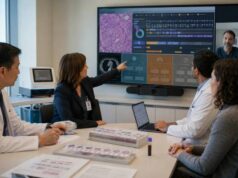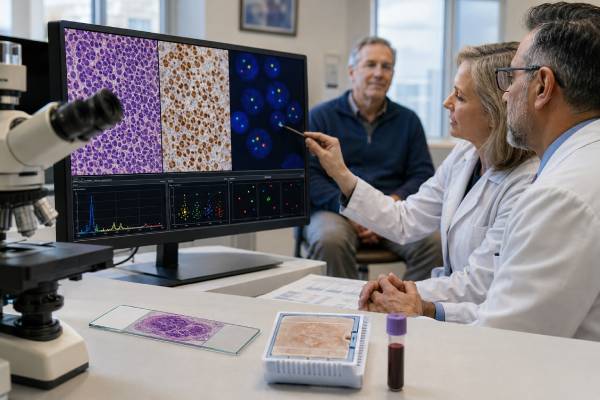
A CCND1/IGH fusion test looks for the chromosome translocation t(11;14)(q13;q32), which places CCND1 next to the immunoglobulin heavy-chain gene IGH. The change causes excess cyclin D1 protein and is the defining genetic hallmark of most mantle cell lymphomas. It is usually detected by fluorescence in situ hybridization, chromosome analysis, or next-generation sequencing.
A positive fusion strongly supports mantle cell lymphoma when the cell appearance, immunophenotype, and clinical pattern fit. It is not completely specific: t(11;14) also occurs in a subgroup of plasma cell myeloma and can rarely appear as a secondary change in other B-cell lymphomas. Conversely, a small minority of mantle cell lymphomas lack CCND1 rearrangement and instead involve CCND2 or CCND3. The report must therefore be integrated with cyclin D1 and SOX11 staining, CD5 and other B-cell markers, morphology, and the disease site. The fusion is acquired in the tumor and is not ordinarily inherited.
- CCND1::IGH detected in a compatible mature B-cell tumor strongly supports mantle cell lymphoma.
- The fusion is usually t(11;14)(q13;q32) and drives cyclin D1 overexpression.
- Cyclin D1 protein staining supports the diagnosis but is not the same as a genomic fusion test.
- A negative CCND1/IGH result does not fully exclude mantle cell lymphoma, because rare cases use CCND2 or CCND3.
- t(11;14) can occur in plasma cell myeloma, so lineage and morphology must be confirmed before naming the disease.
Table of Contents
- What the CCND1/IGH Fusion Is
- When the Test Is Ordered
- How Fusion Testing Works
- How to Read the Results
- Confirming Mantle Cell Lymphoma
- Disease Subtypes and Prognosis
- Treatment Implications
- Limitations and Next Steps
What the CCND1/IGH Fusion Is
CCND1 encodes cyclin D1, a protein that helps cells move from the G1 phase into the DNA-copying S phase of the cell cycle. Normal mature B cells usually keep cyclin D1 low. In t(11;14), regulatory elements from IGH on chromosome 14 are placed near CCND1 on chromosome 11, causing persistent cyclin D1 production and abnormal cell-cycle entry.
The fusion is commonly written as IGH::CCND1 under current gene-fusion notation, although CCND1/IGH and CCND1-IGH are also widely used. The chromosome finding is t(11;14)(q13;q32). These descriptions refer to the same core event but may come from different methods.
The translocation usually develops early in the pathogenesis of mantle cell lymphoma. Additional changes involving ATM, TP53, cell-cycle genes, chromatin regulators, and DNA-damage pathways shape the eventual tumor behavior. The fusion is therefore foundational but not sufficient by itself to explain why one patient has indolent disease and another has blastoid, rapidly progressive lymphoma.
Most mantle cell lymphomas express cyclin D1 and carry a CCND1 rearrangement with an immunoglobulin gene, usually IGH and less often an immunoglobulin light-chain partner. A break-apart CCND1 test may detect any partner, while an IGH/CCND1 dual-fusion assay specifically evaluates the classic combination.
The alteration is somatic. It is present in the abnormal B-cell clone rather than in all body cells and does not generally create a 50% inheritance risk for children. A family history of lymphoma can still be clinically relevant, but relatives are not tested for the tumor fusion as an inherited variant.
The same t(11;14) can appear in plasma cell neoplasms, including multiple myeloma and primary plasma cell leukemia. In those diseases, the abnormal cell is a plasma cell, the immunophenotype differs, and the clinical implications are not the same. A gene fusion result must always be assigned to the correct cell lineage.
Rare aggressive large B-cell lymphomas can acquire CCND1 rearrangement secondarily. They may resemble mantle cell lymphoma genetically at one locus but lack the broader mantle cell molecular program. This is another reason that t(11;14) cannot replace expert morphology and immunophenotyping.
When the Test Is Ordered
Pathologists order CCND1/IGH testing when tissue, blood, or marrow suggests mantle cell lymphoma or when cyclin D1 expression creates a diagnostic question. The test can confirm an otherwise typical case, resolve an unusual immunophenotype, or distinguish mantle cell lymphoma from chronic lymphocytic leukemia, marginal zone lymphoma, follicular lymphoma, lymphoplasmacytic lymphoma, and other small B-cell neoplasms.
Common indications include:
- A mature B-cell lymphoma expressing CD5 but not the typical chronic lymphocytic leukemia profile
- Cyclin D1-positive small or medium B cells
- Mantle-zone or diffuse lymph-node growth
- Blood lymphocytosis with an atypical B-cell clone
- Bone-marrow involvement by a CD5-positive B-cell neoplasm
- Multiple lymphomatous polyposis in the gastrointestinal tract
- Suspected blastoid or pleomorphic mantle cell lymphoma
- A cyclin D1-negative tumor with morphology strongly suggesting mantle cell lymphoma
- A plasma cell neoplasm in which t(11;14) is being assessed for classification or treatment research
A typical mantle cell immunophenotype is CD20 positive, PAX5 positive, CD5 positive, cyclin D1 positive, and usually CD23 and LEF1 negative or weak. Exceptions are common enough that no single marker should be used mechanically. SOX11 is positive in most conventional mantle cell lymphomas and many cyclin D1-negative cases, but it is often absent in leukemic non-nodal disease.
Testing may be reflexively performed after cyclin D1 immunohistochemistry or included in a small B-cell lymphoma FISH panel. In a strongly typical case, some centers accept cyclin D1 expression with the full phenotype without fusion confirmation. Molecular confirmation becomes especially useful when staining is weak, focal, technically questionable, or unexpected.
The test is not used to screen a person with no evidence of lymphoma. Symptoms such as enlarged nodes, fatigue, night sweats, weight loss, abdominal fullness, anemia, or lymphocytosis first require clinical assessment. An excisional or core biopsy often provides the architecture needed for a reliable diagnosis.
At relapse, repeating the fusion test is rarely necessary if the original diagnosis was secure because the founding event usually persists. A new biopsy may still be needed when the disease changes behavior, transformation is suspected, or a new lesion could represent another cancer.
How Fusion Testing Works
Dual-fusion FISH
An IGH/CCND1 dual-fusion FISH assay uses differently colored probes for IGH and CCND1. Normal cells show separate signals. Cells with t(11;14) show fused or closely paired signals representing the joined regions. Because reciprocal translocations create derivative chromosomes, a classic abnormal pattern often includes two fusion signals plus one remaining signal from each gene, although variant patterns occur.
The laboratory counts a defined number of nuclei and compares the abnormal percentage with a validated cutoff. The percentage reflects the sampled nuclei, not the probability of having lymphoma or the percentage of the body affected.
CCND1 break-apart FISH
Break-apart probes sit on opposite sides of CCND1. Separation shows that the gene is rearranged but does not identify the partner. This method can detect IGH and non-IGH partners. If a break-apart assay is positive and the partner matters, an IGH fusion assay or sequencing can provide more detail.
FISH works on formalin-fixed tissue, fresh cell suspensions, blood, marrow, and validated cytology preparations. A FISH test is fast and targeted but does not reveal the entire lymphoma genome.
Chromosome analysis
Karyotyping can show t(11;14) and additional chromosome changes in dividing cells. It provides a broad view but requires viable cells and may fail when the tumor does not grow in culture. Fixed lymph-node tissue cannot be used for conventional culture.
Next-generation sequencing
DNA or RNA sequencing can identify CCND1 rearrangements, partners, breakpoints, and cooperating mutations. RNA sequencing confirms an expressed fusion. DNA panels may miss the event if large intronic regions are not adequately covered. Optical genome mapping and whole-genome approaches can resolve complex or cryptic events in specialized settings.
Cyclin D1 and SOX11 immunohistochemistry
Cyclin D1 staining is rapid and strongly positive in most mantle cell lymphomas. SOX11 is a nuclear transcription-factor stain that supports conventional mantle cell lymphoma and is valuable in many cyclin D1-negative cases. Neither method shows the exact chromosome fusion, and SOX11 can be expressed in a few other tumors.
Most patients need no preparation for the molecular test itself. Preparation relates to the biopsy or bone-marrow procedure. Turnaround is often three to ten working days for FISH, while comprehensive sequencing may take two to four weeks.
How to Read the Results
A useful report states the specimen, assay type, abnormal signal pattern, percentage of abnormal nuclei, cutoff, and interpretation. It may use “IGH::CCND1 fusion detected,” “CCND1 rearrangement detected,” or “t(11;14) positive.” These are related but not always identical findings.
| Result | Usual meaning | Important caution |
|---|---|---|
| IGH::CCND1 detected | The classic t(11;14) fusion is present in the tested clone. | Confirm whether the cells are mantle B cells or plasma cells. |
| CCND1 rearranged, partner unknown | CCND1 is structurally altered. | Additional testing may be needed to identify the partner and disease. |
| Not detected | No covered fusion or rearrangement was found above the cutoff. | Rare cyclin D1-negative MCL with CCND2 or CCND3 remains possible. |
| Equivocal or atypical | The signal pattern is near cutoff or structurally complex. | Repeat FISH, another block, karyotype, or sequencing may clarify it. |
| Insufficient or failed | The sample did not support a reliable result. | This is not a true negative. |
A positive result in a CD5-positive, cyclin D1-positive mature B-cell neoplasm usually confirms mantle cell lymphoma. In a CD138-positive plasma cell neoplasm, the same fusion supports a t(11;14)-positive plasma cell myeloma instead. The report should not use the genetic result without identifying which cells carry it.
A negative result requires review of the assay. An IGH/CCND1 dual-fusion test may miss a CCND1 rearrangement with a light-chain partner. A CCND1 break-apart assay is broader for partners but still will not detect CCND2 or CCND3 rearrangements. If morphology and SOX11 strongly support mantle cell lymphoma, RNA sequencing or targeted FISH for other cyclin D genes may be appropriate. Integrated pathology review is especially important when the classic fusion is not detected.
An atypical pattern can reflect extra copies of CCND1 or IGH, a complex translocation, amplification, deletion of one probe region, or overlapping nuclei. The laboratory should distinguish a true fusion from copy-number gain. Extra CCND1 copies alone are not the classic defining event.
The finding does not predict a specific survival time. Proliferation rate, morphology, TP53 status, stage, disease subtype, and response to treatment provide more prognostic information than the fusion alone.
Confirming Mantle Cell Lymphoma
Mantle cell lymphoma arises from mature B cells related to the mantle-zone compartment. The typical cells are small to medium sized with irregular nuclear contours. Growth can be mantle-zone, nodular, diffuse, or mixed. Blastoid and pleomorphic variants have larger or more immature-looking cells and usually behave more aggressively.
The diagnosis integrates:
- Tissue architecture and cell appearance
- Mature B-cell markers such as CD20 and PAX5
- CD5 expression in most cases
- Cyclin D1 protein
- SOX11 when useful
- CCND1 rearrangement or another cyclin D alteration
- Flow cytometry, often showing a light-chain-restricted B-cell population
- Clinical distribution in nodes, spleen, marrow, blood, or gastrointestinal tract
Chronic lymphocytic leukemia can also be CD5 positive, but it usually expresses CD23, LEF1, and dim CD20 and surface immunoglobulin, and it lacks the classic CCND1 fusion. Some CLL cases show cyclin D1 in proliferation centers, so staining location and intensity matter.
Cyclin D1-positive diffuse large B-cell lymphoma can mimic blastoid mantle cell lymphoma. Large B-cell lymphomas with secondary CCND1 rearrangements often carry rearrangements or mutations more typical of DLBCL and lack the mantle cell molecular profile. Reviewing SOX11, CD5, morphology, prior history, and broader genomic findings prevents misclassification.
Cyclin D1-negative mantle cell lymphoma is rare but recognized. Most cases overexpress cyclin D2 or D3 through CCND2 or CCND3 rearrangements and have a gene-expression profile similar to conventional MCL. SOX11 is frequently positive. These tumors should not be excluded simply because t(11;14) is absent.
In situ mantle cell neoplasia consists of rare cyclin D1-positive B cells restricted to the inner mantle zones of otherwise reactive follicles. It is often incidental and has a low rate of progression. It should not be called overt lymphoma without expansion, dissemination, or other diagnostic features.
Disease Subtypes and Prognosis
Mantle cell lymphoma has at least two major biological pathways. Conventional MCL usually arises from naïve-like B cells, expresses SOX11, and often presents with nodal and extranodal disease. Leukemic non-nodal MCL derives from more antigen-experienced cells, is often SOX11 negative, and may initially involve blood, marrow, and spleen with little lymph-node enlargement.
Leukemic non-nodal disease can follow an indolent course, but it is not always harmless. Acquisition of TP53 abnormalities or other changes can lead to progression. Some asymptomatic patients with low tumor burden can be observed closely rather than treated immediately, regardless of the presence of the fusion.
The Mantle Cell Lymphoma International Prognostic Index uses age, performance status, lactate dehydrogenase, and white blood cell count. Adding the Ki-67 proliferation index improves risk assessment. Blastoid or pleomorphic morphology, high Ki-67, complex karyotype, and TP53 mutation or deletion are associated with poorer outcomes.
TP53 status deserves particular attention because standard chemoimmunotherapy often produces less durable control in TP53-abnormal disease. Clinical trials and non-chemotherapy approaches may be prioritized. The CCND1/IGH fusion itself does not identify TP53 risk, so broad molecular or targeted testing may be needed.
Stage is commonly advanced at diagnosis because marrow, blood, spleen, and gastrointestinal involvement can occur. Advanced stage does not have the same meaning as in many solid tumors; some patients have years of disease control with modern therapy. Prognosis depends on biology, fitness, treatment response, and access to later-line options.
Minimal residual disease assays can track the immunoglobulin clone or a tumor-specific sequence in research and selected clinical protocols. The CCND1 fusion can sometimes be used as a molecular marker, but routine monitoring generally relies on examination, blood counts, imaging, and disease-specific response criteria rather than serial FISH percentages.
Treatment Implications
The fusion confirms a therapeutic disease category but is not a direct target of a routinely approved fusion inhibitor. Treatment is selected for mantle cell lymphoma as a whole and depends on symptoms, age, fitness, stage, TP53, proliferation, prior therapy, and patient goals.
Some asymptomatic patients with low-burden, indolent disease can begin with active surveillance. Treatment starts for symptoms, progressive cytopenias, threatened organs, bulky or rapidly growing disease, or other evidence that benefit outweighs risk.
First-line options may include anti-CD20 antibody-based combinations, bendamustine-containing regimens, cytarabine-containing intensive therapy for selected fit patients, Bruton tyrosine kinase inhibitors in certain modern regimens, and maintenance rituximab. Autologous stem-cell transplantation has been used as consolidation in younger fit patients, although its role is evolving as effective targeted combinations move earlier.
Relapsed disease can be treated with covalent BTK inhibitors, the noncovalent BTK inhibitor pirtobrutinib in appropriate settings, CAR T-cell therapy, bispecific antibodies where available, venetoclax-containing approaches, lenalidomide, and clinical trials. Drug approval and preferred sequence differ by country and change over time.
The presence of t(11;14) in plasma cell myeloma has a separate treatment context. It is associated with BCL2 dependence in many myelomas, and venetoclax has shown activity, but use depends on approval, patient selection, infection risk, and current evidence. A mantle cell report should not import a myeloma-specific interpretation, and vice versa.
Treatment response can reduce the abnormal clone below FISH detection, but a negative post-treatment FISH result is not by itself proof of cure. Imaging, marrow evaluation when indicated, flow cytometry, and clinical status provide a fuller response assessment.
Limitations and Next Steps
FISH can miss disease when few tumor cells are present, tissue is crushed or decalcified, or the breakpoint lies outside probe coverage. It can also detect a real fusion in a sample whose lineage is unclear. Selecting the tumor-rich region and correlating with immunostains reduce these errors.
A small blood clone with t(11;14) should not be assumed to represent symptomatic lymphoma. Monoclonal B-cell populations and very early mantle cell–like clones can exist without treatment indications. The clinical and tissue context determines whether the person has overt mantle cell lymphoma.
Questions to ask include:
- Was the assay dual-fusion IGH/CCND1 or CCND1 break-apart FISH?
- Which cells carry the abnormality: mature B cells or plasma cells?
- Are cyclin D1, SOX11, CD5, CD23, and LEF1 results available?
- Does the morphology fit conventional, leukemic non-nodal, blastoid, or pleomorphic MCL?
- Was TP53 tested, and what is the Ki-67 proliferation index?
- If the fusion is negative, were CCND2 and CCND3 considered?
- Does the result change treatment timing or clinical-trial eligibility?
- Is expert lymphoma pathology review warranted?
Urgent care is appropriate for breathing or swallowing difficulty from a mass, severe abdominal pain or rapidly increasing fullness, new neurologic symptoms, major bleeding, or fever during treatment. Tumor lysis can occur when a bulky lymphoma responds quickly, so high-risk patients may need preventive hydration, laboratory monitoring, and uric-acid treatment.
The final report should name both the disease and the evidence: “mantle cell lymphoma with IGH::CCND1 fusion” is clearer than “t(11;14) positive.” That wording prevents a hallmark alteration from being mistaken for a diagnosis outside its cellular context.
Why the original biopsy should be preserved
Mantle cell lymphoma often needs several tests from the same block: immunohistochemistry, FISH, TP53 analysis, and sometimes broad sequencing. Repeated unstained sections can gradually exhaust a small core. The pathology team may prioritize studies that establish the diagnosis first, then reserve tissue for prognostic or trial testing. When a referral center requests review, sending slides plus the block or representative unstained sections allows confirmation without a new biopsy. A repeat procedure is most justified when the original tissue is truly inadequate, the current disease looks biologically different, or the new sample could change treatment rather than merely duplicate a secure result.
References
- The International Consensus Classification of Mature Lymphoid Neoplasms: a report from the Clinical Advisory Committee 2022 (Position Statement)
- A comparison of the International Consensus and 5th World Health Organization classifications of mature B-cell lymphomas 2023 (Review)
- B- and T-/NK-Cell Lymphomas in the 2022 International Consensus Classification of Mature Lymphoid Neoplasms and Comparison with the WHO Fifth Edition 2024 (Review)
- Large B-cell lymphomas with CCND1 rearrangement have a germinal center phenotype and distinct genomic features 2024
- Updates on the Biological Heterogeneity of Mantle Cell Lymphoma 2025 (Review)
- Cyclin D1-negative mantle cell lymphoma 2025 (Review)
Disclaimer
This article is educational and does not replace diagnosis by a hematopathologist or treatment advice from a lymphoma specialist. CCND1/IGH results must be interpreted with cell lineage, morphology, cyclin D1 and SOX11 staining, flow cytometry, TP53 and proliferation findings, specimen quality, and clinical presentation. Seek urgent care for breathing difficulty, neurologic symptoms, severe abdominal pain, major bleeding, or fever during treatment.