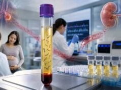
Digital PCR measures a specific DNA or RNA target by dividing a sample into thousands of tiny reactions and counting how many contain the target. This partition-and-count approach can detect rare mutations, quantify low concentrations, measure gene copy number, and track small changes over time without relying on a standard curve. Droplet digital PCR is the most familiar format, but chip- and well-based systems use the same basic principle. The method is highly precise when the target is already known. It is not a broad discovery test: primers and fluorescent probes must be designed for the exact mutation, fusion, pathogen, transcript, or gene region being measured. Results may be reported as copies per microliter, copies per reaction, mutant allele fraction, a copy-number ratio, or detected versus not detected. Those numbers only become clinically useful when the laboratory’s limit of blank, limit of detection, limit of quantification, sample input, and quality controls are considered.
- dPCR is a targeted counting method: It is ideal for known low-level mutations, selected copy-number changes, and precise molecular monitoring.
- Partitioning improves rare-target detection: A few mutant molecules can be separated from a large background of normal DNA.
- “Not detected” does not mean zero molecules: The target may be absent, outside the assay, degraded, or below the validated detection limit.
- More input can improve sensitivity: The number of DNA molecules tested often matters as much as the instrument’s advertised performance.
- Copy number is measured against a reference gene: Poor reference selection, DNA fragmentation, or linked targets can distort the ratio.
- Serial results require the same method and specimen type: A change is easiest to interpret when collection, extraction, assay, and reporting units remain consistent.
Table of Contents
- How Digital PCR Counts Molecules
- Clinical Uses of dPCR
- Assay Design, Controls, and Thresholds
- Reading Low-Level Mutation Results
- Understanding Copy-Number Results
- dPCR Compared With qPCR and NGS
- False Positives, False Negatives, and Uncertainty
- Preparation and Follow-Up
How Digital PCR Counts Molecules
A conventional PCR reaction amplifies all target molecules together in one tube. Digital PCR first divides the mixture into many small partitions. Depending on the platform, those partitions are droplets, microscopic wells, chambers, or channels. Ideally, each partition contains zero, one, or only a few target molecules.
After endpoint amplification, the instrument measures fluorescence in every partition. Partitions above the assay threshold are called positive; those below it are called negative. The fraction of positive partitions is converted into a concentration with Poisson statistics, which corrects for the fact that some positive partitions contain more than one target molecule.
The term “digital” refers to classifying partitions into discrete positive and negative events. The final result is not simply a count of bright droplets. Software estimates the original target concentration from the total number of accepted partitions, positive partitions, partition volume, dilution, and statistical model.
A two-color assay can distinguish a mutant target from the corresponding wild-type sequence. Another design may measure a gene of interest in one channel and a stable reference gene in the other. Multiplex systems can use additional colors or different fluorescence amplitudes, but the number of targets remains far smaller than in next-generation sequencing.
Why partitioning helps with rare targets
Suppose one mutant DNA molecule is mixed with 10,000 normal molecules. In a single bulk reaction, the normal signal can dominate. In dPCR, the molecules are distributed across separate compartments. Some partitions contain only normal DNA, a very small number contain the mutant target, and many contain neither. The rare signal becomes easier to distinguish when the probes are highly specific.
Sensitivity still depends on how many molecules enter the assay. If only 1,000 genome equivalents are tested, a true variant present at 0.01% may not be represented in the reaction at all. Running more wells, using more plasma, improving extraction, or testing replicate reactions can increase the number of opportunities to capture the target.
The basic workflow is:
- Extract DNA or RNA from the specimen.
- Convert RNA to complementary DNA when reverse transcription is required.
- Combine the sample with primers, probes, polymerase, and controls.
- Partition the reaction.
- Amplify to endpoint.
- Read fluorescence and exclude poor-quality partitions.
- Apply thresholds and Poisson correction.
- Report concentration, fraction, or copy number with assay limits.
The same principle supports several clinical uses, but each assay requires its own validation. A mutation assay validated in plasma cannot automatically be applied to bone marrow, urine, or tissue without evidence that extraction, inhibitors, background, and expected target levels are suitable.
Clinical Uses of dPCR
Digital PCR is most valuable when the laboratory knows exactly what to search for and needs high analytical sensitivity or precision.
Cancer mutation detection and monitoring
DPCR can detect known tumor variants in tissue, blood plasma, bone marrow, cerebrospinal fluid, urine, or other specimens. Common applications include tracking BCR::ABL1 or other fusion transcripts, monitoring NPM1 or JAK2 variants, measuring selected EGFR or ESR1 resistance mutations, and confirming a low-frequency finding from sequencing.
For measurable residual disease, the assay may search for one mutation that was documented at diagnosis. A falling mutant level after treatment can support molecular response; a rising level may precede clinical relapse. The interpretation is disease-specific. A technically detectable signal does not always meet a validated threshold for changing treatment.
In solid tumors, dPCR may analyze circulating tumor DNA. It can measure a known mutation at fractions well below those usually reported by routine sequencing, provided enough DNA is tested. This targeted strength is also a limitation: a new resistance mutation elsewhere will be missed unless a separate assay is designed.
Copy-number analysis
Digital PCR can measure whether a selected gene or exon is deleted, duplicated, or amplified by comparing it with a reference locus. It is used for constitutional CNVs, tumor amplifications, transgene copy number, viral integration, and assay confirmation. Precision can be excellent for distinguishing two from three copies, but DNA quality, restriction digestion, reference choice, and genomic linkage influence the result.
Transplant and chimerism applications
Donor- and recipient-specific DNA markers can be quantified after transplantation. Some assays measure donor-derived cell-free DNA or low-level chimerism. The biological meaning depends on the transplanted organ or cell type and cannot be inferred from analytical sensitivity alone.
Infectious disease
DPCR can quantify low pathogen loads, detect resistance mutations, or measure viral reservoirs. It is relatively tolerant of some inhibitors and does not require a calibration curve. Clinical use still depends on specimen-specific validation, contamination control, and evidence linking concentration to diagnosis or treatment response.
Prenatal and reproductive applications
Targeted dPCR can measure fetal or placental alleles in maternal plasma, confirm selected copy-number changes, or test known familial variants in appropriate specimens. It is not a substitute for comprehensive prenatal screening or diagnostic chromosome analysis unless specifically validated for that purpose.
Research and assay validation
Because dPCR provides absolute or near-absolute target quantification, it is used to assign values to reference materials, validate NGS findings, quantify gene-editing outcomes, measure vector copy number, and check standards used by other molecular tests. Research-grade performance should not be assumed to equal clinical diagnostic validation.
Assay Design, Controls, and Thresholds
A reliable dPCR result depends on more than the number of positive partitions. The primers must amplify the intended target efficiently, the probe must distinguish it from closely related sequences, and controls must define background noise.
Limit of blank
The limit of blank describes the highest apparent signal expected when no true target is present. Low-level fluorescence can arise from nonspecific probe binding, polymerase errors, contamination, instrument noise, or misclassification of borderline partitions. Laboratories test many blank or wild-type samples to estimate this background.
Limit of detection
The limit of detection is the lowest concentration or fraction detected with a stated probability, often 95%, under validated conditions. It is not necessarily the lowest signal the instrument has ever seen. A result below the formal limit may be described as detected below quantifiable range, indeterminate, or not reportable.
Limit of quantification
The limit of quantification is the lowest level that can be measured with acceptable precision and bias. A target can sometimes be detected without being quantified reliably. For example, two positive partitions may support a trace signal but produce a very wide confidence interval.
Positive, negative, and no-template controls
A positive control confirms that the assay can detect the target. A wild-type control tests specificity. A no-template control checks for reagent or environmental contamination. Extraction controls can identify specimen-processing failures. Reverse-transcription assays also need controls for RNA quality and genomic DNA contamination.
Partition count and rain
A run must generate enough accepted partitions. Too few partitions widen uncertainty and reduce dynamic range. “Rain” refers to partitions with intermediate fluorescence between clearly positive and negative clusters. It can result from partial inhibition, damaged templates, poor annealing, suboptimal probe design, or genuine sequence variation under a primer or probe.
Thresholds may be set automatically or manually under laboratory rules. Moving the threshold can materially change a low-level result, so validated procedures and blinded review are important. The updated digital MIQE recommendations emphasize transparent reporting of assay design, partition volume, thresholds, controls, precision, and analytical limits.
| Term | What it describes | Why it matters |
|---|---|---|
| Limit of blank | Expected background without target | Helps separate rare signal from noise |
| Limit of detection | Lowest level detected with defined probability | Sets the meaning of “not detected” |
| Limit of quantification | Lowest level measured with acceptable accuracy | Explains why some trace signals are not given a precise number |
| Accepted partitions | Usable reaction compartments | Affects precision and statistical confidence |
| Input molecules | Total analyzable target opportunities | Limits achievable rare-variant sensitivity |
Reading Low-Level Mutation Results
Mutation assays commonly report mutant copies, wild-type copies, and mutant allele fraction. Mutant allele fraction is calculated as mutant molecules divided by total mutant plus wild-type molecules, with method-specific corrections.
A result of 0.5% means roughly five mutant copies per 1,000 measured copies at that locus in the analyzed DNA. It does not mean that 0.5% of the patient’s cells are cancerous. Tumor cells may carry one or several copies of the locus, normal DNA dilutes the specimen, and cell-free DNA shedding differs across tissues.
The confidence interval matters at low counts. If three mutant-positive partitions are detected, the estimated fraction may have wide statistical uncertainty. Replicate consistency, total input, blank performance, and the laboratory’s decision threshold should be reviewed before interpreting a very small number.
Possible report terms include:
- Detected and quantifiable: The signal exceeds the assay’s quantification limit.
- Detected below the quantifiable range: Target-like partitions are present, but the number is too low for precise measurement.
- Detected below the validated clinical threshold: Analytical signal exists, but its medical meaning is not established.
- Not detected: No reportable signal was found above the laboratory’s limit under the tested conditions.
- Indeterminate: Controls, partition quality, inhibition, or borderline fluorescence prevent a confident call.
Serial trends should use the same mutation, assay, laboratory, specimen, and unit whenever possible. A change from 0.2% to 0.4% may not be clinically meaningful if both values have overlapping confidence intervals or differ in input. A change from undetectable to repeatedly rising measurable levels may be more concerning, but disease-specific guidance determines action.
For cancer, dPCR results belong within the broader framework of somatic genetic testing. A plasma variant can also come from clonal hematopoiesis rather than the solid tumor. Comparison with white blood cell DNA or tissue findings may be needed, particularly for genes frequently altered in blood-cell clones.
Understanding Copy-Number Results
A dPCR copy-number assay usually measures a target and a reference locus known to be present at a stable number of copies. The target-to-reference ratio is converted into an estimated copy number.
For an autosomal locus in a constitutional sample, expected results may center near:
- 1 copy for a heterozygous deletion.
- 2 copies for the usual diploid state.
- 3 copies for a heterozygous duplication.
- 4 or more copies for higher gains.
Real values rarely appear as exact integers. A report may show 1.02, 1.96, or 3.08 with a confidence interval. The laboratory uses validated ranges to call deletion, normal dosage, or duplication.
Reference-gene choice
The reference should be stable in the tested specimen and not located in a region commonly gained or lost. This is especially difficult in tumors, where aneuploidy and broad chromosome changes are common. A reference assumed to have two copies may itself be altered, producing a misleading target ratio.
DNA fragmentation and restriction digestion
If the target and reference are physically linked on the same long DNA molecule, they may partition together rather than independently. Restriction enzymes can cut the DNA between targets to improve random distribution. Over-fragmented or degraded DNA can damage primer sites, while incomplete digestion can affect copy-number estimates.
Mosaic and tumor copy number
A constitutional duplication present in only 30% of cells may produce an average value between two and three copies. A tumor amplification is diluted by normal cells and affected by tumor ploidy. Therefore, an estimated copy number of 3 in a low-purity tumor could represent a stronger amplification within the cancer cells.
DPCR is excellent for targeted confirmation but does not show how large the duplicated region is, where the extra copy is located, or whether the duplication is inverted. A positive result may require microarray, MLPA, FISH, or sequencing to define structure. More detail about gene dosage interpretation appears in deletion/duplication genetic testing.
dPCR Compared With qPCR and NGS
Digital PCR and quantitative real-time PCR both amplify selected targets, but they measure them differently. qPCR records fluorescence during amplification and usually compares the cycle threshold with a standard curve or reference. DPCR reads endpoint partitions and applies counting statistics.
| Feature | dPCR | qPCR | Targeted NGS |
|---|---|---|---|
| Target scope | One to a few known targets | One to a few known targets | Dozens to hundreds of genes |
| Quantification | Partition-based absolute or relative counting | Relative or standard-curve based | Read-based, often semi-quantitative |
| Rare-variant sensitivity | Very high for a validated known target | Moderate to high | Broad but depends on depth and error suppression |
| Discovery ability | None outside designed targets | None outside designed targets | Can discover multiple covered variants |
| Best role | Precise targeted detection and monitoring | Fast routine quantification | Broad molecular profiling |
DPCR may outperform qPCR when small concentration differences, rare variants, or standard-curve uncertainty are important. QPCR may be faster, cheaper, and sufficient for high-level targets. NGS is preferable when the relevant mutation is unknown or many genes must be assessed at once.
The methods are often complementary. NGS discovers a tumor mutation; dPCR tracks it during treatment. Microarray identifies a suspected duplication; dPCR confirms the selected locus. QPCR screens a large number of routine samples; dPCR resolves borderline cases.
Claims that dPCR is universally more sensitive should be treated cautiously. A well-designed qPCR assay with abundant input may outperform a poorly designed dPCR assay. Error-corrected NGS can also reach very low variant fractions while examining many targets. Clinical value depends on assay validation, not the generation label.
False Positives, False Negatives, and Uncertainty
Rare-target testing is vulnerable to contamination because one stray molecule may create a positive partition. Laboratories separate pre- and post-amplification areas, use unidirectional workflow, include blank controls, and repeat unexpected low-level results when appropriate.
False-positive or misleading signals can result from:
- Nonspecific probe binding or primer dimers.
- Polymerase errors during early amplification.
- Carryover contamination.
- Fluorescence spillover between channels.
- Poorly placed thresholds.
- Sequence variants under a probe that alter cluster position.
- Clonal hematopoiesis in plasma cancer testing.
- A reference locus altered in a tumor copy-number assay.
False negatives can result from:
- Too few input molecules.
- Degraded DNA or RNA.
- Inhibitors in the specimen.
- A mutation adjacent to the target that disrupts primer binding.
- Target evolution, such as a cancer clone losing the monitored variant.
- Testing the wrong specimen or an anatomic compartment with little target release.
- Reverse-transcription inefficiency in RNA assays.
A high number of partitions does not compensate for absent template. Twenty thousand empty droplets cannot detect a mutation that never entered the reaction. Reports should therefore state the analyzed DNA amount or estimated genome equivalents when rare-event sensitivity is important.
Statistical confidence is another source of uncertainty. At low counts, random sampling creates large relative variation. Two aliquots from the same specimen may not contain the same number of rare molecules. Replicate testing and larger input reduce—but do not eliminate—this sampling effect.
Clinical false reassurance can occur when “not detected” is interpreted as cure or absence of disease. DPCR evaluates one molecular marker. A tumor can persist without releasing detectable DNA, or a different clone can emerge. Imaging, pathology, blood counts, organ function, and symptoms remain essential.
Preparation and Follow-Up
Preparation depends on the specimen. Blood-based DNA testing usually requires no fasting. Plasma assays need dedicated cfDNA tubes or prompt processing to limit white blood cell lysis. Bone marrow, tumor tissue, urine, cerebrospinal fluid, and respiratory samples have their own collection instructions. RNA assays require careful stabilization because RNA degrades quickly.
Before testing, confirm the exact target. A report from an earlier tumor or genetic test should include standard variant notation, such as gene name, transcript, nucleotide change, and protein change. “KRAS positive” is not enough to design a mutation-specific assay because different KRAS variants require different probes.
Ask the ordering team:
- Is the assay diagnostic, confirmatory, or intended for serial monitoring?
- What specimen type was clinically validated?
- How much DNA or RNA was analyzed?
- What are the limit of blank, detection, and quantification?
- Is the result reported as copies, fraction, ratio, or a qualitative call?
- What change is considered clinically meaningful?
- Should a borderline result be repeated from the same extract or a new specimen?
- Could another method find variants outside this target?
For serial monitoring, collect at comparable clinical time points when possible. Treatment, transfusion, surgery, infection, exercise, and specimen handling can change nucleic-acid levels. A trend should not be interpreted without knowing whether the assay and reporting units changed.
Keep the full report with the assay name and target sequence. DPCR is highly assay-specific; results from two laboratories may not be directly interchangeable even when both report a percentage. The clearest interpretation combines the molecular trend with the disease’s validated response criteria and the patient’s overall clinical course.
References
- Digital PCR: from early developments to its future application in clinics 2025 (Review)
- The Digital MIQE Guidelines Update: Minimum Information for Publication of Quantitative Digital PCR Experiments for 2020 2020 (Guideline)
- Application of droplet digital PCR in minimal residual disease monitoring of rare fusion transcripts and mutations in myeloid malignancies 2024 (Study)
- Present and Future Applications of Digital PCR in Infectious Diseases 2024 (Review)
- Multi-gene measurable residual disease assessed by droplet digital polymerase chain reaction and outcomes in acute myeloid leukemia treated with venetoclax and azacitidine 2024 (Study)
Disclaimer
Digital PCR results are target-, specimen-, and assay-specific. This article is educational and does not replace laboratory interpretation, cancer care, infectious disease evaluation, transplant management, or genetic counseling. Do not change treatment based on a trace or borderline result without clinical review.