
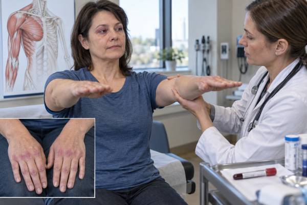
The anti-Mi-2 antibody test looks for an immune protein strongly associated with dermatomyositis, an autoimmune disease that can inflame skin and skeletal muscle. A positive result can make dermatomyositis more likely when a person has a characteristic rash, progressive muscle weakness, elevated muscle enzymes, or supportive imaging or biopsy findings. It does not diagnose the disease by itself. The result must be interpreted with the symptoms, physical examination, testing method, and other laboratory findings because commercial myositis panels can produce weak or isolated positives that are not clinically meaningful. Anti-Mi-2–positive dermatomyositis often causes prominent classic skin findings and substantial muscle inflammation, yet many affected people respond well to treatment. Lung disease and cancer appear less frequent than in several other dermatomyositis antibody groups, but neither risk is zero. The most useful question is therefore not simply whether the test is positive, but whether the entire clinical picture fits anti-Mi-2 dermatomyositis and what organ checks are needed now.
- What it measures: antibodies against Mi-2 proteins, which are part of a nuclear chromatin-remodeling complex.
- What a positive result suggests: classic dermatomyositis is more likely when compatible skin and muscle findings are present.
- What it does not prove: an isolated or weak positive result cannot establish dermatomyositis or explain unrelated fatigue and pain.
- Typical clinical pattern: striking dermatomyositis rash, proximal muscle weakness, and elevated creatine kinase, often with a favorable treatment response.
- Important follow-up: confirm the phenotype, review the assay strength, assess swallowing and breathing, and complete age-appropriate cancer screening.
Table of Contents
- What the Anti-Mi-2 Test Detects
- What a Positive Result Means
- Typical Symptoms and Clinical Pattern
- How Doctors Confirm the Diagnosis
- Treatment and Expected Response
- Lung, Cancer, and Other Organ Risks
- Practical Next Steps After Testing
What the Anti-Mi-2 Test Detects
Anti-Mi-2 is a myositis-specific autoantibody that mainly points toward dermatomyositis. Autoantibodies are immune proteins that recognize the body’s own molecules. In this case, the targets are Mi-2 proteins within the nucleosome-remodeling and deacetylase complex, a group of proteins involved in controlling how DNA is packaged and how genes are expressed.
Clinical laboratories may report anti-Mi-2 as a single result or separate it into Mi-2 alpha and Mi-2 beta. The exact antigen displayed on a commercial test varies. Some laboratories use line blot or dot blot panels, while reference laboratories may use immunoprecipitation or another confirmatory method. These platforms do not perform identically, so a result from one laboratory is not always directly interchangeable with a result from another.
Doctors usually order anti-Mi-2 as part of a broader myositis antibody panel. Testing is most useful when there is a meaningful pretest probability of inflammatory myopathy. Examples include:
- New weakness when rising from a chair, climbing stairs, lifting the arms, or holding the head up
- A violet or reddish rash around the eyelids, knuckles, upper chest, shoulders, back, or outer thighs
- Unexplained creatine kinase elevation
- Muscle edema on magnetic resonance imaging
- Swallowing trouble, nasal speech, or neck weakness
- Skin biopsy or muscle biopsy findings compatible with dermatomyositis
No fasting or special preparation is generally needed. A blood sample is drawn, and the laboratory reports the result as negative, borderline, weak positive, moderate positive, strong positive, or a numerical value with a laboratory-specific cutoff. There is no universal “normal range” across all platforms. The reference interval printed on the report is therefore more useful than comparing numbers from different laboratories.
A negative result does not rule out dermatomyositis. Only a portion of people with dermatomyositis have anti-Mi-2, and some patients have a different myositis-specific antibody or no currently identifiable antibody. A clinician should not stop the evaluation when the rash, weakness, muscle enzymes, imaging, or biopsy strongly supports disease.
What a Positive Result Means
A convincing positive anti-Mi-2 result supports a dermatomyositis diagnosis, but its value depends heavily on the clinical context and assay quality. The result is most persuasive when the patient has classic dermatomyositis skin disease, objective proximal weakness, elevated muscle enzymes, and no more likely explanation.
Anti-Mi-2 has relatively high disease specificity when confirmed by a reliable method. That means a true positive is uncommon in people without an inflammatory myopathy. However, the positive predictive value in everyday practice can be lower than the specificity reported in specialty cohorts. This happens because commercial panels are sometimes ordered for nonspecific symptoms such as fatigue, diffuse pain, or a positive antinuclear antibody. When the starting likelihood of myositis is low, even a technically uncommon false positive becomes important.
Strong, weak, and isolated results are not equivalent
A strong result that matches the phenotype deserves more weight than a barely positive band in someone with normal strength, normal creatine kinase, and no dermatomyositis rash. Line blot studies have shown that weak myositis-specific antibody results are more likely to be false positives than strong results. Results also become less convincing when a panel reports several unrelated myositis-specific antibodies at once, because most patients have one dominant antibody pattern rather than numerous competing specificities.
Some commercial tests separately report Mi-2 alpha and Mi-2 beta. Positivity to both components may increase confidence in a true anti-Mi-2 antibody, although laboratories differ and this rule cannot replace clinical assessment. When a result is unexpected, useful options include reviewing the raw band strength, repeating the test at a laboratory that uses a different method, or asking a myositis specialist whether immunoprecipitation-based confirmation is available.
A positive anti-Mi-2 test does not automatically mean:
- Every symptom is caused by dermatomyositis
- Muscle damage is severe at that moment
- Treatment must begin before objective evaluation
- Lung disease or cancer is absent
- The antibody level should be followed as the main measure of disease activity
Antibody concentrations may move with disease activity in some patients, but anti-Mi-2 is not a standardized stand-alone monitoring test. Clinicians usually track strength, function, rash, swallowing, creatine kinase, aldolase, liver-associated muscle enzymes, imaging, and treatment side effects instead.
Typical Symptoms and Clinical Pattern
Anti-Mi-2 dermatomyositis usually produces a recognizable combination of prominent skin disease and inflammatory muscle weakness. The pattern is helpful, but no single rash or symptom is unique enough to confirm the diagnosis alone.
Common skin findings include:
- Gottron papules: raised red, violet, or scaly areas over the knuckles
- Gottron sign: flat redness over finger joints, elbows, knees, or ankles
- Heliotrope rash: violet discoloration and swelling around the eyelids
- Shawl sign: sun-sensitive redness across the upper back and shoulders
- V-sign: rash over the upper chest in a V-shaped distribution
- Holster sign: rash along the outer thighs
- Scalp redness, scale, itch, or hair thinning
- Ragged cuticles and enlarged capillary loops around the nails
Anti-Mi-2 disease is often described as “classic” dermatomyositis because these visible findings can be especially pronounced. The rash may burn or itch and can leave color changes after inflammation settles. In darker skin tones, active disease may appear violaceous, brown-red, gray, or hyperpigmented rather than bright red. A clinician experienced with inflammatory skin disease can help distinguish dermatomyositis from eczema, psoriasis, allergic contact dermatitis, rosacea, lupus rash, and medication reactions.
Muscle symptoms usually affect the proximal muscles closest to the trunk. A person may struggle to stand from a low chair without using the arms, climb stairs, wash or comb the hair, lift dishes to a shelf, or carry a child. The weakness is often symmetrical. Muscle aching can occur, but weakness and loss of function matter more diagnostically than pain alone. Severe disease can involve neck flexors, breathing muscles, or the muscles used for swallowing.
Unlike anti-MDA5 dermatomyositis, anti-Mi-2 disease usually has clear muscle involvement rather than an amyopathic pattern. Creatine kinase can be markedly elevated, sometimes into the thousands of units per liter, although the number varies with disease stage, muscle mass, treatment, exercise, and laboratory method. A normal creatine kinase does not completely exclude active skin-predominant disease or treated myositis.
Constitutional symptoms such as fatigue, reduced appetite, fever, or weight change may occur. Joint pain is possible, but erosive arthritis is not the defining feature. Raynaud phenomenon and severe interstitial lung disease are generally less characteristic than they are in antisynthetase syndrome or certain overlap myositis patterns.
How Doctors Confirm the Diagnosis
Doctors confirm anti-Mi-2 dermatomyositis by showing that the antibody result fits objective evidence of characteristic skin or muscle inflammation. No one test is sufficient in every case, and the diagnostic pathway changes according to whether skin, muscle, swallowing, or lung symptoms dominate.
The evaluation commonly includes:
- Focused history and strength examination. The clinician checks shoulder, hip, neck, hand, and swallowing function and documents what has changed over time. Functional examples are often more informative than a general report of “weakness.”
- Muscle enzymes. Creatine kinase is central, but aldolase, lactate dehydrogenase, aspartate aminotransferase, and alanine aminotransferase may also rise. AST and ALT can come from muscle, so an elevated “liver test” should be interpreted with creatine kinase and other liver markers rather than assumed to represent liver disease.
- Skin examination and biopsy when needed. A biopsy can support connective-tissue-disease skin inflammation, although lupus and dermatomyositis can overlap microscopically. The clinical distribution remains important.
- Muscle magnetic resonance imaging. Fluid-sensitive MRI sequences can identify muscle edema, help select a biopsy site, and distinguish active inflammation from fatty replacement or chronic damage.
- Electromyography or nerve studies. These may support an irritable myopathy and help exclude neuropathy, motor neuron disease, or neuromuscular-junction disorders.
- Muscle biopsy in selected cases. Dermatomyositis may show perifascicular atrophy, perivascular inflammation, capillary injury, and characteristic immune staining. Biopsy is especially useful when the antibody is uncertain, the presentation is atypical, or another muscle disease is possible.
Clinicians also review alternative causes of weakness and enzyme elevation. These include thyroid disease, electrolyte disturbance, infection, medication toxicity, strenuous exercise, muscular dystrophy, metabolic muscle disease, immune-mediated necrotizing myopathy, and inclusion body myositis. Statin exposure alone does not explain every elevated creatine kinase; persistent severe weakness after stopping a statin may prompt testing for anti-HMGCR autoimmune myopathy.
Classification criteria can support research consistency, but real-world diagnosis still requires judgment. A person can have clinically important dermatomyositis without satisfying every classification item early in the course. Conversely, a positive antibody does not convert nonspecific symptoms into inflammatory myopathy.
Treatment and Expected Response
Anti-Mi-2 dermatomyositis often responds well to immunosuppressive treatment, but early control and careful rehabilitation are still important. “Favorable prognosis” is a group-level description, not a guarantee that the disease will be mild or brief in an individual patient.
Initial therapy depends on disease severity and organ involvement. Systemic glucocorticoids are commonly used for meaningful muscle disease because they act quickly. Clinicians often add a steroid-sparing medicine early to improve control and reduce long-term glucocorticoid exposure. Options may include methotrexate, azathioprine, mycophenolate mofetil, tacrolimus, or another immunomodulator selected for the patient’s skin, muscle, lung, pregnancy, liver, kidney, infection, and medication history.
Intravenous immune globulin can be particularly useful for severe or treatment-resistant dermatomyositis, major swallowing difficulty, or situations in which a faster steroid-sparing effect is needed. Rituximab and other targeted therapies may be considered for refractory disease. Treatment choices continue to evolve, and some therapies are used off label based on specialty experience and published evidence.
Skin disease needs direct treatment even when muscle strength improves. A practical plan may include:
- Daily broad-spectrum sun protection, protective clothing, and shade
- Topical corticosteroids or calcineurin inhibitors for selected areas
- Antimalarial therapy in some patients, with awareness that it does not always control dermatomyositis skin disease
- Systemic treatment adjustment when rash remains active
- Measures for itch, scalp inflammation, and secondary infection
Exercise is part of treatment, not something that must always be avoided. Once the medical team has assessed disease activity and safety, supervised aerobic and resistance exercise can improve strength, endurance, and function without worsening controlled inflammatory myopathy. During a severe flare, the program may start with gentle range-of-motion work, breathing exercises, fall prevention, and carefully dosed activity. Physical and occupational therapists can translate muscle recovery into daily tasks.
Response is measured across several domains. Creatine kinase may fall before full strength returns, because muscle repair and reconditioning take time. A falling enzyme level with worsening function should trigger evaluation for steroid myopathy, chronic muscle damage, neuropathy, infection, or another complication. Likewise, normal enzymes do not prove that an active rash has resolved.
Relapses can occur during dose reduction or after treatment stops. Patients should know their personal early warning signs, such as renewed trouble on stairs, more scalp inflammation, new eyelid swelling, reduced swallowing stamina, or rising enzymes. The anti-Mi-2 test itself is usually not repeated frequently unless a specialist has a specific reason.
Lung, Cancer, and Other Organ Risks
Anti-Mi-2 generally carries a lower interstitial lung disease and malignancy burden than several other dermatomyositis antibody profiles, but standard organ assessment remains necessary. Lower risk should guide proportional screening, not create false reassurance.
Interstitial lung disease is less common in anti-Mi-2 dermatomyositis than in anti-Jo-1 antisynthetase syndrome or anti-MDA5 disease. Clinicians still ask about dry cough, breathlessness, reduced exercise tolerance, and oxygen desaturation. Baseline pulmonary function tests may be reasonable when symptoms, examination, imaging, or the broader antibody profile raises concern. New shortness of breath at rest, chest pain, blue lips, coughing blood, or rapidly falling oxygen saturation needs urgent evaluation.
Adult-onset dermatomyositis as a whole is associated with increased cancer risk, especially near the time myositis begins. Anti-Mi-2 is not considered the highest-risk antibody; anti-TIF1-gamma and anti-NXP2 in some adult cohorts raise more concern. Even so, an anti-Mi-2 result does not remove the need for age- and sex-appropriate cancer screening and a careful review for warning signs.
The screening plan should reflect age, smoking and family history, previous screening, unexplained weight loss, anemia, lymph-node enlargement, abnormal bleeding, and local recommendations. Tests may include breast, cervical, colorectal, lung, prostate, or other screening as appropriate. Broad repeated scanning is not automatically necessary for every anti-Mi-2 patient, but clinicians may intensify the initial evaluation when the disease starts later in life, symptoms are concerning, or routine screening is overdue.
Swallowing involvement deserves special attention because aspiration can be serious even without obvious choking. Warning signs include coughing with meals, a wet voice after drinking, food sticking, prolonged mealtimes, nasal regurgitation, recurrent pneumonia, or unintended weight loss. A speech-language pathologist and an instrumental swallow study can define the problem and recommend safer textures and techniques.
Cardiac involvement is uncommon but possible in inflammatory myopathy. Palpitations, fainting, persistent chest discomfort, unexplained ankle swelling, or disproportionate breathlessness may prompt an electrocardiogram, cardiac biomarkers interpreted carefully, echocardiography, or cardiac MRI. Troponin T can rise from skeletal muscle in active myositis; troponin I is often more cardiac-specific, though the clinical context still controls interpretation.
Practical Next Steps After Testing
The best next step after an anti-Mi-2 result is to match the laboratory finding to objective skin and muscle evidence before labeling or treating disease. The urgency depends on symptoms rather than the antibody name alone.
For a positive result, ask the ordering clinician:
- Was the result weak, moderate, or strong, and which testing platform was used?
- Were Mi-2 alpha, Mi-2 beta, or both detected?
- Does my examination show objective proximal weakness?
- Are creatine kinase, aldolase, AST, ALT, LDH, blood counts, and inflammatory markers abnormal?
- Do I need dermatology, rheumatology, neurology, or a dedicated myositis clinic?
- Should the result be confirmed by another method?
- Do I need lung tests, a swallowing evaluation, MRI, electromyography, or biopsy?
- Is my routine cancer screening current, and is any extra screening justified?
Seek prompt medical review for rapidly progressive weakness, repeated falls, inability to lift the head, dark urine after severe muscle symptoms, dehydration, or marked reduction in food intake. Emergency assessment is appropriate for breathing difficulty, choking with inability to clear secretions, chest pain, fainting, confusion, or signs of aspiration.
For a weak positive result with no compatible findings, avoid assuming that a permanent autoimmune diagnosis has been established. The clinician may repeat the strength examination, check enzymes, review medications and exercise, inspect the skin, and observe over time. Confirmation is especially valuable before exposing someone to long-term immunosuppression.
For a negative result with convincing symptoms, the workup should continue. Dermatomyositis can be antibody-negative, or another antibody may define the phenotype. A broader myositis autoantibody evaluation can help, but imaging, examination, and tissue findings often matter more than expanding panels indefinitely.
Keep copies of the original laboratory report because the platform, cutoff, and exact components affect interpretation. Record functional measures that are meaningful in daily life, such as the time needed to rise from a chair five times, the number of stairs tolerated, or whether hair washing requires rest. These details help the medical team distinguish active inflammation from fixed damage, medication effects, and deconditioning.
Most importantly, treat the result as one part of a coherent diagnosis. In the right setting, anti-Mi-2 can identify a well-recognized dermatomyositis subtype and help clinicians anticipate prominent skin and muscle disease with a generally treatment-responsive course. Outside that setting, cautious confirmation prevents a laboratory label from overshadowing the actual patient.
References
- Treatment guidelines for idiopathic inflammatory myopathies in adults: a comparative review 2025 (Review)
- Predictive value of myositis antibodies: role of semiquantitative classification and positivity for more than one autoantibody 2025
- Updates in Dermatomyositis: Newer Treatment Options and Outcome Measures From Dermatologic Perspectives 2024 (Review)
- Comparison of Lineblot and Immunoprecipitation Methods in the Detection of Myositis-Specific and Myositis-Associated Antibodies in Patients with Idiopathic Inflammatory Myopathies 2024
- Validation of anti-Mi2 autoantibody testing by line blot 2024
Disclaimer
This article provides general education about anti-Mi-2 testing and does not diagnose dermatomyositis or replace care from a qualified clinician. Seek urgent medical attention for breathing difficulty, severe swallowing problems, chest pain, fainting, or rapidly worsening weakness. Test interpretation and treatment should be individualized by a clinician familiar with inflammatory muscle disease.





