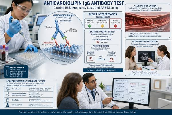
The anticardiolipin IgA antibody test measures IgA-class antibodies that react with cardiolipin-associated antigen complexes. It is sometimes added when a person has thrombosis, pregnancy morbidity, lupus, or another pattern suggestive of antiphospholipid syndrome but standard antiphospholipid tests are negative or incomplete. IgA anticardiolipin antibodies may also appear alongside IgG or IgM antibodies.
IgA anticardiolipin is not included in current APS classification laboratory criteria. A positive result therefore does not diagnose APS, and an isolated result has uncertain predictive value. Laboratories also use different methods, units, and cutoffs, so there is no universal positive range. The result carries more clinical weight when it is clearly elevated, persistent, accompanied by other antiphospholipid antibodies, and linked to an objectively confirmed clot or well-documented obstetric event. It carries less weight when it is weak, detected once, or found in a person without APS-related symptoms.
- The test detects IgA-class anticardiolipin antibodies in a blood sample.
- IgA anticardiolipin is a non-criteria marker; IgG and IgM are the anticardiolipin classes used in APS classification.
- A positive result alone cannot confirm APS or determine treatment.
- Reference intervals vary by assay, so the laboratory’s own cutoff must be used.
- Isolated low-positive IgA may be incidental or temporary, especially during infection or inflammation.
- Emergency symptoms of a blood clot require immediate care regardless of antibody results.
Table of Contents
- What IgA Anticardiolipin Measures
- Why IgA Is Different From IgG and IgM
- When IgA Testing Is Used
- Interpreting Negative, Borderline, and Positive Results
- APS Risk and Clinical Meaning
- Pregnancy and Placental Complications
- Test Preparation and Repeat Testing
- Next Steps After IgA Positivity
What IgA Anticardiolipin Measures
Cardiolipin is a phospholipid found mainly in mitochondrial membranes. In laboratory anticardiolipin assays, clinically relevant antibody binding often depends on beta-2 glycoprotein I, a plasma protein that attaches to negatively charged phospholipid surfaces. The test is therefore not simply detecting an immune response to a free fat molecule; it may detect antibodies recognizing a cardiolipin-protein arrangement created by the assay.
IgA is one of the major immunoglobulin classes. It is best known for protecting mucosal surfaces such as the respiratory and gastrointestinal tracts, but IgA also circulates in blood. Autoimmune IgA antibodies can form against several targets, including cardiolipin and beta-2 glycoprotein I.
A laboratory usually performs an enzyme immunoassay or chemiluminescent immunoassay. Results may be given as arbitrary units, IgA phospholipid units, an index, or a qualitative category. The report should identify:
- the immunoglobulin class as IgA;
- the numerical result and unit;
- the negative, equivocal, and positive cutoffs;
- the assay manufacturer or method when available;
- whether the test was part of a broader antiphospholipid panel.
A result from one platform should not be converted to another platform’s scale. Cardiolipin concentration, beta-2 glycoprotein I dependence, calibrators, and control populations differ. Even two assays labeled “anticardiolipin IgA” may not have identical analytical sensitivity.
This test is different from an anti-beta-2 glycoprotein I IgA test. The two can overlap biologically, but they use different antigen presentations and can produce discordant results.
Cardiolipin binding is often protein-dependent
The name “anticardiolipin” can suggest that the antibody targets cardiolipin alone. In APS, many clinically relevant anticardiolipin antibodies bind most effectively when beta-2 glycoprotein I is present. Infection-related antibodies may bind cardiolipin in a less protein-dependent way and are often temporary. Commercial assays differ in how beta-2 glycoprotein I becomes available in the reaction, which is one reason results vary.
This distinction cannot usually be read directly from the numerical value. It is reflected indirectly in assay design and in the clinical setting. A persistent result in a patient with lupus and other APS antibodies is more likely to represent autoimmune antiphospholipid reactivity than a low result found during a short infection. The laboratory report alone cannot prove pathogenicity.
IgA antibodies may circulate as monomers in blood or form larger immune complexes. Current clinical assays measure binding quantity, not whether those antibodies activate complement, platelets, or endothelial cells. Functional differences between IgA antibodies remain an area of research.
Why IgA Is Different From IgG and IgM
Current APS laboratory criteria recognize anticardiolipin IgG and IgM, not IgA. The same is true for anti-beta-2 glycoprotein I: classification systems use IgG and IgM. This does not mean IgA antibodies are biologically impossible or always irrelevant. It means the evidence, assay harmonization, and independent predictive value have not been strong enough for inclusion.
IgG anticardiolipin is generally the best-established solid-phase antibody class for APS, especially at moderate or high levels and when persistent. IgM can be relevant but often has lower specificity when isolated or low. IgA has been studied in selected populations, including people with lupus, thrombosis, or clinical APS features who are negative for IgG and IgM.
| Class | APS classification role | Typical interpretation |
|---|---|---|
| IgG | Included | Strong evidence when moderate/high and persistent |
| IgM | Included | Context-dependent; isolated low values are less specific |
| IgA | Not included | Possible supplementary marker, especially in selected seronegative or lupus cases |
An isolated IgA result should not be called “triple positivity.” In APS, triple positivity refers to lupus anticoagulant, anticardiolipin, and anti-beta-2 glycoprotein I positivity in the recognized testing framework, not three immunoglobulin classes of one antibody.
IgA interpretation also depends on total immunoglobulin status. Selective IgA deficiency can make IgA antibody testing uninformative, while polyclonal IgA elevation from chronic inflammation, liver disease, or infection may contribute to nonspecific assay reactivity. Total IgA is not required for every anticardiolipin test, but it may help in unusual cases.
When IgA Testing Is Used
IgA anticardiolipin is usually a supplementary test, not the first laboratory step. It may be considered when the clinical history remains concerning after standard testing.
Examples include:
- an unprovoked deep vein thrombosis, pulmonary embolism, stroke, or unusual-site clot;
- recurrent fetal loss or severe placental disease with negative criteria antibodies;
- systemic lupus erythematosus with thrombotic, obstetric, platelet, or vascular features;
- a possible seronegative APS phenotype under specialist review;
- discordant prior laboratory findings;
- a laboratory panel designed to measure IgA anticardiolipin and IgA anti-beta-2 glycoprotein I together.
Testing healthy people without relevant clinical features is unlikely to help. The positive predictive value falls when the pretest probability is low, so a weak positive is more likely to be incidental.
A clinician should confirm that the event being investigated fits the question. A suspected venous clot should be verified by imaging. Pregnancy records should identify gestational age, fetal viability, placental function, blood pressure, and pathology when available. A laboratory result cannot repair an uncertain clinical history.
IgA testing may be particularly considered in populations where studies have reported a higher frequency of IgA antiphospholipid antibodies. However, ethnicity and geographic variation in published cohorts do not create a universal rule to test or treat based on ancestry alone.
The complete standard workup is summarized in the antiphospholipid antibody panel. IgA should be viewed as an addition when there is a reason, not a replacement for criteria assays.
Interpreting Negative, Borderline, and Positive Results
Negative result
A negative IgA anticardiolipin test means no IgA binding was detected above that assay’s threshold. It does not rule out APS. A patient may still have lupus anticoagulant, IgG or IgM anticardiolipin, IgG or IgM anti-beta-2 glycoprotein I, or a non-antibody cause of symptoms.
Borderline result
A borderline value sits near the cutoff where normal analytical variation matters most. It may change category on repeat testing without a true biological shift. Borderline results should be interpreted from the actual number and reference interval, not upgraded automatically to “positive.”
Positive result
A positive result means the laboratory detected IgA anticardiolipin above its cutoff. The clinical weight depends on several features:
- distance above the cutoff;
- persistence on a later sample;
- isolated versus combined antibody positivity;
- objectively documented thrombosis or pregnancy morbidity;
- lupus or another autoimmune disease;
- recent infection, inflammation, or medication exposure;
- assay quality and laboratory experience.
There is no internationally accepted IgA anticardiolipin threshold equivalent to the criteria definitions used for IgG and IgM. A laboratory may label a value “high,” but that label does not automatically map to a validated APS risk category.
| Pattern | Usual clinical weight |
|---|---|
| One low-positive IgA result, no APS event | Often limited; consider temporary or incidental positivity |
| Persistent isolated IgA with confirmed thrombosis | May support specialist concern but does not satisfy criteria laboratory classification |
| IgA plus persistent criteria antibodies | Shows a broader antibody profile; risk is driven mainly by criteria markers and history |
| IgA positive during acute infection | Repeat may be appropriate after recovery if the answer would affect care |
| IgA negative, lupus anticoagulant positive | APS risk can still be substantial; IgA negativity does not reduce the lupus anticoagulant finding |
APS Risk and Clinical Meaning
Studies have linked IgA anticardiolipin or IgA anti-beta-2 glycoprotein I with thrombosis in some cohorts, particularly when IgA antibodies accompany other antiphospholipid markers. Other studies have found little added diagnostic value from isolated IgA testing. The inconsistency reflects small numbers of isolated-positive patients, population differences, assay variation, and overlap with IgG or IgM positivity.
A 2022 study of IgA antiphospholipid antibodies reported associations with vascular events, but isolated IgA positivity was uncommon. A 2024 cohort also found persistent isolated moderate-to-high IgA antiphospholipid positivity to be rare. Rare findings are difficult to study with enough statistical power to define precise risk.
Current classification criteria therefore remain centered on lupus anticoagulant and IgG/IgM solid-phase assays. The 2023 ACR/EULAR criteria do not award laboratory points for IgA anticardiolipin.
The presence of IgA cannot predict the timing or location of a clot. Total risk depends on prior thrombosis, lupus anticoagulant, antibody persistence, smoking, estrogen exposure, surgery, immobility, pregnancy, cancer, age, obesity, and cardiovascular risk factors.
Risk cannot be inferred from the word “positive”
Suppose two reports are both marked positive. One is 1 unit above the cutoff in a patient tested during pneumonia. The other is several times the cutoff, remains positive after 12 weeks, and accompanies lupus anticoagulant in a patient with an unprovoked pulmonary embolism. Treating those findings as equivalent would ignore nearly all clinically relevant information.
The first result may be repeated only if the clinical history justifies it. The second belongs to a broader high-risk evaluation, although the lupus anticoagulant and criteria antibodies carry the formal weight. This example shows why clinicians need complete numbers, dates, and clinical records rather than a verbal history of “my APS test was positive.”
A person with an isolated IgA result and no clinical event may need no drug treatment. A person with a confirmed unprovoked clot may require anticoagulation based on the clot itself, even while the exact APS label remains uncertain. A third person with persistent criteria antibodies may have established APS whether IgA is positive or negative.
Treatment decisions also depend on bleeding risk and on the type of event. Prevention after an arterial stroke differs from prevention after a provoked leg clot, and pregnancy treatment differs from long-term treatment outside pregnancy. IgA anticardiolipin does not select a medication or dose. Direct oral anticoagulants, warfarin, heparin, and antiplatelet drugs have different roles and limitations in APS; the choice belongs to a clinician who has reviewed the complete diagnosis.
Risk-reduction advice may still be appropriate when medication is not. Avoiding smoking, maintaining mobility during long travel, managing blood pressure and cholesterol, and discussing estrogen-containing hormones can reduce preventable clot risks. These measures address the whole risk profile rather than trying to “lower” the antibody number. No diet or supplement has been proven to remove IgA anticardiolipin or prevent APS-related events.
The triple-positive antiphospholipid profile has a stronger established relationship with recurrent thrombosis than isolated IgA anticardiolipin.
Pregnancy and Placental Complications
IgA anticardiolipin has been investigated in recurrent miscarriage, fetal death, preeclampsia, fetal growth restriction, and placental insufficiency. Results are mixed, and most treatment evidence for obstetric APS is based on criteria antibodies rather than isolated IgA.
Pregnancy loss has many causes. Early losses often involve fetal chromosome abnormalities. Other possibilities include uterine abnormalities, endocrine disease, parental chromosome changes, and age-related factors. A positive IgA antibody should not halt the rest of the evaluation.
Obstetric interpretation improves when the history is specific:
- number and timing of losses;
- whether cardiac activity was documented;
- fetal chromosome or pregnancy tissue results;
- fetal death after 10 weeks;
- delivery before 34 weeks because of severe preeclampsia or placental insufficiency;
- placental pathology and fetal growth pattern;
- standard APS antibody values and repeat dates.
For established obstetric APS, low-dose aspirin and heparin may improve outcomes when prescribed appropriately. There is no equivalent high-quality evidence that every person with isolated IgA anticardiolipin benefits from the same regimen. Bleeding risk, prior events, and competing causes must be considered.
A maternal-fetal medicine specialist may decide that persistent IgA positivity adds supportive evidence in a highly suggestive case. That individualized judgment should not be generalized to all positive results. Pregnancy planning should also address blood pressure, smoking, diabetes, weight, estrogen exposure, mobility, and lupus activity.
Urgent pregnancy symptoms include heavy bleeding, severe headache, visual changes, upper abdominal pain, marked swelling, or reduced fetal movement. These symptoms require assessment regardless of antibody status.
Test Preparation and Repeat Testing
The test uses a routine venous blood sample. Fasting is usually unnecessary. Tell the clinician about recent infection, vaccination, surgery, pregnancy, autoimmune flare, and all medicines.
Anticoagulants can disrupt lupus anticoagulant testing, but they usually have less direct analytical effect on anticardiolipin immunoassays. Never stop an anticoagulant without a safe plan from the prescriber.
Temporary anticardiolipin antibodies can appear after infections or inflammatory illnesses. When persistence matters, clinicians may repeat testing after at least 12 weeks, using the same laboratory if possible. The 12-week interval is an established concept for criteria antibodies; applying it to IgA can help distinguish persistent from transient positivity, but it does not turn IgA into a criteria marker.
Repeat testing is most useful when it will change a decision. It may clarify whether a weak result was temporary, document a broader persistent profile, or support pregnancy planning. Routine frequent monitoring is not recommended because antibody levels do not reliably measure day-to-day disease activity.
Keep the full report. The numerical value, unit, cutoff, method, and date are essential. A later report from a different platform may not be directly comparable.
Factors that can complicate the sample or interpretation
Severe lipemia, hemolysis, contamination, and poor storage can affect immunoassays. Laboratories usually have rejection rules for visibly unsuitable samples, but smaller effects may be less obvious. Rheumatoid factor, heterophile antibodies, and marked polyclonal immunoglobulin elevation can sometimes contribute to nonspecific binding. When a result conflicts sharply with the clinical picture, repeating the sample or discussing the assay with the laboratory may be more useful than assuming the result is definitive.
Pregnancy itself changes plasma volume and immune function, but there is no validated pregnancy-specific IgA anticardiolipin range. Serial IgA measurement during pregnancy has not been shown to guide dose changes. Clinical surveillance, blood pressure, fetal growth, and the established antibody profile are more important than small titer movements.
Next Steps After IgA Positivity
A positive result should trigger an orderly review:
- Confirm the test identity. Make sure the result is anticardiolipin IgA and not anti-beta-2 glycoprotein I IgA.
- Review criteria tests. Check lupus anticoagulant, anticardiolipin IgG/IgM, and anti-beta-2 glycoprotein I IgG/IgM.
- Verify persistence. Decide whether repeat testing after at least 12 weeks would change care.
- Document clinical events. Obtain clot imaging and detailed obstetric records.
- Assess alternative explanations. Consider infection, inflammation, inherited or acquired clot risks, and other causes of pregnancy complications.
- Use specialist input. Hematology, rheumatology, or maternal-fetal medicine may be appropriate.
Do not self-start aspirin or anticoagulation. Aspirin can cause gastrointestinal bleeding and interact with other medicines. Anticoagulants carry a larger bleeding risk and require a clear indication.
Seek emergency care for sudden chest pain, shortness of breath, coughing blood, fainting, one-sided leg swelling, facial droop, arm weakness, speech difficulty, sudden vision loss, or a severe new headache. These symptoms require immediate evaluation even if every antibody test is negative.
At a routine visit, ask what the result changes. It may affect whether criteria testing is repeated, whether estrogen-containing contraception is appropriate, how pregnancy is monitored, or how clot prevention is handled during surgery. In some cases, the honest conclusion is that the IgA result is supportive but not independently actionable.
Useful questions for the follow-up visit include: Was the IgA result isolated? How far above the cutoff was it? Were IgG and IgM measured on the same day? Was lupus anticoagulant testing reliable while I was taking anticoagulants? Would repeating the IgA result change pregnancy, surgery, or medication planning? Which part of my risk comes from the antibody profile and which part comes from my prior clinical event?
A well-framed answer may prevent both undertreatment and overtreatment. Ignoring a persistent result in a compelling case can miss useful context, while labeling every isolated IgA result as APS can expose people to unnecessary fear and bleeding risk.
References
- An update on laboratory detection and interpretation of antiphospholipid antibodies for diagnosis of antiphospholipid syndrome: guidance from the ISTH-SSC Subcommittee on Lupus Anticoagulant/Antiphospholipid Antibodies 2025 (Guidance)
- The 2023 ACR/EULAR Antiphospholipid Syndrome Classification Criteria 2023 (Classification Criteria)
- IgA Antiphospholipid Antibodies in Antiphospholipid Syndrome and Systemic Lupus Erythematosus 2022 (Study)
- Persistent Moderate-to-High Levels of Isolated Anticardiolipin or Anti-β2-glycoprotein I IgA Isotype in a Cohort of Patients with Clinical Manifestations of Antiphospholipid Syndrome 2024 (Study)
- Antiphospholipid Syndrome: A Comprehensive Clinical Review 2025 (Review)
- Antiphospholipid Antibody Testing 2025 (Review)
Disclaimer
This article is for general education and does not diagnose antiphospholipid syndrome or prescribe treatment. IgA anticardiolipin results must be interpreted with the performing laboratory’s method, standard APS tests, persistence, and a documented clinical history. Do not begin or stop aspirin or anticoagulants without medical guidance.