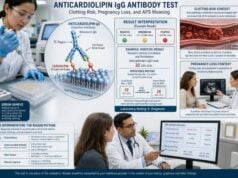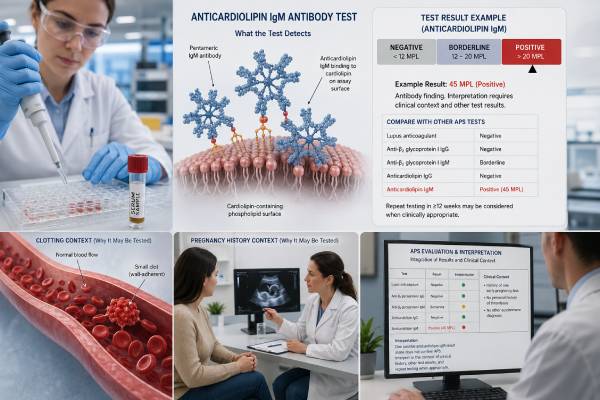
The anticardiolipin IgG antibody test measures IgG autoantibodies that bind cardiolipin-associated targets in the blood. It is one of the main laboratory tests used when clinicians evaluate antiphospholipid syndrome, an autoimmune disorder linked to venous and arterial blood clots, recurrent pregnancy loss, fetal death, and placental complications. IgG anticardiolipin is generally more clinically specific than an isolated low IgM result, especially when the level is moderate or high and remains positive on repeat testing.
A positive test does not diagnose APS by itself. The result must be paired with a compatible clinical event and confirmed persistence, usually on a second sample at least 12 weeks later. Risk also depends on whether lupus anticoagulant or anti-beta-2 glycoprotein I antibodies are present. A weak, temporary IgG result after infection has a very different meaning from persistent high IgG anticardiolipin in someone with an unprovoked clot or qualifying pregnancy morbidity.
- The test measures IgG antibodies against cardiolipin-associated antigen complexes.
- Persistent moderate or high IgG levels carry more APS significance than a one-time low-positive result.
- APS assessment usually requires a compatible clinical event plus repeat positivity at least 12 weeks apart.
- Results may be reported in GPL units or assay-specific units; use the laboratory’s own reference interval.
- IgG anticardiolipin is interpreted with lupus anticoagulant and anti-beta-2 glycoprotein I IgG/IgM.
- Sudden shortness of breath, chest pain, one-sided swelling, weakness, or speech trouble needs urgent care.
Table of Contents
- What the Anticardiolipin IgG Test Detects
- Ranges, Units, and Result Levels
- What a Positive Result Means
- How IgG Anticardiolipin Relates to Clotting Risk
- Pregnancy Loss and Placental Risk
- How the Test Fits Into APS Diagnosis
- Testing, Preparation, and Repeat Timing
- Next Steps After an Abnormal Result
What the Anticardiolipin IgG Test Detects
Cardiolipin is a negatively charged phospholipid found mainly in mitochondrial membranes. In an anticardiolipin laboratory assay, clinically relevant IgG antibodies often bind to a complex involving cardiolipin and beta-2 glycoprotein I, a plasma protein that attaches to phospholipid surfaces. The assay therefore detects an autoimmune binding pattern rather than measuring the amount of cardiolipin in the body.
IgG is the most abundant immunoglobulin class in circulation. In APS, persistent IgG antiphospholipid antibodies are often more strongly associated with thrombosis and obstetric complications than isolated weak IgM responses. That does not make every IgG result dangerous. The antibody’s level, persistence, companion antibodies, and clinical history determine its importance.
Most laboratories use an enzyme-linked or chemiluminescent immunoassay. The report should identify:
- anticardiolipin IgG as the analyte;
- the numerical result and unit;
- negative, borderline, moderate, and high categories when the assay provides them;
- the laboratory reference interval;
- the collection date and, ideally, the method.
The result is not the same as a lupus anticoagulant test. Anticardiolipin is a solid-phase antibody assay, while lupus anticoagulant is detected through phospholipid-dependent clotting tests. A patient may be positive on one and negative on the other.
The test is also separate from the beta-2 glycoprotein I IgG antibody test. Although beta-2 glycoprotein I can help present the cardiolipin target, the assays are not interchangeable.
Ranges, Units, and Result Levels
Anticardiolipin IgG results are often reported in GPL units, historically defined using standardized reference material, or in manufacturer-specific units. Assay platforms do not always agree perfectly. A value should be interpreted only with the range printed on the same report.
Older APS classification language commonly described medium or high anticardiolipin as more than 40 GPL units or above the 99th percentile. The 2023 ACR/EULAR classification system uses defined moderate and high laboratory categories for IgG and IgM anticardiolipin, but the exact category still depends on a validated assay and local reporting.
| Result level | Typical meaning | Usual response |
|---|---|---|
| Negative | No IgG antibody detected above the assay cutoff | Review the other APS markers and clinical history |
| Borderline or low positive | Weak reactivity that may be transient or nonspecific | Interpret cautiously; repeat only when clinically justified |
| Moderate positive | More consistent with clinically relevant antiphospholipid autoimmunity | Confirm persistence and assess the complete APS profile |
| High positive | Greater concern when persistent and linked to an APS event | Specialist review and individualized risk assessment |
A numerical change does not always mean the disease has worsened or improved. Immunoassays have analytical variation, and different manufacturers can produce different values. Serial testing should use the same laboratory when possible.
The antibody level is not a treatment target like blood pressure or glucose. Lowering the number has not been established as a way to prevent clots, and small titer changes should not drive anticoagulant dose adjustments.
Why laboratories may classify the same sample differently
Anticardiolipin testing is more standardized than many extended antiphospholipid assays, but important variation remains. Laboratories may use different antigen sources, beta-2 glycoprotein I concentrations, calibrators, incubation conditions, and instruments. The 99th percentile also depends on the healthy control population used to establish it.
A value near a category boundary can therefore shift from low to moderate when tested on another platform. That does not necessarily mean the patient’s immune response changed. For diagnosis and persistence, clinicians should compare like with like and avoid mixing categories from unrelated assays. When a result seems inconsistent with the history or other markers, discussion with the laboratory can clarify whether the method is traceable to recognized standards and whether the sample was repeated.
The report may use terms such as GPL-U/mL, CU, U/mL, or an index. These units are not automatically interchangeable. The printed interpretation takes priority over an internet chart.
What a Positive Result Means
A positive anticardiolipin IgG result means the assay found antibody binding above its threshold. It does not state whether the antibody is temporary, pathogenic, or responsible for a specific event.
Several patterns have different implications:
One low-positive result
A low result detected once may occur after viral or bacterial infection, during inflammation, or without a related disease. It does not meet persistence requirements and often has limited specificity for APS.
Persistent moderate or high positivity
A clearly elevated result that remains positive at least 12 weeks later is more clinically meaningful. If the patient also has a documented clot or qualifying pregnancy morbidity, the result can contribute to APS classification and diagnosis.
Positivity with other APS antibodies
Anticardiolipin IgG carries more concern when lupus anticoagulant or anti-beta-2 glycoprotein I is also present. Persistent positivity in all three major groups is called triple positivity and is associated with a higher risk of recurrent thrombosis than a single antibody alone.
Positive result without symptoms
Some people have persistent antiphospholipid antibodies but have never had thrombosis or obstetric morbidity. They are antibody-positive carriers, not automatically patients with APS. Their preventive plan depends on the antibody profile and conventional clot risks.
Possible temporary or nonspecific settings include acute infection, some medications, malignancy, and other autoimmune conditions. The result should never be interpreted without knowing when the sample was collected and why the test was ordered.
False-positive and clinically weak patterns
Transient anticardiolipin antibodies are well described after infections. They are often low in level, may involve IgM more than IgG, and can disappear after recovery. Even so, a temporary IgG result is possible. Testing during an acute inflammatory illness should be interpreted cautiously unless an urgent clinical reason exists.
Nonspecific binding can also arise from severe polyclonal immunoglobulin elevation, heterophile antibodies, or technical factors. A result that is sharply discordant with repeat measurements or with every other component of the profile may justify laboratory review. The appropriate response is confirmation, not immediate long-term treatment.
Conversely, a normal anticardiolipin IgG result does not cancel a positive lupus anticoagulant or beta-2 glycoprotein I antibody. Each component answers a different laboratory question.
How IgG Anticardiolipin Relates to Clotting Risk
Persistent moderate-to-high anticardiolipin IgG is associated with venous and arterial thrombosis, but it does not predict a clot with certainty. Many antibody-positive people never develop an event, while thrombosis can occur in antibody-negative people for unrelated reasons.
APS-related thrombosis may affect:
- deep veins of the legs;
- pulmonary arteries;
- cerebral arteries, causing ischemic stroke or transient ischemic attack;
- coronary, renal, retinal, or abdominal vessels;
- small vessels in the skin, kidneys, lungs, or other organs.
Risk is highest when several factors combine. Important contributors include prior unprovoked thrombosis, lupus anticoagulant, high and persistent IgG levels, anti-beta-2 glycoprotein I positivity, lupus, pregnancy, the postpartum period, surgery, immobility, estrogen exposure, smoking, obesity, cancer, hypertension, and high cholesterol.
The antibody may promote clotting through platelet activation, endothelial-cell activation, complement pathways, tissue factor expression, and interference with natural anticoagulant systems. APS is better understood as a thrombo-inflammatory condition than as a simple excess of clotting proteins.
A prolonged clotting time in lupus anticoagulant testing does not mean a patient is protected from thrombosis. The laboratory phenomenon and the clinical risk move in opposite directions. Anticardiolipin IgG itself is measured by immunoassay and does not provide a clotting time.
For a patient who has already had a clot, treatment decisions depend on the event type, provoking factors, antibody profile, and bleeding risk. A single positive result cannot select warfarin, heparin, aspirin, or a direct oral anticoagulant. High-risk APS has specific treatment considerations that require specialist guidance.
The thrombosis antiphospholipid panel places anticardiolipin IgG within the larger risk profile.
Risk during high-risk situations
Temporary exposures can turn a stable antibody profile into a more dangerous setting. Major surgery, prolonged hospitalization, long-distance immobility, pregnancy, and the first six weeks after delivery all raise venous clot risk. A person with persistent moderate-to-high anticardiolipin IgG should tell clinicians about the antibody before these events so that standard preventive measures can be considered.
Prevention may include early walking, mechanical compression, hydration, or medication, depending on the overall risk. The antibody result alone does not define the preventive dose. Previous thrombosis, lupus anticoagulant, bleeding history, kidney function, and the procedure or pregnancy plan all matter.
Estrogen-containing contraception and hormone therapy also deserve review. Estrogen can increase thrombosis risk, and alternative options may be preferable for some antibody-positive patients. This decision should be individualized rather than based on a single weak result.
Pregnancy Loss and Placental Risk
Anticardiolipin IgG is one of the established laboratory markers used in obstetric APS assessment. Persistent moderate or high levels can support the diagnosis when a patient has qualifying pregnancy morbidity.
APS-related obstetric patterns include:
- recurrent early pregnancy loss;
- otherwise unexplained fetal death after 10 weeks;
- preterm delivery caused by severe preeclampsia or placental insufficiency;
- fetal growth restriction and other signs of impaired placental function.
The relationship is not limited to visible placental clots. Antiphospholipid antibodies can affect trophoblast cells, complement activation, placental inflammation, and vascular remodeling. These mechanisms may impair implantation or placental development.
A positive anticardiolipin IgG result does not prove that it caused a miscarriage. Early losses commonly result from fetal chromosome abnormalities, and recurrent loss can involve uterine, endocrine, genetic, or other medical factors. The history should include gestational age, ultrasound findings, fetal or pregnancy-tissue chromosome results, placental pathology, blood pressure, and fetal growth.
For patients who meet accepted obstetric APS criteria, clinician-directed low-dose aspirin and heparin are commonly used and can improve pregnancy outcomes. The plan varies when there is also a history of thrombosis. Treatment should begin only after individualized review because aspirin and heparin can cause bleeding and other complications.
One low-positive test is not enough to justify a lifelong diagnosis or automatic treatment. Persistence and the obstetric pattern both matter. The recurrent pregnancy loss antiphospholipid panel should be interpreted as part of a broader loss evaluation.
How the Test Fits Into APS Diagnosis
APS requires more than an antibody. Clinicians look for a compatible clinical manifestation and persistent laboratory evidence.
The principal laboratory groups are:
- lupus anticoagulant;
- anticardiolipin IgG or IgM;
- anti-beta-2 glycoprotein I IgG or IgM.
The 2023 ACR/EULAR classification criteria use weighted clinical and laboratory domains. Moderate or high anticardiolipin IgG can contribute laboratory points. Classification criteria are intended mainly for research, but they help define which findings have the strongest evidence and standardization.
A positive IgG result should be repeated after at least 12 weeks when APS is being assessed. The interval prevents temporary infection-related antibodies from being mistaken for persistent autoimmunity. Two positive samples collected a few days apart do not establish persistence.
Clinical context includes macrovascular venous or arterial thrombosis, microvascular disease, obstetric morbidity, heart-valve findings, and platelet abnormalities. Not every associated feature alone satisfies classification, but it may still influence clinical judgment.
A patient can have APS with one persistent criteria antibody, but the profile affects risk. Isolated moderate anticardiolipin IgG is not the same as triple positivity. Conversely, a patient with a convincing clot and a one-time low result may not meet APS requirements.
The test does not diagnose systemic lupus erythematosus. APS can occur by itself or with lupus. Additional autoimmune evaluation depends on symptoms such as inflammatory joint pain, rashes, kidney abnormalities, low blood counts, or other autoantibodies.
Testing, Preparation, and Repeat Timing
A blood sample is drawn from a vein. Fasting is usually not required. Patients should provide a current medication list and mention recent infection, vaccination, surgery, hospitalization, pregnancy, or autoimmune flare.
Anticoagulants generally interfere less with anticardiolipin immunoassays than with lupus anticoagulant clotting tests. Still, do not stop warfarin, heparin, or a direct oral anticoagulant without explicit instructions. The full APS panel may need careful timing or specialized interpretation when a patient is anticoagulated.
Reasons to repeat the test include:
- confirming persistence after an initial positive result;
- reassessing a result obtained during acute infection;
- completing APS classification after a documented event;
- clarifying a borderline result when the answer would change care.
Frequent serial monitoring is usually not useful. APS treatment is guided by clinical events and the overall antibody profile, not by trying to keep the anticardiolipin number below a target.
Use the same laboratory and method for comparison when possible. Keep copies of the full reports, including units and reference ranges. A later test from another platform may look higher or lower simply because the assay changed.
Mild bruising after the blood draw is common. Persistent bleeding or swelling should be reported, especially in people taking anticoagulants.
Next Steps After an Abnormal Result
A structured review prevents overreaction to one laboratory line.
- Confirm the level and unit. Determine whether the result is low, moderate, or high according to that assay.
- Check the date and clinical setting. Note infection, inflammation, pregnancy, or recent treatment.
- Review the complete APS panel. Include lupus anticoagulant and anti-beta-2 glycoprotein I IgG/IgM.
- Plan repeat testing. When APS is suspected, repeat after at least 12 weeks using the same method if possible.
- Document the clinical event. Bring imaging for clots and complete records for pregnancy complications.
- Assess other risks. Review estrogen, smoking, immobility, surgery, cancer, cardiovascular factors, and family history.
- Discuss treatment with a specialist. Hematology, rheumatology, or maternal-fetal medicine may be appropriate.
Do not start aspirin simply because the test is positive. Aspirin can cause gastrointestinal bleeding and may be inappropriate for some patients. Anticoagulants carry larger bleeding risks and require a clear indication.
Seek emergency care for sudden shortness of breath, chest pain, coughing blood, one-sided leg swelling, facial droop, arm weakness, speech trouble, sudden vision loss, or a severe new headache. During pregnancy, heavy bleeding, severe headache, visual changes, upper abdominal pain, or reduced fetal movement also needs prompt assessment.
At follow-up, ask whether the result meets a moderate or high category, whether it is isolated, and what decision would change after repeat testing. The goal is not merely to label the test positive, but to determine whether the entire clinical and laboratory pattern supports APS and requires prevention or treatment.
Bring a timeline rather than a list of disconnected results. Include the date and type of every clot, provoking factors, pregnancy gestational ages, laboratory values, and medications used at the time. This allows the clinician to see whether antibody positivity preceded, followed, or merely coincided with the event.
Questions worth asking include whether the first sample was collected during infection, whether repeat testing should use the same assay, whether lupus anticoagulant testing was reliable while anticoagulated, and whether the result changes advice about surgery, travel, pregnancy, or hormones. A precise answer may be “persistent criteria antibody but no APS event,” “probable APS requiring specialist judgment,” or “criteria APS.” Those categories lead to different care.
References
- The 2023 ACR/EULAR Antiphospholipid Syndrome Classification Criteria 2023 (Classification Criteria)
- An update on laboratory detection and interpretation of antiphospholipid antibodies for diagnosis of antiphospholipid syndrome: guidance from the ISTH-SSC Subcommittee on Lupus Anticoagulant/Antiphospholipid Antibodies 2025 (Guidance)
- Guidelines on the investigation and management of antiphospholipid syndrome 2024 (Guideline)
- Antiphospholipid Syndrome: A Comprehensive Clinical Review 2025 (Review)
- Antiphospholipid Antibody Testing 2025 (Review)
- Antiphospholipid antibodies in patients with antiphospholipid syndrome 2024 (Review)
Disclaimer
This article provides general information and does not diagnose APS or determine an individual treatment plan. Anticardiolipin IgG results must be interpreted with the assay range, repeat testing, other APS markers, documented clinical events, and bleeding risk. Do not start or stop aspirin or anticoagulants without medical guidance.