The beta-2 glycoprotein 1 IgG antibody test measures IgG autoantibodies against beta-2 glycoprotein I, a major target in antiphospholipid syndrome (APS). It is one of the established blood tests used when a person has an unexplained clot, an APS-type pregnancy complication, or another clinical pattern that raises concern for antiphospholipid antibodies.
A positive result can provide important laboratory evidence, especially when the antibody level is moderate or high and remains positive at least 12 weeks later. It still does not diagnose APS by itself. The clinical event must be confirmed, and the result should be interpreted with lupus anticoagulant, anticardiolipin IgG/IgM, anti-beta-2 glycoprotein I IgM, medication history, and other causes of thrombosis or pregnancy loss. Assay methods and units differ between laboratories, so the report’s own cutoff matters. Persistent high IgG in a multi-marker profile usually carries more concern than a single low value found during temporary immune activation.
- The test detects IgG antibodies to beta-2 glycoprotein I; it does not measure an active blood clot.
- Anti-beta-2 glycoprotein I IgG is an established APS laboratory marker when appropriately measured and persistent.
- Moderate or high values generally carry more significance than a borderline or weak isolated result.
- APS assessment also requires lupus anticoagulant and anticardiolipin IgG/IgM testing.
- A repeat sample at least 12 weeks later is usually needed before the result supports persistent APS antibody positivity.
Table of Contents
- What the Test Detects
- When Anti-Beta-2 Glycoprotein I IgG Is Ordered
- Result Levels, Units, and Cutoffs
- How the Result Supports APS Diagnosis
- Clotting Risk and Antibody Profile
- Pregnancy Loss and Placental Complications
- Repeat Testing, Preparation, and Limitations
- Next Clinical Steps
What the Test Detects
Beta-2 glycoprotein I, abbreviated β2GPI, is a plasma protein that can attach to negatively charged phospholipid surfaces. It has five structural domains and can change shape when it binds to a surface. That change may expose regions recognized by pathogenic antibodies.
The anti-beta-2 glycoprotein I IgG test detects IgG antibodies binding to this protein. It does not measure total beta-2 glycoprotein I, total IgG, blood viscosity, platelet function, or the location of a clot.
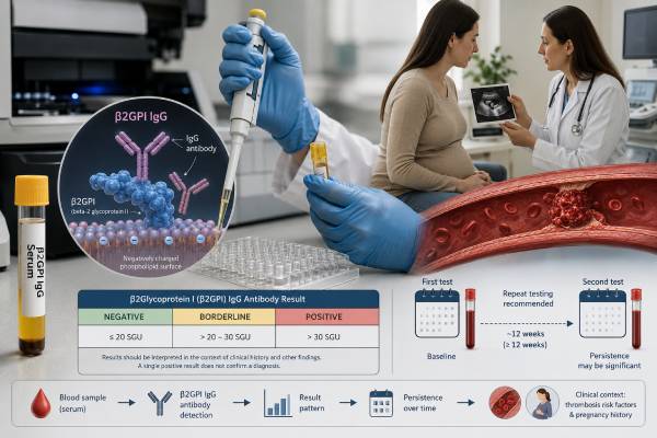
Laboratories use solid-phase immunoassays. In a common format, beta-2 glycoprotein I is attached to a test surface, patient serum is added, and bound IgG is measured through an enzyme or automated light-producing signal. The assay design affects which antibodies are captured.
Anti-beta-2 glycoprotein I IgG is more target-specific than the name “antiphospholipid antibody” may suggest. The immune reaction is primarily directed against a phospholipid-binding protein. Anticardiolipin antibodies often depend on beta-2 glycoprotein I as a cofactor, but anticardiolipin and anti-beta-2 glycoprotein I remain separate laboratory tests.
Research assays may examine antibodies against domain I of beta-2 glycoprotein I. A 2025 meta-analysis found that anti-domain I testing had high specificity with moderate sensitivity for APS and was associated with future thrombosis in prospective cohorts. Domain I testing is not the same as a routine whole-protein anti-beta-2 glycoprotein I IgG assay and is not universally available or required for standard evaluation.
A negative anti-domain I result would not cancel a positive routine whole-protein IgG result, because antibodies can recognize other regions and the assays have different sensitivity. Likewise, a positive domain I test should be described as an additional marker rather than substituted for the established whole-protein result. Its main current role is refinement in selected specialist or research settings.
The routine result is most useful as part of a complete antiphospholipid syndrome blood test panel, not as an isolated screen.
When Anti-Beta-2 Glycoprotein I IgG Is Ordered
Clinicians order this test when the history creates a meaningful possibility of APS. Testing is most informative after a defined event, not as a broad explanation for nonspecific symptoms.
Common indications include:
- Deep vein thrombosis or pulmonary embolism without a sufficient explanation
- Recurrent thrombosis or thrombosis in an unusual vascular site
- Ischemic stroke or another arterial event, especially at a younger age
- Suspected small-vessel thrombosis or APS-related organ involvement
- Recurrent early pregnancy loss under defined circumstances
- Unexplained fetal death
- Delivery before 34 weeks because of severe preeclampsia, eclampsia, or placental insufficiency
- Systemic lupus erythematosus with a relevant thrombotic or obstetric concern
- A positive anticardiolipin or lupus anticoagulant result that needs a complete profile
The event should be verified. Leg pain is not equivalent to an ultrasound-confirmed deep vein thrombosis. A transient neurologic symptom is not automatically an ischemic stroke. “Miscarriage history” should be expanded into gestational ages, number of losses, ultrasound findings, pathology, and other causes assessed.
Testing healthy people without symptoms is generally not useful, except in selected autoimmune settings such as systemic lupus erythematosus where the result may influence risk counseling. Low-pretest-probability screening creates incidental positives that are difficult to interpret and can lead to unnecessary treatment.
The test may be ordered during an acute clot because solid-phase antibody assays are less directly affected by anticoagulant drugs than lupus anticoagulant testing. However, acute illness can accompany temporary immune changes, and the initial result still requires later confirmation if it is being used to support APS.
A complete order should include IgG and IgM anti-beta-2 glycoprotein I, anticardiolipin IgG/IgM, and lupus anticoagulant unless a specialist has a specific reason to narrow the set. Testing only one antibody may miss a higher-risk combined profile.
Result Levels, Units, and Cutoffs
The report may use units per milliliter, standardized units, arbitrary units, or a manufacturer index. The number is interpreted against the reference interval for that exact assay.
The 2023 ACR/EULAR classification criteria define moderate solid-phase anticardiolipin or anti-beta-2 glycoprotein I levels as 40–79 units and high levels as at least 80 units for specified enzyme-linked immunosorbent assays. These thresholds support a research classification framework. They should not be applied automatically to every chemiluminescent or manufacturer-specific method.
Laboratories should establish suitable cutoffs, often using a reference population and method validation. Differences in protein source, surface coating, calibration, instrument signal, and data analysis can produce different values from the same sample.
| Finding | Typical interpretation | What is needed next |
|---|---|---|
| Negative | No IgG detected above this assay’s cutoff | Review other APS markers and the clinical diagnosis |
| Borderline or weak positive | Uncertain significance and greater susceptibility to variation | Check context, method, other markers, and whether repeat testing will matter |
| Moderate positive | More supportive of clinically important antiphospholipid autoimmunity | Confirm persistence and correlate with a documented event |
| High positive | Stronger laboratory evidence, particularly in a persistent combined profile | Complete APS assessment and specialist risk review |
| Positive with lupus anticoagulant and anticardiolipin | Triple established-marker positivity, often considered a high-risk profile | Verify every component, repeat as needed, and review treatment implications |
A result should be described with the actual value, category, unit, and laboratory—not only “positive.” A value of 43 on one method cannot be assumed to equal 43 on another.
The numerical level does not identify where a clot is located and is not an emergency test. It also is not a direct treatment-monitoring marker. A fall after anticoagulation does not show that the medicine is working, and a rise does not prove that a new clot has formed.
How the Result Supports APS Diagnosis
Anti-beta-2 glycoprotein I IgG is an established APS laboratory marker, but APS remains a clinical-laboratory syndrome. The test supports diagnosis only when it is connected to the right kind of clinical evidence.
The 2023 ACR/EULAR criteria use an entry requirement and weighted domains. The entry requirement includes a positive antiphospholipid antibody test within three years of an APS-related clinical criterion. Clinical domains cover venous, arterial, microvascular, obstetric, cardiac valve, and hematologic manifestations. Laboratory domains cover lupus anticoagulant and moderate or high anticardiolipin or anti-beta-2 glycoprotein I IgG/IgM.
These are classification criteria designed for research uniformity. Routine diagnosis can consider a broader clinical picture, but should still be rigorous. Current guidance advises against using the classification score as the only bedside diagnostic method.
A strong diagnostic assessment establishes:
- An objectively confirmed event compatible with APS
- A meaningful antibody level on a validated assay
- Persistence on two occasions at least 12 weeks apart
- The complete antibody profile
- Alternative causes and provoking factors
For example, a persistent high anti-beta-2 glycoprotein I IgG result after an unprovoked pulmonary embolism offers much stronger support than a borderline one-time result during pneumonia in a person without thrombosis. A high result after a clot caused by major trauma may still be relevant, but the clinician must decide how much APS contributed and whether it changes recurrence prevention.
Timing between the clinical event and antibody result also matters. The classification entry rule uses a three-year window, but routine clinical assessment may revisit older events when the original imaging and laboratory records are strong. Confidence falls when the event is remembered only as “a clot years ago,” the site is unknown, or the antibody method cannot be recovered.
Antibody positivity without a clinical event is not APS. Such a person may be described as an antiphospholipid antibody carrier. The risk and need for preventive medication depend on the broader profile and personal factors.
A negative result does not exclude APS if lupus anticoagulant or anticardiolipin is positive. It also does not rule out other causes of thrombosis. The laboratory panel must be interpreted as a whole.
Clotting Risk and Antibody Profile
Persistent anti-beta-2 glycoprotein I IgG can be associated with venous, arterial, and small-vessel thrombosis. The risk is not fixed by one antibody value. It changes with the entire profile and the patient’s history.
Features that increase concern include:
- A previous unprovoked or recurrent clot
- Arterial thrombosis
- Persistent lupus anticoagulant
- High anti-beta-2 glycoprotein I IgG
- Concurrent anticardiolipin IgG
- Double or triple established-marker positivity
- Systemic lupus erythematosus
- Smoking, hypertension, diabetes, high cholesterol, or obesity
- Temporary triggers such as surgery, immobility, pregnancy, estrogen exposure, cancer, or severe infection
The term triple positive means lupus anticoagulant, anticardiolipin, and anti-beta-2 glycoprotein I are all present. It does not mean three immunoglobulin isotypes or three non-criteria tests. Triple positivity is often associated with greater recurrence concern and can influence anticoagulant selection.
An isolated positive anti-beta-2 glycoprotein I IgG result is more difficult to interpret. It may be clinically meaningful, especially if high and persistent, but studies have not shown that every isolated result predicts APS events equally. A specialist should review whether anticardiolipin and lupus anticoagulant were truly negative and whether medication made LA testing unreliable.
People who have never had a clot are considered separately from those receiving secondary prevention after thrombosis. A positive antibody alone does not automatically justify lifelong anticoagulation. Bleeding risk must be balanced against a demonstrated benefit.
After APS-related thrombosis, vitamin K antagonist treatment such as warfarin remains important for many patients. Direct oral anticoagulants may not be suitable for certain high-risk APS profiles, particularly triple positivity or arterial disease. The anti-beta-2 glycoprotein I result contributes to that assessment but does not independently prescribe treatment.
General prevention includes not smoking, managing blood pressure and diabetes, maintaining movement during prolonged immobility, and planning for surgery or hospitalization. Estrogen-containing medication should be discussed in the context of total thrombotic risk.
Emergency evaluation is required for sudden shortness of breath, chest pain, coughing blood, unilateral leg swelling, new weakness or speech difficulty, sudden severe vision loss, or signs of a threatened limb. Antibody testing does not replace imaging for an acute event.
Pregnancy Loss and Placental Complications
Anti-beta-2 glycoprotein I IgG can support obstetric APS when it is persistent and paired with a defined pregnancy complication. The antibody may contribute to placental inflammation, complement activation, abnormal trophoblast function, and impaired blood flow. Placental disease is more complex than small clots alone.
Pregnancy events considered in APS frameworks include:
- Recurrent early pregnancy losses under defined conditions
- Unexplained fetal death after a specified gestational stage
- Delivery before 34 weeks because of severe preeclampsia, eclampsia, or recognized placental insufficiency
Not every miscarriage indicates APS. Chromosomal abnormalities are a common cause of early loss. Uterine anatomy, endocrine conditions, parental genetics, infection, maternal age, and other placental disorders can produce similar outcomes.
The obstetric record should identify gestational age, fetal cardiac activity, ultrasound findings, pathology, maternal blood pressure, fetal growth, and the reason for delivery. One phrase such as “recurrent miscarriage” may hide clinically different events.
For example, three consecutive losses before 10 weeks after other common causes have been excluded differ from one biochemical pregnancy. An unexplained fetal death after 10 weeks differs from a loss with a confirmed chromosomal abnormality. Delivery at 31 weeks for severe preeclampsia and placental insufficiency differs from spontaneous preterm labor. The antibody result can only be connected accurately when those distinctions are preserved.
Risk also depends on whether the patient has previous thrombosis, lupus anticoagulant, high IgG levels, or multiple antibodies. Lupus anticoagulant is often a strong predictor of adverse pregnancy outcome, so a negative or positive anti-beta-2 glycoprotein I IgG result should never be interpreted without it.
Treatment evidence is strongest for established obstetric APS. Low-dose aspirin and heparin are commonly used, with full-dose anticoagulation considered when there is a thrombosis history. These medicines should be prescribed by clinicians who can weigh bleeding, delivery timing, and maternal and fetal risks.
Preconception assessment is valuable. It allows confirmation of the antibody profile, review of prior records, control of blood pressure and lupus activity, and replacement of medications that are unsafe during pregnancy. Warfarin management requires particular planning because it is generally avoided during pregnancy when suitable alternatives are indicated.
Pregnancy monitoring extends beyond antibody tests. Maternal blood pressure, urine protein, blood count, kidney and liver function, fetal growth, amniotic fluid, and placental blood-flow studies may be needed. A falling antibody level does not prove the placenta is safe.
Repeat Testing, Preparation, and Limitations
No fasting or special diet is usually required. A blood sample is collected from a vein, and ordinary hydration is sufficient. The clinician should record recent infection, pregnancy, autoimmune activity, and medication.
Solid-phase anti-beta-2 glycoprotein I IgG testing is not affected by anticoagulant drugs in the same direct way as clot-based lupus anticoagulant assays. This makes it possible to measure the antibody while a patient is receiving warfarin, heparin, or a direct oral anticoagulant. Medication still matters because the accompanying LA result may be unreliable.
A positive established antibody is generally repeated after at least 12 weeks—84 days—to demonstrate persistence. Repeat testing too early does not meet the usual interval. The same laboratory and assay are preferable because platform changes can create artificial differences.
Common limitations include:
- Different assays may recognize different antibody populations.
- Reference intervals and units are not fully harmonized.
- A borderline value may cross the cutoff because of ordinary variation.
- Infection or immune activation may produce transient positivity.
- Acute thrombosis and treatment complicate the overall panel even when the solid-phase result is technically valid.
- Classification thresholds may not transfer cleanly between ELISA and automated chemiluminescent methods.
A laboratory flag should never be read without the method and cutoff. If two platforms disagree, the clinician may repeat with the original method, consult the laboratory, or interpret the result based on the complete profile rather than choosing the higher number.
After APS is established, serial anti-beta-2 glycoprotein I IgG titers are not a routine measure of anticoagulant effectiveness. Warfarin is monitored with INR or an alternative method when needed; heparin monitoring follows drug-specific principles. The APS monitoring blood test panel guide explains those follow-up tests.
Next Clinical Steps
The next step depends on whether the result is negative, newly positive, or persistently positive.
For a negative result, confirm that lupus anticoagulant and anticardiolipin IgG/IgM were also tested. If the clinical event is real, continue evaluating other clotting, cardiac, vascular, obstetric, genetic, inflammatory, or medication-related causes.
For a new positive result, record the numerical value, unit, laboratory category, method, and collection date. Check whether the result occurred during infection and whether the complete APS panel was performed. Plan repeat testing only when it can affect diagnostic confidence or management.
For a persistent moderate or high result, review the clinical event and profile with the appropriate specialist. Hematology often leads thrombosis assessment. Rheumatology helps with lupus or systemic autoimmune disease. Maternal-fetal medicine evaluates obstetric events and future pregnancy plans. Neurology, nephrology, or cardiology may contribute for organ-specific disease.
Questions to ask include:
- Is the value moderate or high on this exact assay?
- Is it isolated, double positive, or part of triple positivity?
- Was lupus anticoagulant testing valid while I was taking my medication?
- Does my clot or pregnancy history meet an APS-associated pattern?
- When should the result be repeated, and should the same laboratory be used?
- Does the profile change anticoagulant choice, treatment duration, pregnancy care, surgery planning, or estrogen use?
- What bleeding precautions apply to the recommended treatment?
Do not start aspirin or an anticoagulant solely because the result is positive. Do not stop prescribed treatment because a later value falls or becomes negative. Both choices require a clinical assessment of thrombosis recurrence and bleeding.
The most accurate summary contains the target, isotype, level, persistence, other markers, and event: for example, “Persistent high anti-beta-2 glycoprotein I IgG with lupus anticoagulant after an unprovoked deep vein thrombosis.” That statement communicates far more than “APS test positive” and supports safer decisions.
References
- An update on laboratory detection and interpretation of antiphospholipid antibodies for diagnosis of antiphospholipid syndrome: guidance from the ISTH-SSC Subcommittee on Lupus Anticoagulant/Antiphospholipid Antibodies 2025 (Guideline)
- 2023 ACR/EULAR antiphospholipid syndrome classification criteria 2023 (Classification Criteria)
- Guidelines on the investigation and management of antiphospholipid syndrome 2024 (Guideline)
- Anti-β2 glycoprotein domain 1 antibody as a diagnostic marker for antiphospholipid syndrome: a systematic review and meta-analysis 2025 (Systematic Review)
- Antiphospholipid syndrome in pregnancy: a comprehensive literature review 2025 (Review)
- Laboratory Diagnosis of Antiphospholipid Syndrome: Insights and Hindrances 2022 (Review)
Disclaimer
This article provides general education and cannot diagnose APS, identify an active clot, or determine an individual treatment plan. Anti-beta-2 glycoprotein I IgG must be interpreted with the assay, repeat result, complete antibody profile, clinical event, and bleeding risk. Seek emergency care for symptoms of a possible clot, stroke, or serious pregnancy complication.