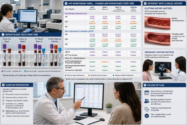
APS monitoring is not one fixed blood panel repeated at every visit. Follow-up for antiphospholipid syndrome usually combines medication-specific tests, blood counts, kidney and liver measures, clinical review for new clots or bleeding, and pregnancy surveillance when relevant. Antiphospholipid antibodies may be repeated to confirm the original diagnosis, but their numerical levels are not a simple measure of whether treatment is working.
The monitoring plan depends on the person’s situation. Someone taking warfarin needs regular international normalized ratio (INR) testing. A pregnant patient using low-molecular-weight heparin usually needs maternal and fetal monitoring that differs from routine warfarin care. A person with antibodies but no prior clot may need risk-factor review rather than frequent laboratory panels. Changes in symptoms, medication, kidney function, pregnancy status, surgery plans, or bleeding risk can all change the schedule. The most useful follow-up separates diagnostic antibody testing from treatment monitoring and assigns each test a clear purpose.
- There is no universal APS monitoring panel; tests are selected according to medication, organ risk, pregnancy, and symptoms.
- Antibody levels usually do not show whether anticoagulation is effective and should not replace medication-specific monitoring.
- Warfarin is generally followed with INR, while heparin monitoring is individualized by drug type and clinical circumstances.
- CBC, kidney function, liver tests, and bleeding review may be needed to monitor treatment safety.
- Pregnancy follow-up includes maternal assessment and fetal/placental surveillance, not antibody levels alone.
Table of Contents
- What APS Monitoring Includes
- Baseline and Routine Blood Tests
- When Antiphospholipid Antibodies Are Repeated
- Monitoring Warfarin and Heparin
- Tracking Clotting and Bleeding Risk
- Monitoring APS During Pregnancy
- How Often Follow-Up Is Needed
- Using Results to Adjust Care
What APS Monitoring Includes
APS follow-up has several separate goals. A test that helps with one goal may be useless for another.
The main monitoring tasks are:
- Confirming persistent antiphospholipid antibodies when the diagnosis is still being established
- Keeping anticoagulant treatment within a safe and effective range
- Detecting medication-related bleeding, low platelets, anemia, or organ dysfunction
- Reviewing new symptoms that could indicate thrombosis or a non-thrombotic APS manifestation
- Managing temporary risks such as surgery, infection, immobility, estrogen exposure, or pregnancy
- Monitoring maternal, placental, and fetal health during pregnancy
- Controlling cardiovascular risks that add to antibody-related risk
This explains why a broad commercial panel may not match the clinical need. Repeating lupus anticoagulant, anticardiolipin, and anti-beta-2 glycoprotein I every few months does not confirm that warfarin is therapeutic. A normal complete blood count does not exclude a new deep vein thrombosis. A therapeutic INR does not assess fetal growth.
A useful monitoring plan states the purpose beside each test. For example:
| Goal | Common test or assessment | What it cannot answer alone |
|---|---|---|
| Confirm persistent antibodies | Repeat established APS markers after at least 12 weeks | Whether anticoagulation is at the correct intensity |
| Monitor warfarin | INR, with method review when lupus anticoagulant interferes | Whether a new symptom is a clot |
| Check treatment safety | CBC, creatinine/eGFR, liver tests when indicated | Placental function or fetal well-being |
| Evaluate suspected recurrence | Urgent clinical assessment and site-specific imaging | Answered by routine antibody titers |
| Monitor pregnancy | Blood pressure, urine protein, blood tests, and ultrasound surveillance | Replaced by a single antibody panel |
Monitoring should also identify who coordinates each part of care. Hematology may oversee thrombosis prevention, rheumatology may manage associated lupus, an anticoagulation clinic may adjust warfarin, and maternal-fetal medicine may direct pregnancy surveillance.
Baseline and Routine Blood Tests
Before starting or changing treatment, clinicians often obtain tests that establish safety and reveal other conditions affecting risk. The exact set is individualized.
A common baseline group includes:
- Complete blood count (CBC): measures hemoglobin and platelets. Anemia may reflect bleeding, while thrombocytopenia can occur with APS, lupus, medication effects, or other disorders.
- Creatinine and estimated glomerular filtration rate (eGFR): help assess kidney function and guide dosing of some anticoagulants, especially low-molecular-weight heparin.
- Liver tests: may be appropriate before or during medications metabolized through the liver or when liver disease affects clotting and bleeding.
- Prothrombin time/INR and aPTT: provide baseline coagulation information but may be influenced by lupus anticoagulant or treatment.
- Urinalysis and urine protein assessment: may be used when kidney involvement, lupus, hypertension, or pregnancy complications are concerns.
These tests are not automatically needed at the same frequency. A stable person on warfarin may need frequent INR measurements but only periodic CBC and kidney/liver review. Someone with changing renal function or recurrent bleeding may need closer safety monitoring.
A falling hemoglobin should not be attributed automatically to anticoagulation. Clinicians need to look for the source, which may include gastrointestinal bleeding, heavy menstrual bleeding, surgery, nutritional deficiency, kidney disease, or another condition. Similarly, a low platelet count can represent APS-associated thrombocytopenia, heparin-induced thrombocytopenia, immune thrombocytopenia, lupus activity, infection, or laboratory error.
Routine inflammatory markers such as C-reactive protein or erythrocyte sedimentation rate do not measure APS clotting activity directly. They may be useful for an associated inflammatory or autoimmune disease, but a normal result does not mean APS risk is absent.
Additional tests are chosen for specific organ concerns rather than added automatically. Persistent high blood pressure, protein in the urine, or declining kidney function may lead to urine protein quantification, blood-smear review, complement tests, or imaging. New shortness of breath may require oxygen assessment and chest imaging. Neurologic symptoms may require brain and vascular imaging. These investigations evaluate a suspected complication; they are not routine components of an antibody monitoring bundle.
D-dimer is not a routine long-term APS monitoring marker. It may help evaluate suspected venous thromboembolism in selected clinical settings, but its interpretation depends on symptoms and pretest probability. It can rise because of infection, pregnancy, surgery, cancer, inflammation, or age. A person with concerning symptoms should be assessed through a validated diagnostic pathway rather than ordering serial D-dimer tests at home or during routine visits.
When Antiphospholipid Antibodies Are Repeated
Antibody retesting is most important during diagnostic confirmation. Established markers include lupus anticoagulant, anticardiolipin IgG/IgM, and anti-beta-2 glycoprotein I IgG/IgM. A positive result generally needs to remain positive on another sample collected at least 12 weeks later.
After APS has been established, routine serial antibody measurement has a limited role. Titers can change, but a fall does not prove that thrombosis risk has disappeared. A patient should not stop warfarin, heparin, or aspirin because an antibody becomes negative without a complete clinical review.
Repeat antibody testing may still be considered when:
- The initial diagnosis was incomplete or based on outside records that cannot be verified
- The first sample was collected during infection, acute illness, or another setting associated with transient positivity
- Lupus anticoagulant testing was distorted by anticoagulant medication
- A specialist needs an updated profile before a major management decision
- The original result was borderline and its persistence would change diagnostic confidence
- Pregnancy planning requires clarification of a previously uncertain antibody profile
The same laboratory and method are preferable when comparing solid-phase antibody values. Different platforms can produce different numbers and cutoffs. The original report should be retained with its units, date, and reference interval.
Lupus anticoagulant requires special caution. Warfarin, unfractionated heparin, low-molecular-weight heparin, and direct oral anticoagulants can interfere with clot-based assays. Retesting should be planned by the treating clinician and laboratory. Stopping anticoagulation solely to obtain a cleaner result can expose a patient to a dangerous untreated interval.
Repeated antibody tests should answer a defined question. Ordering them every month “to watch APS” can create anxiety and lead to inappropriate medication changes without improving outcome. The 12-week antibody confirmation guide explains the diagnostic timing in detail.
Monitoring Warfarin and Heparin
Medication monitoring is determined by the anticoagulant, not by the antibody level.
Warfarin and the INR
Warfarin is usually monitored with the international normalized ratio. The prescriber sets a target based on the type and history of thrombosis. Many venous APS cases use a conventional target range, while selected recurrent or arterial cases may require a different strategy. The individual target should appear clearly in the medical record.
An INR below target may leave insufficient anticoagulant effect. An INR above target increases bleeding risk. Dose changes should consider the trend, not just one number, along with missed doses, illness, alcohol, diet, and interacting medicines.
Lupus anticoagulant can sometimes affect the thromboplastin reagent used for prothrombin time and make the INR less representative of anticoagulation intensity. Venous laboratory INR is often reliable, but unexplained disagreement between the INR and the clinical or dosing picture needs investigation. Point-of-care INR devices may not agree with venous results in every patient with APS.
Specialist options can include comparing point-of-care and venous INR values or using an alternative measure such as chromogenic factor X in selected cases. These are not routine substitutes for everyone; they are troubleshooting tools when standard monitoring appears unreliable.
Warfarin monitoring becomes more frequent when treatment begins, doses change, the INR is unstable, interacting medication is started or stopped, dietary intake changes substantially, or illness occurs. Once stable, testing intervals can often be extended according to the anticoagulation service’s protocol.
Low-molecular-weight and unfractionated heparin
Low-molecular-weight heparin (LMWH) is commonly used in pregnancy and in some patients who cannot use warfarin. Routine anti-Xa monitoring is not required for every person receiving standard LMWH, but it may be considered in selected situations such as severe kidney impairment, extreme body size, recurrent events, unusual dosing, or concern about accumulation.
Unfractionated heparin is often monitored with aPTT or anti-Xa activity. Lupus anticoagulant can prolong baseline aPTT and make aPTT-based heparin monitoring unreliable. An anti-Xa approach may be preferred when baseline aPTT is affected, depending on the clinical setting and laboratory.
Platelet counts may be monitored when heparin-induced thrombocytopenia is a concern. A sudden platelet fall during heparin exposure requires prompt clinical evaluation rather than being assumed to reflect APS.
The monitoring plan should name the anticoagulant, target, responsible clinic, action thresholds, and instructions for missed doses or bleeding.
Tracking Clotting and Bleeding Risk
No routine blood panel can guarantee that a new clot is not forming. Clinical review remains central.
At follow-up, clinicians ask about:
- New unilateral leg swelling, pain, warmth, or discoloration
- Sudden shortness of breath, chest pain, fainting, or coughing blood
- New weakness, numbness, speech difficulty, severe vision change, or unusual neurologic symptoms
- Abdominal pain that could reflect an unusual-site thrombosis
- Skin ulcers, painful discoloration, or signs of impaired limb circulation
- Heavy menstrual bleeding, black stools, blood in urine, frequent nosebleeds, or large unexplained bruises
- Falls, head injury, or procedures while anticoagulated
Suspected recurrence needs urgent imaging and clinical evaluation. Rechecking antibodies, INR alone, or D-dimer without an appropriate diagnostic pathway can delay treatment.
Risk review also includes factors that can be changed:
- Smoking and nicotine exposure
- Blood pressure, diabetes, and cholesterol
- Body weight and regular movement
- Estrogen-containing contraception or hormone therapy
- Dehydration and prolonged immobility during illness or travel
- Planned surgery or hospitalization
- Medication adherence and interactions
A stable anticoagulant result does not remove the need for prevention during high-risk situations. Surgery plans may require temporary interruption, bridging, or mechanical and medication prophylaxis. These decisions balance clotting risk against procedural bleeding and should be documented in advance.
Catastrophic APS is rare but requires immediate hospital care. Rapid development of problems in several organs—such as breathing failure, kidney injury, neurologic changes, skin ischemia, or severe abdominal symptoms—cannot be monitored through a scheduled outpatient panel.
Monitoring APS During Pregnancy
Pregnancy monitoring focuses on maternal thrombosis, blood pressure, placental function, fetal growth, treatment safety, and delivery planning. Antibody titers alone do not show whether the pregnancy is progressing safely.
The plan often begins before conception. Clinicians review prior thrombosis, previous pregnancy outcomes, the antibody profile, current anticoagulant, kidney function, blood pressure, lupus activity, and medications that are unsafe in pregnancy. Warfarin is generally replaced with a pregnancy-compatible anticoagulant when indicated under specialist direction.
Maternal follow-up may include:
- Blood pressure checks and symptom review
- Urine protein assessment
- CBC for hemoglobin and platelets
- Kidney and liver tests when preeclampsia, organ disease, or medication concerns arise
- Review of injection technique, bruising, bleeding, and adherence
- Assessment for symptoms of venous or arterial thrombosis
Fetal and placental surveillance may include ultrasound assessment of growth, amniotic fluid, placental function, and Doppler studies according to the obstetric risk and local protocol. The frequency is individualized rather than determined by one antibody value.
Low-dose aspirin and LMWH are commonly used in established obstetric APS, with the regimen shaped by whether the patient has prior thrombosis. Routine anti-Xa testing is not necessary for every pregnant patient on prophylactic LMWH. It may be used in selected situations, and pregnancy-related changes in weight and kidney function may affect the plan.
Delivery planning should address when to hold anticoagulation, eligibility for neuraxial anesthesia, induction or planned cesarean timing, bleeding response, and postpartum restart. The postpartum period carries increased thrombosis risk, so follow-up does not end at delivery.
Urgent pregnancy assessment is needed for severe headache, visual symptoms, chest pain, shortness of breath, one-sided leg swelling, heavy bleeding, severe upper abdominal pain, markedly reduced fetal movement, or signs of high blood pressure. These symptoms should not wait for the next scheduled panel.
How Often Follow-Up Is Needed
There is no single correct interval for all APS patients. Frequency changes with treatment stage and stability.
| Situation | Why follow-up may be closer |
|---|---|
| New warfarin treatment or dose change | INR can change quickly before a stable dose is established |
| Recent thrombosis or bleeding | Recurrence, complications, and medication safety need reassessment |
| Pregnancy or postpartum | Maternal and fetal risks evolve across gestation and after delivery |
| Kidney or liver dysfunction | Drug handling and bleeding risk may change |
| Stable long-term treatment | Visits and laboratory intervals may be extended within a structured plan |
| Upcoming surgery or prolonged travel | Temporary prevention and medication instructions are needed |
Follow-up should occur sooner than scheduled when a new medicine is prescribed, especially antibiotics, antifungals, anti-inflammatory drugs, antiplatelet drugs, or supplements that may alter bleeding or warfarin effect. Vomiting, diarrhea, major dietary changes, heavy alcohol use, and acute illness can also destabilize anticoagulation.
Transitions between care settings are common points of failure. Hospital discharge instructions should state the last anticoagulant dose, the next dose, the next laboratory date, and which service will review the result. After surgery or childbirth, the written plan should explain when prophylactic or full-dose treatment restarts. A patient should not leave with two teams each assuming the other will manage the INR or injections.
For stable long-term care, an annual or periodic broader review can reassess kidney and liver function, cardiovascular risks, medication interactions, contraception, pregnancy plans, and whether the recorded diagnosis still matches the evidence. This review is separate from the more frequent anticoagulant checks needed to keep treatment in range.
Remote INR services and home point-of-care testing can improve convenience for selected patients, but agreement with venous INR should be established when APS or lupus anticoagulant raises concern about device accuracy. A home number is useful only when the patient knows who receives it and what action to take.
Using Results to Adjust Care
Each abnormal result should trigger the question, “What decision does this change?” A high INR may require dose adjustment or urgent bleeding assessment. A falling platelet count during heparin exposure may require immediate evaluation. A higher antibody titer in an otherwise stable patient may require no change at all.
A result trend is more useful when collection conditions are visible. INR values should be viewed beside dose changes, missed tablets, illness, and interacting drugs. Platelet counts should be viewed beside heparin exposure and the timing of the fall. Creatinine should be viewed beside hydration, blood pressure, and the anticoagulant dose. A computer-generated red flag without this context can lead to the wrong response.
A clear monitoring record should include:
- Confirmed APS manifestations and date of the last event
- Antibody profile and whether persistence was established
- Current anticoagulant, dose, target, and monitoring method
- Recent INR or anti-Xa results when applicable
- Baseline and current CBC, kidney, and liver values when relevant
- Bleeding history and medication interactions
- Pregnancy status and obstetric plan
- Instructions for emergencies, procedures, and missed doses
Patients should avoid changing anticoagulant doses from antibody results, stopping therapy because they feel well, or doubling a missed dose unless their care team has given that exact instruction. Warfarin users should keep vitamin K intake reasonably consistent rather than avoiding nutritious green vegetables completely.
A review is also an opportunity to simplify unnecessary testing. Stable APS does not require every available non-criteria antibody, monthly D-dimer measurements, or repeated inflammatory panels without a clinical reason. Removing low-value tests can make the meaningful signals easier to see.
The strongest monitoring system is coordinated: the patient knows the target and warning signs, the laboratory knows the medication and assay limitations, and each specialist knows who is responsible for adjustments. That structure protects against both recurrent clotting and avoidable bleeding more effectively than any single “APS monitoring panel.”
References
- Warfarin and heparin monitoring in antiphospholipid syndrome 2024 (Review)
- Guidelines on the investigation and management of antiphospholipid syndrome 2024 (Guideline)
- An update on laboratory detection and interpretation of antiphospholipid antibodies for diagnosis of antiphospholipid syndrome: guidance from the ISTH-SSC Subcommittee on Lupus Anticoagulant/Antiphospholipid Antibodies 2025 (Guideline)
- Antiphospholipid syndrome in pregnancy: a comprehensive literature review 2025 (Review)
- Antiphospholipid Syndrome: A Comprehensive Clinical Review 2025 (Review)
- Diagnosis and management of antiphospholipid syndrome 2024 (Review)
Disclaimer
This information is educational and does not provide an individual monitoring schedule or medication instructions. APS follow-up must be tailored to the anticoagulant, prior events, bleeding risk, organ function, pregnancy, and laboratory method. Seek urgent care for symptoms of a possible clot, major bleeding, stroke, or serious pregnancy complication.