
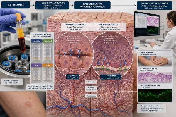
An autoimmune blistering disease antibody panel helps identify immune attacks on proteins that hold skin cells together or anchor the outer skin layer to the tissue below it. These disorders include pemphigus, bullous pemphigoid, mucous membrane pemphigoid, epidermolysis bullosa acquisita, linear IgA disease, and several rarer conditions. Blood tests can detect antibodies such as desmoglein 1, desmoglein 3, BP180, BP230, collagen VII, and others, but the panel is only one part of diagnosis. The most important test is often direct immunofluorescence on a carefully selected skin or mucosal biopsy, paired with a routine biopsy from a fresh lesion. Blister location, itching or pain, mouth and eye involvement, age, medications, and the pattern of antibody binding all guide interpretation. A negative blood panel does not exclude disease, especially when antibodies target an antigen not included in the assay or remain concentrated in tissue. Prompt dermatologic evaluation matters because extensive skin loss, infection, dehydration, airway involvement, or scarring of the eyes and throat can become urgent.
- Pemphigus antibodies usually disrupt connections between epidermal cells; pemphigoid antibodies target the basement membrane zone.
- Desmoglein 1 and 3, BP180 and BP230, and collagen VII are common blood-test targets.
- Direct immunofluorescence of perilesional tissue is a cornerstone test and requires the correct biopsy site and transport medium.
- Salt-split skin testing helps localize basement membrane antibodies to the roof or floor of an artificial split.
- A negative serum panel cannot rule out an autoimmune blistering disease.
- Eye, mouth, throat, genital, or extensive skin involvement needs prompt specialist assessment.
Table of Contents
- How autoimmune blisters form
- Antibodies in pemphigus
- Antibodies in pemphigoid and related diseases
- Blood tests, biopsies, and immunofluorescence
- Reading positive, negative, and mixed results
- Clinical clues and differential diagnosis
- Follow-up, monitoring, and urgent care
How autoimmune blisters form
Skin remains intact because cells adhere to one another and because the epidermis is anchored to the basement membrane zone. Autoimmune blistering diseases develop when antibodies recognize proteins in one of these adhesion systems. Inflammation, complement activation, enzymes, and mechanical stress then create separation within or below the epidermis.
The level of separation helps organize the disorders:
- Intraepidermal blistering occurs within the epidermis. Pemphigus is the major autoimmune group. Blisters are often fragile because only a thin layer covers them.
- Subepidermal blistering occurs below the epidermis at the basement membrane zone. Pemphigoid diseases, epidermolysis bullosa acquisita, dermatitis herpetiformis, and linear IgA disease fall into this broad category. Blisters may be tense because the full epidermis forms the roof.
This distinction is useful but not sufficient. Scratching can remove a tense blister, mucosal lesions may appear as erosions rather than bubbles, and early bullous pemphigoid may present only as itchy plaques. Routine histology shows the level and inflammatory pattern, while immunofluorescence shows where antibodies and complement are deposited.
Autoimmune blistering differs from many more common causes of blisters. Friction, burns, allergic contact dermatitis, eczema, infection, drug eruptions, porphyria, genetic skin fragility, and severe reactions such as Stevens-Johnson syndrome can create similar lesions through different mechanisms. That is why a blood antibody result should never be interpreted without examination and tissue testing.
The panel’s purpose is not merely to label “autoimmune.” It helps identify the target protein because target location can predict the disease family, likely sites of involvement, treatment approach, and need for additional cancer or medication evaluation in selected syndromes.
Antibodies in pemphigus
Pemphigus is characterized by loss of adhesion between epidermal cells, called acantholysis. The principal targets are desmogleins, proteins in desmosomes that help neighboring keratinocytes stay attached.
Desmoglein 3
Anti-desmoglein 3 IgG is strongly associated with pemphigus vulgaris involving mucous membranes. Patients may develop painful mouth erosions that persist for weeks, interfere with eating, and precede skin disease. The nose, throat, eyes, genital area, and esophagus can also be affected.
When anti-desmoglein 3 is present without substantial anti-desmoglein 1, disease may be mainly mucosal. This is a general pattern rather than an absolute rule.
Desmoglein 1
Anti-desmoglein 1 IgG is associated with superficial skin involvement. In pemphigus foliaceus, desmoglein 1 antibodies produce scaly, crusted, easily eroded lesions on the scalp, face, upper chest, or back, usually without mouth involvement.
Patients with both anti-desmoglein 1 and 3 may have mucocutaneous pemphigus vulgaris. Antibody levels often parallel disease activity, but clinical lesions remain the main treatment guide. A patient can have residual antibodies while clinically controlled, or active disease with a value that does not change as expected.
Other pemphigus antibodies
Expanded testing may include desmocollins, envoplakin, periplakin, and other epithelial proteins. These are especially relevant when the presentation suggests paraneoplastic pemphigus, now often termed paraneoplastic autoimmune multiorgan syndrome. This severe disorder can cause stubborn oral erosions, varied skin lesions, eye or genital disease, and lung involvement. It may be associated with an underlying lymphoid or other neoplasm.
IgA pemphigus involves IgA rather than classic IgG deposition and may target desmocollins or other epidermal antigens. Standard IgG-only panels can miss it.
| Pemphigus pattern | Common serum target | Typical clinical emphasis | Direct immunofluorescence clue |
|---|---|---|---|
| Pemphigus vulgaris, mucosal | Desmoglein 3 | Painful oral or other mucosal erosions | Intercellular IgG/C3 within epidermis |
| Pemphigus vulgaris, mucocutaneous | Desmoglein 3 and 1 | Mucosa plus fragile skin blisters/erosions | Intercellular epidermal staining |
| Pemphigus foliaceus | Desmoglein 1 | Superficial crusted skin lesions, little or no mucosal disease | Superficial intercellular staining |
| Paraneoplastic pemphigus | Envoplakin, periplakin and others | Severe stomatitis, polymorphic eruption, possible tumor | Intercellular and/or basement membrane findings |
| IgA pemphigus | Desmocollins or undetermined targets | Pustules or annular lesions | Intercellular IgA |
A positive desmoglein ELISA supports pemphigus only when the phenotype and tissue findings agree. Low-positive values can occur without active classic disease, and assay thresholds differ. The complete immunodermatology report is more informative than one red numerical flag.
Antibodies in pemphigoid and related diseases
Pemphigoid diseases target components of the basement membrane zone. Although their skin findings can overlap, the antigen and binding location help separate them.
BP180 and BP230
Bullous pemphigoid most often involves antibodies to BP180, also called type XVII collagen, and BP230. BP180 spans the cell membrane and extends into the basement membrane; its NC16A region is the target used by many commercial ELISAs. BP230 is an intracellular hemidesmosomal protein.
BP180 antibodies are generally more sensitive for active bullous pemphigoid than BP230 antibodies. BP230 can add support when BP180 is negative or may occur alongside it. Some patients have antibodies against BP180 regions not represented in a standard NC16A test, so a negative ELISA cannot exclude disease.
Bullous pemphigoid often affects older adults with intense itching, urticarial plaques, and tense blisters. A nonbullous phase can last weeks or months. Mucosal involvement is less dominant than in pemphigus vulgaris but can occur.
Mucous membrane pemphigoid
Mucous membrane pemphigoid is a clinical group characterized by predominant mucosal disease and risk of scarring. Targets can include BP180, BP230, laminin 332, integrin alpha-6 beta-4, and other basement membrane proteins. Serum tests are less sensitive than in classic bullous pemphigoid, making biopsy particularly important.
Eye involvement can scar the conjunctiva, and disease in the nose, throat, larynx, esophagus, or genital area can cause permanent structural damage. Laminin 332 antibodies may prompt a clinician to consider evaluation for an associated malignancy, although risk assessment should be individualized.
Epidermolysis bullosa acquisita
Epidermolysis bullosa acquisita, or EBA, is associated with antibodies to type VII collagen, the main protein of anchoring fibrils below the basement membrane. It may cause trauma-prone blisters, scarring, and tiny white cysts called milia, or it may resemble inflammatory bullous pemphigoid.
On salt-split skin, EBA antibodies usually bind the dermal floor, whereas classic bullous pemphigoid antibodies usually bind the epidermal roof. Other diseases can also produce floor binding, so collagen VII-specific testing and clinical features are needed.
Linear IgA disease and dermatitis herpetiformis
Linear IgA bullous dermatosis shows linear IgA along the basement membrane. Targets often include portions of BP180. It may occur spontaneously or after medications, particularly vancomycin. Adults and children can be affected.
Dermatitis herpetiformis is the blistering skin manifestation of celiac disease. It commonly causes intensely itchy grouped papules and small blisters on extensor elbows, knees, buttocks, scalp, or back. Granular IgA deposition in dermal papillae on direct immunofluorescence is characteristic. Blood evaluation uses celiac antibodies, including tissue transglutaminase and sometimes epidermal transglutaminase, rather than a standard pemphigoid panel. The celiac disease antibody panel guide explains the serologic pathway.
Blood tests, biopsies, and immunofluorescence
The strongest diagnosis usually combines morphology, two biopsies, and serum studies.
Routine histology biopsy
A biopsy from the edge of a fresh blister or active lesion is placed in formalin for routine microscopy. The pathologist looks for the level of skin separation, acantholysis, eosinophils, neutrophils, and other clues. An old eroded lesion may have lost the structures needed for interpretation.
Direct immunofluorescence biopsy
A separate biopsy is taken from normal-appearing skin or mucosa close to an active lesion, not from the eroded center. This perilesional sample is placed in a special transport medium or fresh-frozen according to laboratory instructions—not routine formalin. Formalin can destroy the immunoreactants needed for standard direct immunofluorescence.
Direct immunofluorescence, or DIF, detects antibodies and complement already deposited in the patient’s tissue. Common patterns include:
- intercellular “chicken-wire” IgG/C3 in pemphigus;
- smooth linear IgG and/or C3 at the basement membrane in pemphigoid;
- linear IgA at the basement membrane in linear IgA disease;
- granular IgA in dermal papillae in dermatitis herpetiformis.
A negative DIF can result from the wrong site, treated skin, an old lesion, poor transport, or a genuinely different diagnosis. If suspicion remains high, repeat biopsy from a better site may be more useful than repeatedly expanding blood panels.
Indirect immunofluorescence
Indirect immunofluorescence, or IIF, incubates patient serum with a substrate such as monkey esophagus, human skin, or salt-split skin. It detects circulating antibodies and shows a binding pattern. In salt-split testing, normal skin is separated within the basement membrane zone:
- roof binding supports BP180/BP230-type pemphigoid;
- floor binding suggests collagen VII, laminin 332, p200/laminin gamma-1, or another dermal target.
ELISA and antigen-specific assays
ELISA provides target-specific values for desmoglein 1, desmoglein 3, BP180, BP230, and collagen VII. Immunoblot, immunoprecipitation, or multiplex biochip assays may identify additional antigens. These tests can support diagnosis when biopsy is delayed, help distinguish related diseases, and provide a baseline for selected monitoring.
| Test | Best contribution | Main limitation |
|---|---|---|
| Clinical examination | Defines morphology and involved sites | Different diseases can look alike |
| Routine histology | Shows level of blister and inflammatory pattern | Does not reliably identify the precise antibody target |
| Direct immunofluorescence | Demonstrates tissue-bound immune deposits | Sensitive to biopsy site, treatment, and specimen handling |
| Indirect immunofluorescence | Shows circulating antibody pattern and salt-split location | May be negative when antibodies are tissue-bound or low level |
| ELISA/antigen assay | Identifies and quantifies a specific target | Misses targets not included and can yield isolated low positives |
Reading positive, negative, and mixed results
A result should answer three linked questions: Does the antibody match the clinical appearance? Does it match the tissue pattern? Is the antigen known to cause this disease phenotype?
Concordant results
A patient with painful oral erosions, fragile skin blisters, suprabasal acantholysis on histology, intercellular IgG on DIF, and high desmoglein 3 plus desmoglein 1 antibodies has a coherent pemphigus vulgaris profile. Agreement across methods makes each result more persuasive.
Likewise, an older adult with severe itch and tense blisters, subepidermal eosinophilic separation, linear C3/IgG at the basement membrane, roof-binding serum antibodies, and elevated BP180 has a coherent bullous pemphigoid profile.
Positive blood test with negative DIF
Possible explanations include a false-positive or clinically irrelevant serum result, biopsy from an unsuitable site, treatment before biopsy, low tissue deposition, or an evolving disorder. The strength of the serum result and phenotype matter. Repeating DIF correctly is often preferable to diagnosing solely from a weak blood value.
Positive DIF with negative blood panel
This is not rare. Antibodies may be below serum detection, bound mainly in skin, directed at an untested antigen, or poorly recognized by the commercial assay. The DIF pattern may still establish the disease family. Specialist testing can then identify the target if doing so will alter management.
Multiple positive antibodies
Epitope spreading, overlapping disease, assay cross-reactivity, and nonspecific low-level bands can produce multiple results. A panel showing several unrelated weak positives should be interpreted cautiously. The dominant clinical and tissue pattern should lead.
Antibody value and disease activity
Desmoglein 1/3 and BP180 levels often fall with clinical improvement and may rise before relapse in some patients. The correlation is imperfect. A numerical change should not override active blister counts, healing, mucosal examination, infection, medication toxicity, and patient function.
A negative result after treatment can be reassuring, but it is not a guarantee that therapy can be stopped. Persistent positivity does not always mean treatment failure. Decisions should use disease-specific clinical remission definitions and the medication’s risks.
Clinical clues and differential diagnosis
The history narrows the testing pathway before the first biopsy.
Fragile blisters and painful mouth erosions: Pemphigus vulgaris is a major concern, but erosive lichen planus, mucous membrane pemphigoid, paraneoplastic pemphigus, herpes infection, drug reactions, and other inflammatory disorders can look similar.
Intense itch before tense blisters: Bullous pemphigoid becomes more likely, especially in an older adult. Eczema, urticaria, scabies, drug eruption, and neurologic-associated scratching remain alternatives.
Scarring eye or mouth disease: Mucous membrane pemphigoid needs urgent consideration. Ocular cicatricial disease can progress even when skin involvement is mild. Ophthalmology assessment should not wait for a complete serum panel.
Trauma-site blisters with scarring and milia: EBA or a genetic fragility disorder may be considered. Salt-split skin and collagen VII testing can help.
Grouped itchy bumps on elbows and knees: Dermatitis herpetiformis suggests celiac-related testing and granular IgA DIF. Starting a gluten-free diet before evaluation can reduce the sensitivity of blood and tissue tests.
New blistering after a medication: Drug-induced bullous pemphigoid, linear IgA disease, pemphigus, or a severe non-autoimmune drug reaction may be possible. DPP-4 inhibitors used for diabetes have been associated with bullous pemphigoid, and vancomycin is a recognized trigger of linear IgA disease. Medication causality should be assessed by the treating clinicians; essential drugs should not be stopped casually.
Blistering during pregnancy or postpartum: Pemphigoid gestationis often involves BP180 antibodies and typically begins around the abdomen, sometimes spreading widely. It requires coordinated obstetric and dermatologic care.
Severe stomatitis plus varied skin lesions and systemic symptoms: Paraneoplastic pemphigus should be considered, particularly when there is a known or possible lymphoproliferative disorder. A broader antibody and malignancy evaluation may be required.
A broad autoantibody blood test panel cannot replace this morphology-based approach. Immunodermatology tests are most accurate when the clinician tells the laboratory the suspected disease, involved sites, treatments, and biopsy location.
Follow-up, monitoring, and urgent care
Once diagnosed, follow-up addresses disease control, treatment toxicity, infection risk, nutrition, pain, wound care, and threatened organs. The antibody panel is only one part.
Clinical monitoring may include:
- number of new blisters or erosions;
- healing of existing lesions;
- mouth, eye, throat, genital, and esophageal symptoms;
- ability to eat, drink, speak, and sleep;
- signs of skin infection;
- blood counts, liver and kidney tests, glucose, and bone health depending on therapy;
- desmoglein or BP180 trends when useful for that individual;
- repeat DIF in selected remission or diagnostic situations, not routinely for everyone.
Photographs taken under similar lighting can document intermittent lesions. Patients should avoid deliberately breaking blisters, use wound care recommended by dermatology, and report fever, pus, spreading redness, or rapidly increasing pain.
Immediate or urgent evaluation is needed for:
- eye redness, pain, light sensitivity, scarring, or vision change;
- hoarseness, trouble breathing, noisy breathing, or throat tightness;
- inability to swallow fluids, dehydration, or major weight loss;
- widespread skin detachment or rapidly expanding blistering;
- fever, confusion, low blood pressure, or signs of infection;
- genital or urinary scarring symptoms;
- severe weakness or breathing symptoms in suspected paraneoplastic disease.
Useful questions for the dermatologist and laboratory include:
- Was the DIF biopsy taken from the correct perilesional site and transported properly?
- What did routine histology show about the level of the blister?
- Which immunoglobulin and complement pattern was found?
- Did salt-split skin bind to the roof, floor, or both?
- Which specific antigens were included in the blood panel?
- Does my antibody profile match the skin and mucosal pattern?
- Will serial antibody levels change treatment decisions?
- Do I need ophthalmology, oral medicine, ear-nose-throat, gastroenterology, or cancer evaluation?
The safest diagnostic sequence preserves tissue evidence before treatment whenever clinically possible, but treatment should not be dangerously delayed in severe disease. A well-planned biopsy and focused antibody strategy can prevent weeks of uncertainty and distinguish diseases that require different monitoring, counseling, and therapy.
Because systemic corticosteroids and immune-suppressing treatments can quickly change lesions and laboratory findings, patients should tell the dermatologist about every recent cream, tablet, injection, or infusion. The date of the last new blister and the exact site selected for biopsy are useful details for the pathology requisition. When local testing is inconclusive, referral tissue and serum can sometimes be sent to a specialist immunodermatology laboratory for additional substrates, antigen mapping, immunoblotting, or immunoprecipitation. These advanced tests are most useful when they answer a specific unresolved question, such as separating EBA from another floor-binding pemphigoid or identifying paraneoplastic pemphigus.
References
- State-of-the-art diagnosis of autoimmune blistering diseases. 2024. Frontiers in Immunology review.
- Direct Immunofluorescence in Immunobullous Disorders of Skin With Histopathological Correlation. 2025. Original clinical study.
- Insights into bullous pemphigoid: A comprehensive review of diagnosis and management. 2025. JAAD Reviews review.
- Updated S2 K guidelines for the management of bullous pemphigoid initiated by the European Academy of Dermatology and Venereology (EADV). 2022. European clinical guideline.
- Antidesmoglein 1 and 3 serum IgG and positivity by direct immunofluorescence in pemphigus patients during clinical remission. 2025. JAAD International original research.
Disclaimer
This article provides general education and cannot diagnose a blistering disorder. Proper diagnosis may require urgent specialist examination, correctly located biopsies, immunofluorescence, and targeted serum testing. Seek immediate care for eye symptoms, breathing or swallowing difficulty, widespread skin loss, dehydration, fever, infection signs, or rapidly progressive blistering.





