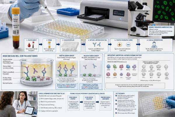
An ANA screen by ELISA checks blood for antibodies against a selected group of cellular targets linked to systemic autoimmune rheumatic diseases. It is an automated alternative to microscope-based ANA testing and may be used as the first step in evaluating lupus, Sjögren syndrome, systemic sclerosis, mixed connective tissue disease, or inflammatory myositis. The test is convenient, consistent, and often faster than indirect immunofluorescence, but its result depends on which antigens the manufacturer placed in the assay. A positive ELISA screen shows that the sample reacted above the laboratory cutoff; it does not identify a disease by itself. A negative result lowers the likelihood of the conditions the assay is designed to detect, yet it can miss clinically important antibodies that are absent or poorly represented in that test kit. The report should therefore be read alongside symptoms, the assay name, the numerical index or ratio, and any reflex testing. When clinical suspicion remains high, an ANA by IFA or targeted antibody test may still be appropriate.
- What it measures: Reactivity against a manufacturer-defined mixture of nuclear and sometimes cytoplasmic antigens.
- Positive result: Suggests one or more included autoantibodies may be present, but confirmation and clinical correlation are usually needed.
- Negative result: Makes several connective tissue diseases less likely, but does not exclude antibodies the assay does not contain.
- Result units vary: Laboratories may report positive/negative, an index, ratio, units, or an equivocal zone rather than an ANA titer.
- No fasting is usually required: A routine blood draw is generally sufficient unless other tests ordered at the same time require preparation.
- Best use: Screening people with compatible symptoms, not testing healthy people without a specific clinical reason.
Table of Contents
- How an ANA ELISA Screen Works
- Which Antibodies the Test May Detect
- Reading Positive, Equivocal, and Negative Results
- ANA ELISA Versus ANA by IFA
- When the Test Is Useful
- False Results and Important Limitations
- Follow-Up After an ANA ELISA
- Preparation and Questions to Ask
How an ANA ELISA Screen Works
ELISA stands for enzyme-linked immunosorbent assay. In an ANA screening ELISA, selected cellular antigens are attached to a solid surface, usually a well in a microplate. The laboratory adds a patient’s serum. If antibodies in the serum bind to those antigens, an enzyme-linked detection antibody produces a measurable color or signal. The analyzer compares the signal with calibrators and the laboratory’s cutoff.
Unlike the ANA by IFA test, ELISA does not require a technologist to inspect fluorescent staining on whole cells. It is highly automatable, can process many samples at once, and usually gives a straightforward numerical or qualitative result. Those features reduce reader-to-reader variation and help laboratories manage high testing volumes.
The tradeoff is that ELISA can detect only the antigens built into the assay. Some kits use purified or recombinant versions of individual proteins. Others combine defined antigens with extracts from cultured cells. Two products both labeled “ANA ELISA” may therefore have different target lists, cutoffs, sensitivity, and specificity.
A result is often calculated as an index or ratio. One laboratory might define values below 0.7 as negative, 0.7–0.99 as equivocal, and 1.0 or above as positive. Another might use arbitrary units with completely different thresholds. These values cannot be converted into IFA titers such as 1:80 or 1:320, and they should not be compared across manufacturers as though they are the same scale.
The test is generally ordered as a screening assay. Some laboratories automatically perform additional testing when the screen is positive. That may include ANA by IFA, an ENA panel, anti-dsDNA, or individual disease-associated antibodies. The exact reflex pathway should appear in the test description or laboratory report.
Which Antibodies the Test May Detect
Commercial ANA ELISA screens commonly include antigens associated with lupus, Sjögren syndrome, systemic sclerosis, mixed connective tissue disease, and myositis. A broad assay may contain targets such as:
- Double-stranded DNA
- Smith antigen
- U1-RNP proteins
- SSA/Ro52 and SSA/Ro60
- SSB/La
- Centromere B
- Scl-70, also called topoisomerase I
- Jo-1
- Histones
- Ribosomal P
- RNA polymerase III
- Fibrillarin
- PM/Scl
- Mi-2
- PCNA
The list is not universal. One assay may include 17 antigens, another fewer than 10, and another may use a mixed nuclear extract that contains a broader but less precisely defined range. The package insert or laboratory test directory is the only reliable way to know what a specific screen can detect.
This matters because autoimmune diseases do not produce one uniform antibody profile. A patient with systemic sclerosis may have antibodies to centromere proteins, Scl-70, RNA polymerase III, fibrillarin, Th/To, or other targets. An assay containing only one centromere protein and Scl-70 will not represent the full disease spectrum. A patient with inflammatory myositis may have MDA5, TIF1-gamma, NXP2, HMGCR, SRP, or an antisynthetase antibody other than Jo-1; many ANA screens include few or none of these.
Even when an antigen name appears on the list, its biochemical form can affect performance. Recombinant proteins may not fold or undergo post-translational modifications exactly as they do in human cells. Purified native antigens may preserve some epitopes better but can vary in purity. Antibodies may recognize a complex of proteins rather than one isolated component. These technical differences help explain why one ELISA can be positive while another assay is negative.
A positive screen also does not reveal which included antigen caused the signal unless the laboratory follows it with antigen-specific testing. A general autoantibody blood test panel or targeted confirmatory assays can identify the actual specificity and make the result more clinically useful.
Reading Positive, Equivocal, and Negative Results
Positive
A positive ANA ELISA means the measured signal exceeded the assay cutoff. It supports the possibility that an antibody to one or more included targets is present. It does not prove lupus or any other autoimmune disease.
The chance that a positive is clinically meaningful rises when the person has characteristic findings such as inflammatory joint swelling, photosensitive rash, persistent dry eyes and dry mouth, Raynaud phenomenon with skin changes, objective muscle weakness, unexplained low blood counts, kidney abnormalities, or inflammation around the lungs or heart. A positive screen found during broad testing for isolated fatigue or nonspecific pain has a lower positive predictive value.
The magnitude of the index may provide some context, but it is not a disease-activity score. A strongly positive ratio can still reflect an antibody unrelated to current symptoms, and a modest positive can be important if it identifies a highly disease-specific antibody. The confirmatory result matters more than assuming that “higher” always means “sicker.”
Equivocal or borderline
An equivocal result falls near the cutoff where analytical variation and weak reactivity make classification uncertain. The laboratory may repeat the measurement, ask for a new specimen, reflex to another method, or recommend clinical correlation. A borderline result should not be treated as a mild autoimmune diagnosis.
Whether to pursue it depends on the pretest probability. With convincing symptoms, targeted antibody testing or IFA may be reasonable. Without suggestive findings, repeated broad screening can generate more ambiguity than clarity.
Negative
A negative result means the assay did not detect reactivity above its threshold against the antigens it contains. It lowers the probability of ANA-associated connective tissue disease, especially when symptoms are nonspecific and the assay has been well validated in the relevant population.
A negative ELISA does not exclude:
- An antibody absent from the kit
- An antibody poorly recognized in its recombinant or purified form
- A low concentration below the cutoff
- A disease with mostly cytoplasmic or organ-specific antibodies
- Early or evolving autoimmune disease
- An autoimmune condition that is not usually ANA-associated
Rheumatoid arthritis, spondyloarthritis, celiac disease, autoimmune thyroid disease, type 1 diabetes, vasculitis, and autoimmune liver disease may occur with a negative ANA screen. When a clinician strongly suspects Sjögren syndrome, systemic sclerosis, or myositis, individual tests may be appropriate even after a negative general ELISA.
ANA ELISA Versus ANA by IFA
ELISA and indirect immunofluorescence answer related but different questions. ELISA asks whether antibodies bind to a defined antigen mixture. IFA asks whether antibodies bind anywhere on intact HEp-2 cells and, if so, what staining pattern appears.
| Feature | ANA ELISA | ANA by IFA |
|---|---|---|
| Target range | Defined by the antigens included in the kit | Broad cellular antigen display on HEp-2 cells |
| Typical report | Negative, equivocal, positive, index, or ratio | Positive or negative, titer, and fluorescence pattern |
| Automation | Highly automated and high-throughput | More labor-intensive, though automated imaging is available |
| Reader subjectivity | Low | Pattern and endpoint interpretation can vary |
| Pattern information | Usually none | Provides nuclear, cytoplasmic, and mitotic patterns |
| Main blind spot | Antibodies not represented in the antigen mixture | Weak, substrate-dependent, or difficult-to-recognize reactivity |
Studies comparing the methods do not produce one universal winner. Performance changes with the assay, cutoff, disease mix, laboratory expertise, and the population being tested. Some modern ELISA panels perform well because they include many clinically relevant antigens. Other solid-phase assays gain specificity by using a narrower set but lose sensitivity for uncommon antibodies. IFA can detect broad reactivity, yet it also finds low-level antibodies in healthy people and depends on skilled pattern interpretation.
Discordant results are therefore expected. ELISA-positive/IFA-negative results may occur with antibodies such as SSA/Ro or certain dsDNA and Scl-70 reactivities, depending on the substrate and assay. IFA-positive/ELISA-negative results may represent an antibody not included in the ELISA, a dense fine speckled pattern, a rare antigen, or nonspecific fluorescence.
Some laboratories use a combined strategy: ELISA first, then IFA and specific antibodies after a positive result. Others begin with IFA and use an ENA panel for identification. A dual-testing approach can improve detection in selected patients, but routinely ordering both methods in everyone increases cost and incidental findings.
When the Test Is Useful
ANA ELISA is most useful when a clinician is evaluating symptoms that suggest a systemic connective tissue disease and the laboratory’s assay has been validated for that purpose. It can be an efficient first-line screen in primary care, rheumatology, dermatology, nephrology, neurology, or other specialties.
Reasonable indications include combinations of:
- Objective inflammatory arthritis
- Photosensitive or otherwise characteristic autoimmune rash
- Recurrent mouth or nasal ulcers with systemic features
- Raynaud phenomenon plus abnormal nailfold capillaries, skin thickening, or fingertip injury
- Persistent dry eyes and dry mouth with glandular or systemic signs
- Proximal muscle weakness, high muscle enzymes, or a myositis-type rash
- Unexplained protein in the urine, blood-cell abnormalities, serositis, or multisystem inflammation
It is less appropriate as a screening test for the general population. ANA-associated antibodies are not rare in people without systemic disease. Testing a group with very low disease prevalence creates many positives that do not lead to a diagnosis.
The test may also be used within a laboratory cascade. A positive result can trigger IFA to obtain a titer and pattern, followed by specific antibodies based on the pattern. Another cascade may identify the antigen directly before or instead of IFA. The ANA with reflex test article explains why the order of these steps can change what appears on the final report.
False Results and Important Limitations
False-positive ANA ELISA results can occur when an assay detects low-level or cross-reactive antibodies that are not causing systemic autoimmune disease. Infections, other autoimmune disorders, some medications, malignancy, and normal immune variation can contribute. Older age increases the background prevalence of several autoantibodies.
False-negative results are a particular concern when clinicians assume every ANA ELISA contains every relevant antigen. Important causes include:
- Restricted antigen selection. The target antibody is simply not in the kit.
- Antigen presentation. The manufactured protein does not display the epitope recognized in the patient.
- Cutoff choice. A higher cutoff improves specificity but may miss weak true positives.
- Disease-specific gaps. Myositis and systemic sclerosis include numerous antibodies beyond common screening panels.
- Isotype or technical differences. Assays may focus mainly on IgG and use different detection chemistry.
- Treatment or timing. Antibody concentrations can change, although many remain detectable for long periods.
A numerical result may look precise while still being method-dependent. Ratios from different manufacturers cannot be trended as if they were the same biomarker. Even repeat measurements on the same platform are not usually used to monitor disease activity. Treatment decisions should rely on clinical status and organ-specific measures rather than serial ANA screen indexes.
Another limitation is loss of pattern information. A centromere, nucleolar, cytoplasmic reticular, or dense fine speckled pattern may provide a clue that a simple positive/negative ELISA does not. Pattern information can be especially useful when deciding whether to investigate systemic sclerosis, autoimmune liver disease, myositis, or isolated DFS70 antibodies.
Finally, a test can be analytically accurate but clinically unhelpful. When the original symptom is unlikely to represent systemic autoimmune disease, a technically true antibody result may still lead to anxiety, referrals, and unnecessary repeat testing without improving care.
Why two laboratories may disagree
A changed result does not always mean the immune system changed. One laboratory may use a broad extract-based ELISA, another a panel of recombinant antigens, and a third a chemiluminescent or multiplex assay that is marketed under a similar “ANA screen” name. Their calibrators, signal calculations, and cutoffs may differ. A sample near one cutoff can reasonably fall on opposite sides of another.
Specimen handling can add smaller differences. Delayed separation, storage conditions, repeated freezing and thawing, or interference from unusual serum proteins may affect some assays. Quality-controlled laboratories minimize these problems, but no method is entirely free from analytical variation.
When reports conflict, first compare the method and antigen list rather than choosing the newest result automatically. A clinician may ask the laboratory whether the tests are directly comparable and whether confirmatory testing was performed. Repeating the sample on the same platform can help when a labeling or handling error is suspected. Testing by IFA or ordering the disease-specific antibody can be more informative when the disagreement reflects different assay designs.
The timing of symptoms also matters. A person can develop a specific autoantibody before, during, or after recognizable clinical disease, and treatment may reduce some reactivities. Even so, dramatic interpretation of a small index change is rarely justified. A meaningful change is one that fits a change in symptoms, examination, or organ findings and is confirmed with an appropriate method.
Follow-Up After an ANA ELISA
The next step depends on the result, the test design, and the clinical picture.
After a positive result, common follow-up options include:
- ANA by IFA for a titer and pattern
- Anti-dsDNA and anti-Sm when lupus is suspected
- SSA/Ro and SSB/La for Sjögren syndrome, lupus, or specific pregnancy concerns
- RNP for mixed connective tissue disease or overlap features
- Centromere, Scl-70, and RNA polymerase III for systemic sclerosis
- A dedicated myositis panel for objective weakness, rash, or interstitial lung disease
- Complement C3 and C4, blood counts, urinalysis, urine protein, creatine kinase, liver tests, CRP, or ESR depending on the organs involved
After an equivocal result, the clinician may repeat the same assay after an appropriate interval, use IFA, or order one specific antibody suggested by the symptoms. There is little value in repeatedly checking a borderline number without a clinical change.
After a negative result, no further ANA testing may be needed when symptoms are nonspecific and examination is reassuring. High clinical suspicion changes that calculation. A person with severe dry eyes, parotid swelling, neuropathy, and inflammatory features may still need SSA/Ro antibody testing. A person with Raynaud phenomenon and skin thickening may need systemic sclerosis-specific antibodies. A patient with muscle weakness and lung disease may need a comprehensive myositis evaluation.
Urgent symptoms should be assessed directly rather than waiting for antibody confirmation. Severe shortness of breath, chest pain, coughing blood, seizure, sudden neurologic deficit, rapidly worsening weakness, very low urine output, or signs of a blood clot require prompt medical attention.
Preparation and Questions to Ask
Fasting is usually unnecessary for ANA ELISA. The blood draw itself is routine. Tell the clinician about current medications, recent infections, pregnancy, and previous autoimmune test results. Do not stop a medication because it might be associated with ANA positivity unless the prescriber gives specific instructions.
When reviewing the report, ask:
- What exact ANA method was used?
- Which antigens are included in this ELISA?
- What are the laboratory’s negative, equivocal, and positive cutoffs?
- Did the laboratory perform reflex IFA or specific antibody tests?
- Which symptoms or examination findings prompted the order?
- Would a positive result change treatment, referral, or organ testing?
- If the result is negative, is there a specific disease-associated antibody that still needs testing?
A useful report should be interpreted as one part of a diagnostic sequence, not a final label. The most reliable approach connects the test to a defined clinical suspicion, identifies the specific antibody when needed, checks for objective organ involvement, and avoids repeated screening when no new evidence has appeared.
Keep a copy of the complete report rather than recording only “positive” or “negative.” The assay name, date, numerical value, reference interval, and reflex results can prevent unnecessary duplication later. When seeing a specialist, bring prior reports from the original laboratories because patient-portal summaries sometimes omit method details. This is especially helpful when symptoms have evolved or when results from different health systems appear inconsistent.
References
- Antinuclear Antibodies (ANAs) by ELISA 2026 (Test Fact Sheet)
- Is there an alternative to the indirect immunofluorescence ANA HEp-2 assay for the diagnosis of connective tissue diseases? 2025 (Original Research)
- Comparison of ANA testing by indirect immunofluorescence and solid phase assays in a Spanish multicenter cohort 2023 (Original Research)
- Comparison of Indirect Immunofluorescence and Enzyme Immunoassay for the Detection of Antinuclear Antibodies 2022 (Original Research)
- Clinical utility of ANA-ELISA vs ANA-immunofluorescence in connective tissue diseases 2021 (Original Research)
Disclaimer
An ANA ELISA result cannot diagnose or exclude a systemic autoimmune disease by itself. Interpretation depends on the assay’s antigen list, cutoff, symptoms, examination, and confirmatory testing. Seek urgent care for severe breathing problems, chest pain, sudden neurologic symptoms, suspected clotting, or rapidly worsening weakness.