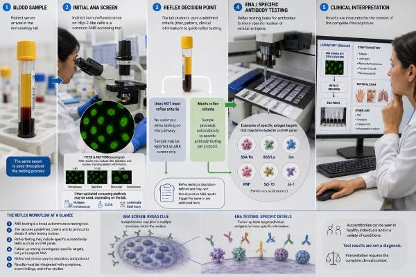
An ANA with reflex test begins with an antinuclear antibody screen and automatically adds selected tests when the first result meets the laboratory’s rules. The purpose is to move from a broad clue to more specific autoimmune antibodies without requiring a separate blood draw or a second order. A reflex pathway may add an ANA titer and pattern, an extractable nuclear antigen panel, anti-double-stranded DNA, or antibodies associated with systemic sclerosis and myositis. The exact sequence is not standardized. One laboratory may reflex only after a positive ANA by IFA, while another starts with ELISA and confirms positive samples by IFA. Some test specific antibodies even when the ANA screen is negative. Because the panel name alone does not reveal what will be performed, interpretation starts with the laboratory’s algorithm. A positive reflex antibody can support a diagnosis when symptoms fit, but no cascade diagnoses autoimmune disease on its own. A negative cascade lowers the chance of the conditions it covers, not every autoimmune disorder.
- What “reflex” means: The laboratory automatically performs additional tests only when predefined results or patterns trigger them.
- Common second-line tests: ANA titer and pattern, anti-dsDNA, SSA/Ro, SSB/La, Sm, RNP, Scl-70, centromere, and Jo-1.
- Panels differ substantially: The initial method, positivity threshold, reflex antigens, and reporting rules depend on the laboratory.
- A positive ANA may stop short of diagnosis: Disease-specific antibodies, symptoms, examination, and organ testing determine clinical meaning.
- A negative reflex panel is not universal clearance: It does not exclude rheumatoid arthritis, vasculitis, celiac disease, autoimmune thyroid disease, or antibodies outside the panel.
- Preparation is simple: Fasting is usually unnecessary, and reflex tests are generally performed from the original blood sample.
Table of Contents
- What Reflex Testing Does
- Common ANA Reflex Pathways
- ENA Antibodies in the Panel
- How Results Are Interpreted
- When Reflex Testing Helps
- Gaps, Pitfalls, and Unexpected Results
- Clinical Follow-Up Beyond the Antibody Panel
- Before and After the Blood Draw
What Reflex Testing Does
Reflex testing is a laboratory decision pathway. The clinician orders one named test, and the laboratory applies if-then rules to the sample. For example, if an ANA screen is negative, testing may stop. If it is positive at or above a specified threshold, the laboratory may determine an IFA titer and pattern. A homogeneous pattern may trigger anti-dsDNA testing, while a speckled pattern may trigger an ENA panel.
The main advantages are speed, specimen conservation, and diagnostic stewardship. The patient often avoids another blood draw, and the clinician receives more specific information in one report. A well-designed pathway also limits indiscriminate panel testing. Instead of measuring many low-yield antibodies in every person, it reserves them for samples that show a relevant screening signal.
Reflex is different from a “panel performed in full.” In a fixed panel, all listed tests are run regardless of the first result. In a reflex panel, the final charge and number of reported tests may vary from patient to patient. A test catalog may list 15 possible components even though only two are performed on a particular sample.
The starting assay may be ANA by IFA, an ELISA, a chemiluminescent assay, or another solid-phase screen. That choice affects which samples move forward. IFA can reveal broad nuclear, cytoplasmic, and mitotic staining but produces more low-level positives. Solid-phase screens are automated and may be more specific for the antigens they contain, yet they can miss antibodies outside that selection.
A reflex algorithm is therefore a local testing strategy, not a universal medical standard. It reflects the laboratory’s instruments, validation data, expertise, patient population, and agreements with clinicians. The ordering clinician must still decide whether ANA testing is appropriate in the first place.
Common ANA Reflex Pathways
Several designs are used in practice. Understanding the design prevents false assumptions about a short or unexpectedly long report.
IFA-first pathway
The laboratory screens serum on HEp-2 cells. If no qualifying fluorescence is seen, it reports a negative ANA and stops. If positive, it reports a titer and pattern and may add antigen-specific tests selected by pattern.
A possible pattern-based sequence is:
- Homogeneous pattern → anti-dsDNA, chromatin, or histone antibodies
- Speckled pattern → SSA/Ro, SSB/La, Sm, and RNP
- Centromere pattern → centromere antibody confirmation
- Nucleolar pattern → systemic sclerosis-associated antibodies
- Cytoplasmic speckled pattern → myositis or antisynthetase antibodies
- Cytoplasmic reticular pattern → antimitochondrial antibody testing
Not every laboratory follows these exact branches. Mixed patterns may require manual review because one staining pattern can hide another.
Solid-phase-first pathway
The laboratory begins with an ANA ELISA screen or similar automated assay. A positive result may reflex to IFA for a titer and pattern, then to an ENA panel. This design can reduce microscope workload and may filter out some nonspecific IFA positives. Its weakness is dependence on the antigen mixture in the first assay. If the screen does not contain the relevant target, the cascade may stop too early.
Parallel or broad cascade
Some systems perform IFA and selected disease-specific antibodies at the same time. Others reflex to specific antibodies even after a negative screen when the ordered panel is designed to compensate for known blind spots. This can detect SSA/Ro, Jo-1, Scl-70, or other reactivities that may not produce a typical screening result. It also increases the chance of isolated positives that require careful confirmation.
| Order label | Possible first step | Possible reflex step | Information that may be absent |
|---|---|---|---|
| ANA IFA with reflex | HEp-2 IFA | Titer, pattern, then selected antibodies | Antibodies unrelated to the observed pattern |
| ANA ELISA with reflex | Defined antigen screen | IFA and/or ENA confirmation | Targets not included in the first screen |
| ANA comprehensive cascade | IFA plus specific immunoassays | Additional confirmatory methods | Rare antibodies outside the cascade |
| ANA titer and pattern reflex only | Qualitative IFA screen | Endpoint titer and pattern | ENA or disease-specific identification |
The test name should be read together with the “components,” “testing algorithm,” and “reflex criteria” sections in the laboratory directory. That information is more reliable than assuming that every ANA with reflex order includes an ENA panel.
ENA Antibodies in the Panel
ENA stands for extractable nuclear antigen, a historical laboratory term. Modern panels measure antibodies to named cellular proteins or protein-RNA complexes. The standard group often includes SSA/Ro, SSB/La, Smith, RNP, Scl-70, and Jo-1, although the composition varies.
| Antibody | Common clinical associations | Interpretive caution |
|---|---|---|
| SSA/Ro | Sjögren syndrome, lupus, subacute cutaneous lupus, neonatal lupus risk | Can occur with a negative or weak ANA screen |
| SSB/La | Sjögren syndrome and lupus | Isolated low-level positivity is less persuasive than combined SSA/SSB findings |
| Sm | Systemic lupus erythematosus | Specific but not sensitive; many people with lupus are negative |
| U1-RNP | Mixed connective tissue disease, lupus, overlap syndromes | Level, assay, and clinical features affect meaning |
| Scl-70 | Diffuse systemic sclerosis and interstitial lung disease risk | Unexpected low positives may need confirmation by another method |
| Jo-1 | Antisynthetase syndrome, myositis, interstitial lung disease | It represents only one of several antisynthetase antibodies |
Many laboratories add anti-dsDNA even though it is not always classified as an ENA. Anti-dsDNA can support lupus diagnosis and may be useful in evaluating kidney involvement and disease activity when interpreted with complement, urine findings, and clinical status. The assay method matters because ELISA, fluorescence-based tests, and the Crithidia luciliae method do not have identical specificity.
Expanded cascades may include centromere B, chromatin, ribosomal P, RNA polymerase III, PM/Scl, Ro52, DFS70, or myositis antibodies. A dedicated ENA 10 panel may offer broader coverage than a six-antigen panel, but more analytes also create more opportunities for weak or incidental positives.
The result should be interpreted antibody by antibody. “ENA panel positive” is incomplete without the specific target, numerical value, cutoff, and clinical context. Different antibodies within the same panel have very different diagnostic value.
How Results Are Interpreted
A reflex report is best read in sequence rather than as a list of unrelated numbers.
ANA negative and no reflex performed
This usually means the initial screen did not meet the laboratory’s threshold. It lowers the likelihood of lupus and several other ANA-associated rheumatic diseases, but it does not exclude all autoimmune disease. It may also fail to exclude an antibody not well detected by the starting method.
When symptoms strongly suggest Sjögren syndrome, systemic sclerosis, or inflammatory myositis, clinicians may order specific antibodies despite a negative screen. An algorithm that stops after a negative ELISA is especially dependent on the ELISA antigen list.
ANA positive but specific antibodies negative
This is common. It may reflect low-level ANA in a healthy person, an antibody not included in the reflex panel, a DFS70-associated pattern, an infection, medication exposure, another autoimmune condition, or an early/undifferentiated connective tissue disease. The titer, pattern, and symptoms determine whether further evaluation is justified.
A positive ANA with a negative ENA panel does not mean the first result was “wrong.” The tests measure different layers of information. IFA detects cellular binding; the ENA panel searches for selected identities.
ANA positive and one disease-associated antibody positive
This combination is more informative, especially when the antibody is specific and the clinical picture fits. Anti-Sm in a person with compatible lupus manifestations has different weight from a weak isolated RNP result in someone without systemic findings. The antibody supports a diagnosis; it does not replace classification criteria, examination, or organ assessment.
Several antibodies positive
Multiple reactivities can occur in lupus or overlap syndromes, but broad low-level positivity may also reflect assay interference or nonspecific binding. The laboratory may confirm selected findings with another method. A mixed clinical picture deserves specialist review rather than an automatic assumption that several separate diseases are present.
Numerical values from different platforms should not be compared directly. A value of 3.0 on one assay is not equivalent to 30 units on another. In most cases, serial ANA and ENA levels are not useful treatment targets.
When Reflex Testing Helps
Reflex testing works best when the initial ANA order is clinically justified. Appropriate reasons include persistent inflammatory joint swelling, photosensitive rash, Raynaud phenomenon with systemic features, objective muscle weakness, unexplained dry eyes and dry mouth, characteristic skin thickening, recurrent serositis, unexplained low blood counts, or kidney abnormalities suggesting immune-mediated disease.
It can shorten the time from suspicion to a useful antibody profile. A person with a high-titer speckled ANA and dry mouth may receive SSA/Ro and SSB/La results without returning for another specimen. A patient with a homogeneous pattern and inflammatory urine findings may receive anti-dsDNA results while the clinician arranges kidney evaluation.
Reflex rules also reduce low-value testing. Antigen-specific subserologies have a higher chance of false-positive interpretation when ordered in people with no symptoms and no screening evidence. Restricting them to appropriate settings can reduce unnecessary referrals and repeat tests.
However, reflex testing should not be used as a substitute for history and examination. A large cascade ordered for fatigue, brain fog, or widespread pain without objective inflammatory features may produce incidental results that do not explain the symptoms. The systemic autoimmune rheumatic disease panel is most useful when a clinician has already identified a plausible disease category.
Reflex testing is also not designed to monitor a known disease automatically. Once lupus, systemic sclerosis, or myositis is diagnosed, follow-up testing should be selected for the disease and organs involved rather than repeatedly running the full diagnostic cascade.
How the clinical question changes the useful branch
The same positive ANA can lead to different follow-up because symptoms change the probability of each disease. A person with sun-sensitive rash, low white blood cells, and protein in the urine may need anti-dsDNA, anti-Sm, complement, and urgent urine assessment. A person with dry eyes, dry mouth, and parotid swelling may need SSA/Ro, SSB/La, objective tear testing, and salivary evaluation.
Raynaud phenomenon plus puffy fingers or skin thickening makes centromere, Scl-70, and RNA polymerase III more relevant than a broad lupus-only branch. Proximal muscle weakness, high creatine kinase, mechanic’s hands, or unexplained interstitial lung disease may justify a dedicated myositis panel even if the routine reflex cascade reports only Jo-1.
This symptom-led approach also helps interpret negative branches. If a standard ENA panel is negative but the clinical picture strongly suggests systemic sclerosis or myositis, stopping the workup would be inappropriate because many important antibodies are not standard ENAs. By contrast, a low-titer positive ANA followed by a negative ENA panel in someone with no objective inflammatory findings often needs observation rather than an expanded commercial panel.
Pregnancy creates another targeted situation. ANA itself is not a general pregnancy-risk test, but known or suspected maternal autoimmune disease may prompt SSA/Ro, SSB/La, or antiphospholipid antibody testing because those antibodies have specific fetal or obstetric implications. The relevant branch is chosen from history, not from ANA positivity alone.
Gaps, Pitfalls, and Unexpected Results
The largest pitfall is assuming that “ANA with reflex” has one fixed meaning. Laboratories may differ in all of the following:
- Starting method and cutoff
- Whether cytoplasmic patterns count as positive
- Minimum IFA titer required for reflex
- Which patterns trigger which tests
- Number of ENA antigens
- Whether anti-dsDNA is included
- Whether weak positives are confirmed
- Whether reflex testing occurs after a negative screen
- Whether the report includes comments or disease associations
Pattern-based rules are helpful but imperfect. A speckled pattern can represent many antibodies, and mixed patterns may obscure clinically important staining. Some antibodies do not produce the expected pattern. Rigidly limiting follow-up to one pattern can miss disease.
Assay-specific false positives deserve attention. Low-positive Scl-70 results on some multiplex methods, for example, may conflict with the clinical picture and IFA pattern. Confirmation by a different method can prevent an incorrect systemic sclerosis label. Isolated SSB/La or RNP findings also need cautious interpretation when ANA is negative and symptoms are absent.
Conversely, a negative reflex panel can create false reassurance. Standard ENA panels do not cover the full range of systemic sclerosis or myositis antibodies. They also do not evaluate rheumatoid factor, anti-CCP, ANCA, antiphospholipid antibodies, thyroid antibodies, celiac antibodies, or organ-specific autoimmunity.
Reflex testing may create additional charges because each triggered assay is billed separately. Patients can ask the ordering clinic or laboratory how billing works, particularly when the order includes a large cascade.
Repeated cascades are another common problem. ANA and ENA results usually remain stable enough that frequent retesting adds little unless symptoms change, the original method was inadequate, or a specific new disease manifestation appears. An international analysis of repeated ANA/ENA testing has reinforced the need to reduce redundant orders and focus on clinical change.
Clinical Follow-Up Beyond the Antibody Panel
Antibodies identify immune patterns; they do not show the full extent of organ injury. Follow-up tests should match the suspected disease and symptoms.
For possible lupus, clinicians may order a complete blood count, metabolic panel, urinalysis, urine protein-to-creatinine ratio, complement C3 and C4, and anti-dsDNA. For Sjögren syndrome, evaluation may include objective tear and saliva testing, dental assessment, salivary gland imaging, or biopsy. For systemic sclerosis, lung function tests, chest imaging, echocardiography, blood pressure monitoring, and kidney tests may be needed. For myositis, creatine kinase, aldolase, electromyography, muscle imaging, lung assessment, or biopsy may follow.
Inflammatory markers can add context but are not specific. A CRP test or ESR may be normal in some autoimmune diseases and elevated for many nonautoimmune reasons.
A rheumatology referral is reasonable when a disease-associated antibody accompanies compatible symptoms or objective abnormalities. Referral may also help when the ANA titer is high, the pattern is strongly suggestive, or several systems appear involved. A weak isolated result without characteristic findings can often be reviewed by the original clinician before specialist referral.
Urgent care should be based on symptoms, not on waiting for the final reflex report. Severe shortness of breath, chest pain, coughing blood, seizure, sudden weakness, rapidly declining urine output, a suspected blood clot, or quickly progressive muscle weakness needs prompt assessment.
Before and After the Blood Draw
Fasting is generally not required. One blood sample is usually enough for the full cascade, although the laboratory may request another specimen if the volume is inadequate or a specialized confirmatory method is performed elsewhere. Tell the clinician about medications, recent infections, pregnancy, and prior positive autoimmune tests.
Before ordering, useful questions include:
- Which disease is being considered?
- What initial ANA method does the laboratory use?
- What threshold triggers reflex testing?
- Which ENA and related antibodies are included?
- Will positive findings be confirmed by another method?
- Are additional reflex charges possible?
After results return, review the report in order: initial method, positive or negative screen, titer and pattern if available, specific antibody identity, numerical value, and reference range. Keep the complete report because summaries in patient portals may omit the algorithm or method. Record the laboratory name and collection date as well, since later comparison is difficult when a different health system uses another cascade.
The most informative outcome is not the largest panel. More testing does not automatically produce more certainty or better care. It is a coherent result that fits the symptoms, directs targeted organ evaluation, and avoids unnecessary testing. When the laboratory pathway and clinical question align, ANA reflex testing can turn a broad autoimmune screen into a focused next step.
References
- Antinuclear Antibody (ANA) With HEp-2 Substrate by IFA 2026 (Test Fact Sheet)
- Antinuclear Antibody Testing 2025 (Guideline)
- An international multicenter retrospective analysis of repeated anti-ENA testing in ANA-associated rheumatic diseases, a data-driven proposal to increase testing efficacy 2025 (Original Research)
- Evaluation of Antinuclear Antibody and Subserology Reflex Testing for the Diagnosis of Systemic Autoimmune Rheumatic Disorders in an Academic Teaching Hospital 2023 (Original Research)
- Why should we implement algorithmic testing for systemic autoimmune rheumatic diseases? 2023 (Review)
Disclaimer
ANA reflex panels differ by laboratory and cannot diagnose an autoimmune disease without clinical evaluation. Results should be interpreted with symptoms, examination, the testing algorithm, and organ-specific studies. Seek urgent care for severe breathing, neurologic, kidney, clotting, or rapidly progressive symptoms.