
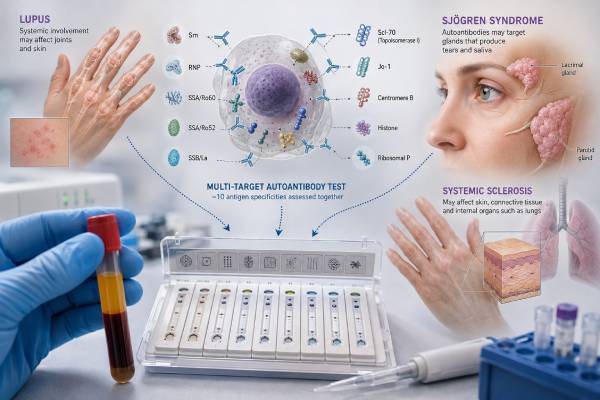
An ENA 10 panel is a group of blood tests that looks for autoantibodies directed against specific cellular proteins. These antibodies can help distinguish among systemic autoimmune rheumatic diseases after symptoms, examination findings, and an antinuclear antibody test raise concern. The panel may support the evaluation of lupus, Sjögren disease, systemic sclerosis, mixed connective tissue disease, inflammatory myositis, and overlap syndromes. “ENA 10” is not a universal recipe, however. Laboratories may use the same name for panels with different combinations of antibodies, methods, units, and cutoffs. A positive result therefore has meaning only after the exact analyte, strength, ANA pattern, and clinical setting are reviewed. Some antibodies are strongly associated with one disease, while others appear in several conditions or occasionally in people who never develop a defined connective tissue disease. The panel can sharpen a diagnosis, but it cannot replace a medical history, physical examination, organ assessment, or longitudinal follow-up.
- An ENA 10 panel identifies specific autoantibodies: it is more detailed than a general ANA screen.
- The ten antibodies are not standardized: always check the analyte list printed on the laboratory report.
- A positive antibody is evidence, not a diagnosis: symptoms and organ findings determine its clinical importance.
- A negative panel does not exclude autoimmune disease: some patients are seronegative or have antibodies outside the panel.
- No fasting is usually needed: results can take several days, especially when testing is sent to a specialty laboratory.
Table of Contents
- What an ENA 10 Panel Tests
- Common Antibodies and Their Disease Associations
- When the Panel Is Most Useful
- How to Interpret a Positive ENA Result
- Negative, Borderline, and Conflicting Results
- Testing Process, Methods, and Preparation
- Next Steps After the Results
What an ENA 10 Panel Tests
ENA stands for extractable nuclear antigen. The name comes from older laboratory techniques that extracted salt-soluble proteins from cell nuclei. Many modern assays use purified or manufactured antigens rather than the original extraction process, but the historical term remains.
The immune system normally makes antibodies to recognize outside threats. Autoantibodies recognize the body’s own molecules. ENA panels look for antibodies against named targets such as Ro/SSA, La/SSB, Smith, RNP, topoisomerase I, or Jo-1. These targets are found in the nucleus, cytoplasm, or complexes that help cells process RNA and proteins.
A broad antinuclear antibody test usually serves as an initial screen when a systemic connective tissue disease is suspected. An ENA panel then identifies specific antibody targets that may clarify the likely disease, expected organ risks, or need for additional testing. This sequence is useful because ANA positivity is common and can occur with infections, medications, other illnesses, increasing age, and even in healthy people.
The phrase “ENA 10 panel” describes panel size, not a fixed international standard. One laboratory may include SSA, SSB, Sm, RNP, Scl-70, Jo-1, centromere B, chromatin, ribosomal P, and dsDNA. Another may divide SSA into Ro52 and Ro60, include PM/Scl or RNA polymerase III, or omit dsDNA because it is tested separately. Some panels marketed as ENA 10 include antibodies that are not traditionally classified as extractable nuclear antigens.
Before interpreting a result, confirm:
- The exact ten antibodies tested
- Whether SSA was reported as one result or split into Ro52 and Ro60
- The assay method and reference interval
- Whether the panel was performed automatically after a positive ANA
- Whether results are qualitative, numerical, or graded
- Whether weak positives were repeated or confirmed by another method
The test does not measure the amount of inflammation, prove that symptoms are autoimmune, or show how much organ damage is present. It provides a serologic pattern that must be fitted to the person’s clinical picture.
Common Antibodies and Their Disease Associations
The following antibodies frequently appear in extended ENA panels. The report may contain a different set, so this table is a guide rather than a definition of every ENA 10 product.
| Antibody | Strongest common associations | Important clinical context |
|---|---|---|
| SSA/Ro | Sjögren disease, lupus, subacute cutaneous lupus | May relate to photosensitive rash, neonatal lupus, or fetal conduction problems during pregnancy |
| SSB/La | Sjögren disease and lupus | Usually carries more weight when SSA is also positive |
| Smith (Sm) | Systemic lupus erythematosus | Relatively specific for lupus but not present in most patients |
| U1-RNP or RNP | Mixed connective tissue disease, lupus, overlap disease | High-level anti-U1-RNP with a matching phenotype supports MCTD |
| Scl-70 or topoisomerase I | Systemic sclerosis | Associated with diffuse skin disease and interstitial lung disease risk |
| Centromere B | Limited cutaneous systemic sclerosis | Can be associated with later pulmonary arterial hypertension risk |
| Jo-1 | Antisynthetase syndrome and inflammatory myositis | Prompts attention to muscle weakness and interstitial lung disease |
| Chromatin or nucleosome | Lupus | May support lupus when interpreted with ANA, dsDNA, complement, and symptoms |
| Ribosomal P | Lupus | May be considered in selected neuropsychiatric or hepatic presentations, but is not diagnostic alone |
| dsDNA | Lupus, especially possible renal activity | Method and titer matter; many laboratories test it outside the ENA panel |
Lupus-associated antibodies
Anti-Sm is one of the most disease-specific antibodies for lupus, yet many people with lupus never test positive for it. Anti-dsDNA is also strongly associated with lupus and may track kidney involvement in some patients, although assays differ in sensitivity and specificity. Chromatin, ribosomal P, SSA, SSB, and RNP can add support but are not unique to lupus.
A positive lupus-associated antibody should be considered with ANA titer and pattern, blood counts, urine protein or blood, kidney function, complement levels, skin findings, joint symptoms, and other classification features. An anti-dsDNA result is especially dependent on method; a low positive from a sensitive solid-phase assay may not carry the same meaning as a strong result confirmed with a high-specificity method.
Sjögren-associated antibodies
Anti-SSA/Ro is the main antibody used in current Sjögren classification. It can also appear in lupus, systemic sclerosis, inflammatory myositis, autoimmune liver disease, and people without a defined systemic disease. Anti-SSB/La often accompanies SSA, but isolated SSB has much less diagnostic weight than it once did.
Ro is not one single antigen. Ro60 and Ro52 are different proteins. Some assays report them separately, while others combine them. Ro52 can appear across several autoimmune conditions and is particularly relevant when lung disease or myositis is being evaluated. Ro60 is more closely tied to classic SSA patterns in Sjögren disease and lupus.
Dry eyes and dry mouth have many nonautoimmune causes, including medications, dehydration, diabetes, aging, and local eye or salivary disorders. A Sjögren antibody panel supports evaluation, but objective tear testing, saliva testing, ultrasound, or a minor salivary gland biopsy may still be needed.
Systemic sclerosis-associated antibodies
Anti-Scl-70, also called anti-topoisomerase I, is associated with systemic sclerosis and a higher risk of interstitial lung disease. Anticentromere antibodies are more often linked to limited cutaneous disease, long-standing Raynaud phenomenon, and pulmonary arterial hypertension risk. Some extended panels also include RNA polymerase III, PM/Scl, fibrillarin, or Th/To, which can define other risk patterns.
Commercial assays can occasionally produce false-positive Scl-70 results, especially when the clinical picture and ANA immunofluorescence pattern do not fit. Confirmation with another method may prevent unnecessary anxiety and testing.
Myositis and overlap antibodies
Anti-Jo-1 targets histidyl-tRNA synthetase and is the most familiar antisynthetase antibody. It may occur with inflammatory muscle disease, interstitial lung disease, arthritis, Raynaud phenomenon, fever, or rough cracked skin on the hands called mechanic’s hands. A negative Jo-1 result does not exclude antisynthetase syndrome because antibodies to PL-7, PL-12, EJ, OJ, and other synthetases may not be included.
RNP, PM/Scl, Ku, and Ro52 can appear in overlap syndromes where features of lupus, systemic sclerosis, myositis, or Sjögren disease occur together. An ENA 10 panel can point toward overlap, but a dedicated myositis autoantibody panel is often more complete when weakness, elevated muscle enzymes, or interstitial lung disease is central.
When the Panel Is Most Useful
ENA testing is most informative when the pretest probability of systemic autoimmune disease is meaningful. That means symptoms and examination findings already suggest a connective tissue disease. Ordering a large antibody panel for nonspecific fatigue alone increases the chance of finding a weak positive that does not explain the symptoms.
Features that may justify testing include:
- Persistent inflammatory joint swelling or prolonged morning stiffness
- Photosensitive or otherwise characteristic autoimmune rashes
- Recurrent mouth or nasal ulcers
- Raynaud phenomenon with abnormal nailfold capillaries, skin tightening, or fingertip injury
- Unexplained dry eyes and dry mouth with objective gland dysfunction
- Proximal muscle weakness or markedly elevated creatine kinase
- Unexplained interstitial lung disease
- Protein or blood in the urine suggesting inflammatory kidney disease
- Low blood cell counts, serositis, or recurrent unexplained fevers
- A positive ANA with a pattern and clinical features suggesting a systemic rheumatic disease
An ANA with reflex testing is one common laboratory strategy. The laboratory first performs an ANA screen and then adds antigen-specific tests when the screening result meets predefined criteria. This can reduce unnecessary testing, but reflex rules vary. Some clinically important SSA antibodies may occur with a negative or weak ANA by immunofluorescence, so clinicians sometimes order SSA directly when neonatal lupus risk, subacute cutaneous lupus, or Sjögren disease is strongly suspected.
Repeating a stable positive ENA panel at short intervals is usually not useful. Many ENA antibodies remain present for years and do not accurately measure disease activity. Repeat testing is more reasonable when the original result was borderline, a method-related false positive is suspected, the phenotype has changed, or a specialist needs confirmation before making a high-impact diagnosis.
How to Interpret a Positive ENA Result
A positive result means the assay detected antibody binding above its cutoff. It does not automatically mean the patient has the disease most commonly associated with that antibody. Interpretation depends on five layers.
1. The exact antibody
Some antibodies are more disease-specific than others. Strong anti-Sm or a convincingly positive Scl-70 result generally carries more diagnostic weight than isolated low-level Ro52 or RNP. Even highly specific antibodies can occasionally appear outside their classic disease, so the finding still needs clinical confirmation.
2. The strength of the result
Reports may use an index, antibody units, arbitrary units, or categories such as weak, moderate, and strong positive. Values from different laboratories cannot be compared directly. A result just above the cutoff is more vulnerable to analytical variation and nonspecific binding than a result many times above the upper limit.
3. The testing method
ENA antibodies can be measured by ELISA, chemiluminescence, multiplex bead assays, line immunoassays, immunoblotting, immunodiffusion, and other techniques. Each method uses different antigen preparations and cutoffs. A person can test positive by one method and negative by another without either laboratory making a simple error.
When a result would substantially change diagnosis, pregnancy management, cancer screening, or organ surveillance, clinicians may confirm it using a different platform or a more specific assay.
4. The ANA pattern and other antibodies
The antibody combination often matters more than one isolated result. Anti-Sm plus dsDNA, low complement, and a compatible ANA pattern strongly supports lupus. High RNP with Raynaud phenomenon, swollen hands, arthritis, and muscle or lung features can support mixed connective tissue disease. Scl-70 with a nucleolar or speckled ANA pattern and scleroderma features is more convincing than a low isolated Scl-70 in someone without Raynaud phenomenon.
Multiple positives may be biologically linked rather than representing several separate diseases. For example, Sm and RNP antigens are part of related cellular complexes, while SSA and SSB often occur together.
5. The clinical phenotype
The same antibody can lead to different conclusions in different people. SSA in a patient with dry eyes, dry mouth, and abnormal salivary testing supports Sjögren disease. SSA in a pregnant patient matters because of fetal monitoring considerations. SSA in someone with a photosensitive annular rash may support subacute cutaneous lupus. Without matching clinical features, it may represent preclinical autoimmunity or an incidental finding.
A positive antibody can precede symptoms by years. This does not mean disease is inevitable. Follow-up should be proportionate to the specificity and strength of the antibody, the person’s symptoms, and the known organ risks.
Negative, Borderline, and Conflicting Results
A negative ENA 10 panel lowers the likelihood of diseases strongly associated with the tested antibodies, but it does not rule them out. Not every patient makes a recognized autoantibody, and no ten-analyte panel covers the full autoimmune spectrum.
Reasons for a negative result despite genuine disease include:
- The relevant antibody is not included in that laboratory’s panel
- The antibody level is below the assay cutoff
- The test method does not detect the person’s antibody well
- Disease is early or seronegative
- Immunosuppressive treatment has altered antibody levels
- The diagnosis depends mainly on clinical, imaging, or tissue findings
Seronegative Sjögren disease can be diagnosed through objective eye and salivary testing and, when appropriate, salivary gland biopsy. Systemic sclerosis may be diagnosed from skin findings, Raynaud phenomenon, nailfold capillary changes, and organ involvement even when standard antibodies are negative. Inflammatory myositis can be antibody-negative, particularly when only a limited panel was used.
A borderline result needs restraint. Laboratories place cutoffs where sensitivity and specificity are balanced, but biology does not divide neatly into negative and positive. Repeating the same test immediately may reproduce the same uncertainty. A better approach may be to review the ANA pattern, repeat after a clinically meaningful interval, use another method, or order a focused confirmatory test.
Conflicting results are common when different platforms are compared. One laboratory may report combined SSA positivity while another reports Ro52 positive and Ro60 negative. A multiplex ANA screen can be negative even when ANA by indirect immunofluorescence is positive because the assays expose different antigens. The report should be interpreted according to the method actually used rather than assuming all “ANA” or “ENA” tests are interchangeable.
Testing Process, Methods, and Preparation
The panel requires a routine blood draw. No fasting is usually necessary. A clinician may coordinate ENA testing with ANA, anti-dsDNA, complement C3 and C4, rheumatoid factor, urinalysis, blood counts, kidney and liver tests, creatine kinase, or inflammatory markers depending on the suspected disease.
Tell the clinician about pregnancy, recent infections, immune-modifying medications, and any previous autoimmune results. Most medications do not need to be stopped for the blood draw, and stopping prescribed treatment without guidance can be harmful. Biotin can interfere with some laboratory immunoassays, so patients taking high-dose biotin should ask whether it needs to be held before collection.
Results may be reported as individual antibody values or as an initial panel screen followed by specific identifications. Turnaround can range from one day to more than a week. A longer wait does not imply a more serious result; it often means the sample was sent to a reference laboratory or requires confirmatory testing.
Quality varies with antigen design and assay calibration. A panel that offers ten results at once is convenient, but convenience can create unexpected positives. Clinicians often place more weight on antibodies that match the ANA pattern and symptoms and may discount or confirm isolated weak results that do not fit.
Why broad panels can create misleading positives
Testing ten antibodies at once increases the amount of information, but it also increases the chance that at least one result will sit just above a cutoff. This is a basic statistical problem: the more markers tested in a person with a low likelihood of autoimmune disease, the more likely an incidental or method-dependent positive becomes. The result may lead to repeated testing, specialist visits, and worry even when no connective tissue disease is present.
Pretest probability changes the meaning of the same number. A moderate anti-Scl-70 result in a person with Raynaud phenomenon, puffy fingers, abnormal nailfold capillaries, and a matching ANA pattern is far more persuasive than the same result in a person tested only for fatigue. Likewise, SSA positivity in someone with objective dry-eye findings carries different weight from an isolated weak SSA result during an unrelated illness.
This is why clinicians usually begin with a focused history and examination. They look for symptom clusters, duration, objective inflammation, and organ involvement before deciding whether a broad panel is appropriate. A carefully selected test can shorten diagnosis; an indiscriminate panel can lengthen it.
Why ENA antibodies usually do not measure disease activity
Most ENA antibodies are diagnostic or phenotypic markers rather than reliable activity meters. Anti-Sm, RNP, SSA, SSB, Scl-70, centromere, and Jo-1 often remain positive even when symptoms improve. Their numerical values can also shift because of assay variation rather than a true clinical change. Repeating the whole panel every few months rarely shows whether treatment is working.
Monitoring should instead match the organ at risk. Lupus follow-up may emphasize urine protein, kidney function, complement, blood counts, and anti-dsDNA. Systemic sclerosis care may emphasize lung function, imaging, echocardiography, and blood pressure. Myositis care may use muscle strength, creatine kinase, and lung assessment. The ENA result helps define the map; organ-specific tests show what is happening on that map.
Next Steps After the Results
Follow-up should target the disease and organ risks suggested by the complete pattern rather than simply repeating the panel.
For a lupus-like pattern, common next tests include urinalysis, urine protein measurement, kidney function, C3, C4, anti-dsDNA, complete blood count, and antiphospholipid antibodies when clinically indicated. New leg swelling, foamy urine, high blood pressure, chest pain, shortness of breath, or neurologic symptoms may need prompt assessment.
For SSA or SSB positivity, clinicians may assess tear production, ocular surface damage, saliva flow, dental complications, gland enlargement, neuropathy, lung symptoms, and pregnancy status. A pregnant person with anti-SSA or anti-SSB should discuss fetal surveillance with obstetric and rheumatology specialists because a small proportion of exposed fetuses develop conduction abnormalities or neonatal lupus manifestations.
For Scl-70 or another systemic sclerosis-associated antibody, evaluation may include pulmonary function tests, high-resolution chest imaging when indicated, echocardiography, blood pressure monitoring, kidney tests, and nailfold capillaroscopy. Antibody type helps prioritize surveillance but does not predict an individual outcome with certainty.
For Jo-1 or another myositis-associated pattern, clinicians may check creatine kinase, aldolase, liver-associated muscle enzymes, pulmonary function, chest imaging, electromyography, MRI, or muscle biopsy. Lung disease can sometimes dominate even when muscle weakness is mild.
A rheumatology referral is reasonable when a disease-specific antibody is confirmed, multiple related antibodies are present, symptoms affect organs, or interpretation remains unclear. People with an isolated weak positive and no compatible symptoms may need observation rather than a diagnosis. The safest interpretation avoids both extremes: dismissing a meaningful antibody and labeling every positive as active autoimmune disease.
References
- The Relative Timing, Outcomes, and Economic Impact of Anti-Nuclear Antibody and Extractable Nuclear Antigen Testing 2024 (Review)
- Autoantibodies in Sjögren’s syndrome and its classification criteria 2021 (Review)
- Pathogenic role of anti-nuclear autoantibodies in systemic sclerosis 2024 (Review)
- ENA Panel 2024 (Official Page)
- ENA test 2025 (Official Page)
Disclaimer
ENA 10 panels differ among laboratories, and no individual antibody result should be used to diagnose or exclude a connective tissue disease without clinical evaluation. Discuss positive, negative, or conflicting findings with a clinician who can review the exact assay, symptoms, organ findings, medications, and related tests.





