
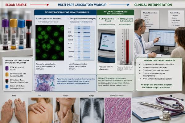
An autoimmune disease blood test panel is not a single universal package. Clinicians build an evaluation from several layers: general tests that reveal inflammation or organ injury, screening antibodies such as ANA, disease-focused antibodies such as an ENA panel, and targeted studies chosen from the person’s symptoms. CRP and ESR can show that inflammation may be present, but they do not identify its cause. ANA can support evaluation for lupus and related connective tissue diseases, yet a positive result is common outside those conditions. ENA antibodies can narrow the possibilities, while blood counts, kidney and liver tests, complement, urinalysis, muscle enzymes, and imaging often show whether an organ is actually involved. A normal panel does not rule out every autoimmune disease, and an abnormal panel does not replace diagnosis. The most useful interpretation asks whether all findings tell one coherent story: the right symptoms, objective examination findings, a plausible antibody pattern, and evidence of inflammation or tissue injury.
- ANA screens for several systemic rheumatic diseases but is not a general proof of autoimmunity.
- ENA panels contain specific antibodies such as SSA/Ro, SSB/La, Smith, RNP, Scl-70, and others; contents vary by laboratory.
- CRP and ESR are nonspecific inflammatory markers and may disagree.
- CBC, metabolic tests, urinalysis, complement, and organ-specific enzymes often determine urgency.
- Broad testing in a low-risk setting increases incidental and borderline findings.
- Diagnosis is based on the combined clinical pattern, not the number of abnormal results.
Table of Contents
- Building an autoimmune blood workup
- ANA and ENA in connective tissue disease
- CRP, ESR, and other inflammation markers
- Tests that look for organ involvement
- Targeted antibodies beyond ANA and ENA
- Interpreting common result combinations
- Follow-up, referral, and urgent findings
Building an autoimmune blood workup
Autoimmune diseases can affect one organ or several systems. No fixed panel can screen equally well for lupus, rheumatoid arthritis, celiac disease, type 1 diabetes, autoimmune thyroid disease, vasculitis, inflammatory myositis, autoimmune hepatitis, and neurologic autoimmunity. The workup should begin with the pattern of illness.
A clinician first defines whether symptoms suggest inflammation rather than a noninflammatory condition. Important clues include visible joint swelling, prolonged morning stiffness, a characteristic rash, Raynaud phenomenon, mouth ulcers, objective muscle weakness, unexplained fevers, recurrent eye inflammation, blood or protein in urine, low blood counts, thrombosis, dry eyes and mouth, or organ-specific abnormalities.
The laboratory strategy commonly has four layers:
- Basic health and organ tests. CBC, comprehensive metabolic panel, urinalysis, and sometimes thyroid, muscle, or coagulation studies identify anemia, cytopenias, kidney injury, liver abnormalities, protein loss, or other objective changes.
- Inflammation tests. CRP and ESR estimate the acute-phase response. They do not show whether inflammation is autoimmune, infectious, malignant, or caused by tissue injury.
- Screening immunology. ANA, RF, and sometimes other broad markers are selected when the phenotype makes them useful.
- Confirmatory or subclassifying tests. ENA antibodies, anti-dsDNA, anti-CCP, ANCA, antiphospholipid antibodies, complement, and organ-specific autoantibodies answer narrower questions.
The order may change in urgent illness. Someone with rapidly rising creatinine, blood in urine, and coughing blood needs immediate kidney-lung evaluation, not a slow stepwise outpatient panel. Someone with mild dry eyes and no objective abnormalities can be assessed more selectively.
Pretest probability governs interpretation. A positive ANA in a person with inflammatory arthritis, photosensitive rash, low complement, and proteinuria is more meaningful than the same result in a person tested for isolated fatigue. This does not mean vague symptoms are unimportant. It means the test should be chosen after common causes and objective clues are considered.
The phrase “autoimmune panel” on an order form can hide major variation. Patients should obtain the component list rather than assuming all autoimmune markers were checked. The related autoantibody blood test panel guide explains why different antibody families cannot be collapsed into one immune score.
ANA and ENA in connective tissue disease
ANA and ENA are often ordered together, but they play different roles.
ANA as a screening marker
Antinuclear antibody testing looks for antibodies that bind cellular structures. HEp-2 indirect immunofluorescence may report a titer and pattern. Solid-phase immunoassays may report an index or units. The methods are not interchangeable.
ANA is highly sensitive for systemic lupus erythematosus and commonly positive in Sjögren disease, systemic sclerosis, mixed connective tissue disease, and some inflammatory myopathies. It is also found in healthy people, particularly at low titers, and can occur with infections, medications, autoimmune thyroid or liver disease, and other conditions.
A positive ANA answers only one question: antibodies to included cellular antigens were detected. It does not prove that inflammation is active, identify the affected organ, or show that treatment is needed. Titer, pattern, method, disease-specific antibodies, and clinical context determine weight. The antinuclear antibody test article provides detailed interpretation.
ENA as a group of more specific antibodies
ENA stands for extractable nuclear antigen. Modern assays do not necessarily use literal extraction methods, but the historical name remains. Common components include:
| ENA or related antibody | Major disease associations | Key caution |
|---|---|---|
| SSA/Ro | Sjögren disease, lupus, neonatal lupus risk, some myositis/lung phenotypes | Can occur with a negative ANA depending on method |
| SSB/La | Sjögren disease and lupus, often with SSA | Isolated low-positive SSB has limited diagnostic value |
| Smith (Sm) | Systemic lupus erythematosus | Specific but not sensitive |
| U1 RNP | Mixed connective tissue disease, lupus, overlap disease | Level and phenotype matter |
| Scl-70/topoisomerase I | Systemic sclerosis, especially diffuse/ILD-associated patterns | Weak multiplex positives may require confirmation |
| Centromere antibodies | Limited systemic sclerosis spectrum | Can precede full clinical disease; not diagnostic alone |
| Jo-1 | Antisynthetase syndrome and inflammatory myositis | A standard ENA panel misses many other myositis antibodies |
Some laboratories include chromatin, ribosomal P, RNA polymerase III, PM/Scl, Ro52, or other targets. Others label a small six-antibody group as “ENA.” Always read the component list and assay method.
Reflex testing
An ANA with reflex test may automatically add ENA or anti-dsDNA after a positive screen. Reflex pathways reduce unnecessary testing when well designed, but they differ. A negative screen can stop testing even when the suspected antibody is poorly captured by that method. Strong Sjögren, myositis, or systemic sclerosis features may justify targeted testing despite a negative initial immunoassay.
Anti-dsDNA and complement
Anti-double-stranded DNA is not always part of ENA. It is a separate lupus-associated antibody. Method matters because assays differ in sensitivity and specificity. Anti-dsDNA trends may contribute to lupus activity assessment in some patients, especially with kidney concern, but should be combined with C3, C4, urinalysis, urine protein, creatinine, blood counts, and symptoms.
Low complement can reflect immune-complex consumption in lupus and other conditions. Complement can also be low because of inherited deficiency, severe infection, liver disease, or protein loss. Normal complement does not rule out lupus.
CRP, ESR, and other inflammation markers
CRP and ESR are often described as “inflammation tests,” but they behave differently.
C-reactive protein
CRP is a protein made by the liver in response to inflammatory cytokines. It can rise within hours and fall relatively quickly when the stimulus resolves. Infection, trauma, surgery, inflammatory arthritis, vasculitis, tissue injury, and many other conditions can elevate it.
A normal CRP does not exclude autoimmune disease. Lupus activity, particularly without major infection or serositis, may produce less CRP elevation than expected. Some inflammatory disorders remain localized or low grade. Medications and timing also affect the result.
High-sensitivity CRP, or hs-CRP, measures lower concentrations mainly for cardiovascular risk assessment. It is not a separate autoimmune marker. A standard CRP is usually more appropriate for active inflammatory evaluation.
Erythrocyte sedimentation rate
ESR measures how far red blood cells settle in a tube over a set time. Inflammation changes plasma proteins and can make cells fall faster. ESR responds more slowly than CRP and is influenced by factors unrelated to inflammation, including age, pregnancy, anemia, red-cell size and shape, kidney disease, and immunoglobulin levels.
An elevated ESR can support an inflammatory picture but cannot locate or identify the cause. A normal ESR does not rule out autoimmune disease. Very high values require evaluation for infection, malignancy, major inflammatory disease, or other causes.
Why CRP and ESR disagree
| Pattern | Possible explanations |
|---|---|
| High CRP, normal or modest ESR | Early acute inflammation, infection, tissue injury, some inflammatory arthritis; ESR may lag |
| High ESR, normal CRP | Anemia, age, pregnancy, high immunoglobulins, kidney disease, some lupus patterns |
| Both high | Significant inflammation, infection, vasculitis, active arthritis, malignancy, or tissue injury |
| Both normal | No strong systemic acute-phase response, treated disease, localized disease, or a condition not reflected by these markers |
Discordance is not automatically a laboratory error. The clinician should compare the time course, symptoms, blood count, immunoglobulins, kidney function, and medications.
Other inflammatory clues
Ferritin can rise with inflammation but is also affected by iron stores, liver disease, infection, and metabolic conditions. Markedly elevated ferritin can occur in macrophage activation syndrome or adult-onset Still disease, but it is not diagnostic alone. Fibrinogen, platelets, albumin, and globulins may provide indirect evidence of inflammation. None identifies autoimmunity by itself.
CRP and ESR are frequently useful for monitoring rheumatoid arthritis, polymyalgia rheumatica, giant cell arteritis, and some infections or vasculitides. Their value in monitoring differs by disease and individual. Treatment should not be escalated solely to normalize a nonspecific number when symptoms and objective findings do not support active inflammation.
Tests that look for organ involvement
The most important abnormality in an autoimmune workup may not be an antibody. Routine tests often reveal which organ is at risk and how urgently the person needs care.
Complete blood count
A CBC can show:
- anemia from inflammation, blood loss, kidney disease, hemolysis, marrow suppression, or nutrient deficiency;
- leukopenia or lymphopenia seen in lupus and other immune conditions;
- neutropenia from disease or treatment;
- thrombocytopenia from immune destruction, APS-related processes, lupus, medication, infection, or marrow disease;
- thrombocytosis as a nonspecific inflammatory response.
Cell counts are not disease-specific. A blood smear, reticulocyte count, iron studies, B12/folate, hemolysis tests, or hematology evaluation may be needed.
Kidney tests and urinalysis
Serum creatinine and estimated filtration rate measure kidney function, but they can remain normal early in glomerular inflammation. Urinalysis may detect blood, protein, or cellular casts. A urine protein-to-creatinine or albumin-to-creatinine ratio quantifies protein loss.
Protein and blood in urine can result from infection, stones, menstruation contamination, diabetes, hypertension, exercise, or kidney inflammation. Repeating a correctly collected sample and examining urine sediment can clarify the pattern. In suspected lupus nephritis, ANCA vasculitis, or anti-GBM disease, biopsy may define the exact lesion and guide treatment.
Liver tests
ALT and AST suggest hepatocellular injury; alkaline phosphatase and bilirubin help identify cholestatic patterns. Albumin and INR reflect liver synthetic function but can be affected by other illnesses. Autoimmune hepatitis and primary biliary cholangitis use different antibody profiles and may require imaging or biopsy. The autoimmune liver disease antibody panel article covers those pathways.
Muscle tests
Creatine kinase, aldolase, AST, ALT, LDH, and urine myoglobin can support muscle injury. A normal CK does not exclude every inflammatory myopathy, especially certain skin- or lung-dominant syndromes. Objective weakness, MRI, electromyography, lung evaluation, and biopsy may matter more than one enzyme.
Complement and immunoglobulins
C3 and C4 can fall with immune-complex activation. Total immunoglobulins and serum protein electrophoresis can reveal polyclonal immune activation, monoclonal proteins, or antibody deficiency. High IgG may support autoimmune hepatitis; high IgG4 is relevant only in an appropriate organ pattern and is not diagnostic of IgG4-related disease alone.
Coagulation and clot evaluation
An unexpectedly prolonged aPTT may reflect a lupus anticoagulant, factor deficiency, heparin, or another inhibitor. APS evaluation requires lupus anticoagulant plus anticardiolipin and anti-beta-2 glycoprotein I antibodies, persistence, and a qualifying clinical event. It is not inferred from ANA positivity.
Targeted antibodies beyond ANA and ENA
The symptom pattern determines whether other antibody systems are appropriate.
Rheumatoid arthritis
RF and anti-CCP support rheumatoid arthritis in a person with inflammatory joint swelling. Anti-CCP is more specific, but seronegative rheumatoid arthritis exists. Ultrasound or examination may demonstrate synovitis even when CRP and ESR are normal.
Vasculitis
PR3-ANCA and MPO-ANCA are central in suspected ANCA-associated vasculitis. Testing should be driven by features such as glomerulonephritis, pulmonary hemorrhage, destructive sinus disease, mononeuritis multiplex, or compatible skin vasculitis. Positive ANCA can occur with infection, drugs, inflammatory bowel disease, and other autoimmune conditions.
Antiphospholipid syndrome
Lupus anticoagulant, anticardiolipin, and anti-beta-2 glycoprotein I antibodies are ordered for selected thrombosis and pregnancy histories. Anticoagulants can interfere with lupus anticoagulant testing. Persistence after at least 12 weeks is generally required.
Thyroid disease
TPO and thyroglobulin antibodies support autoimmune thyroiditis; TSH receptor antibodies support Graves disease. TSH and free thyroid hormone levels show whether thyroid function is abnormal. A positive TPO antibody does not explain every symptom if thyroid function is normal.
Celiac disease
tTG-IgA plus total IgA is a common first-line combination. EMA and DGP are used in selected situations. Testing must usually occur while the person is eating gluten.
Diabetes, liver, skin, and neurology
GAD65, IA-2, ZnT8, and insulin autoantibodies identify beta-cell autoimmunity. AMA, smooth muscle/F-actin, LKM, and SLA support autoimmune liver evaluation. Desmoglein and BP180/BP230 antibodies support autoimmune blistering disorders. Neural antibody panels should be phenotype-specific and often require cerebrospinal fluid.
Broadly ordering all of these groups for nonspecific symptoms creates a high chance of unrelated findings. A positive GAD65 result, for example, means different things in diabetes screening and neurologic autoimmunity depending on concentration, specimen, and syndrome.
Interpreting common result combinations
Patterns across tests are more informative than isolated flags.
Positive ANA, negative ENA, normal CBC and urine
This common pattern may represent an incidental ANA, an early or undifferentiated connective tissue disease, an antibody not included in ENA, or a nonrheumatic association. Titer, IFA pattern, symptoms, examination, and follow-up determine whether more evaluation is needed. Repeating ANA frequently usually adds little.
Positive ANA, disease-specific antibody, and matching organ findings
An ANA with anti-dsDNA, low complement, proteinuria, and active urine sediment strongly raises concern for lupus kidney involvement. Anti-Scl-70 with Raynaud phenomenon, skin thickening, and interstitial lung disease supports systemic sclerosis. The matching phenotype gives the antibody clinical force.
High ESR with normal CRP
Anemia, age, high immunoglobulins, kidney disease, pregnancy, or lupus-related patterns may explain this discordance. It should not automatically be interpreted as severe active autoimmune inflammation.
High CRP with negative autoimmune antibodies
Infection must be considered, along with seronegative inflammatory arthritis, vasculitis, inflammatory bowel disease, tissue injury, and malignancy. Negative ANA or ENA does not convert a high CRP into a noninflammatory result.
Normal CRP and ESR with strong symptoms
Objective synovitis, Raynaud complications, inflammatory myopathy, Sjögren disease, cutaneous lupus, or neurologic autoimmunity can occur with normal markers. The examination and organ-specific studies should lead.
Multiple low-positive antibodies on a broad panel
This may reflect nonspecific binding or cutoff effects rather than several simultaneous diseases. Review the assay, strength, expected antibody combinations, and phenotype. Confirmation with another method is sometimes appropriate before diagnosis or high-risk screening.
Abnormal routine tests with negative serology
Cytopenias, proteinuria, muscle injury, or liver abnormalities deserve evaluation even when antibody tests are negative. Tissue biopsy, imaging, genetics, infection studies, or a different autoimmune pathway may be required.
Classification criteria can organize evidence, but they were often designed for research and should not be used as a do-it-yourself diagnostic checklist. A clinician must exclude mimics and judge whether the findings are attributable to one disease.
Follow-up, referral, and urgent findings
Follow-up should be proportional to clinical risk.
A person with a low-positive ANA and no objective abnormalities may need reassurance, symptom-directed care, and instructions about what changes would justify reassessment. A person with high-specificity antibodies, inflammatory findings, cytopenias, abnormal urine, lung disease, or progressive weakness needs targeted specialist evaluation.
Rheumatology referral is appropriate for persistent inflammatory joint swelling, a convincing connective tissue disease pattern, suspected vasculitis, recurrent unexplained systemic inflammation, or disease-specific autoantibodies with compatible symptoms. Nephrology, dermatology, neurology, hematology, gastroenterology, endocrinology, maternal-fetal medicine, or pulmonology may be more urgent when one organ dominates.
Repeat testing should have a defined purpose. ANA generally does not need serial repetition. ENA antibodies often remain stable. CRP, ESR, CBC, kidney tests, urine protein, complement, liver enzymes, CK, and disease-specific markers may be repeated when they reflect activity or treatment safety. A test should not be repeated simply because it was abnormal; the repeat should answer a management question.
Before the appointment, gather full reports with units and reference ranges, medication and supplement lists, symptom timing, photographs of rashes or Raynaud changes, and prior urine or imaging results. Useful questions include:
- Which diagnosis was each test intended to evaluate?
- Is this antibody sensitive, specific, or mainly prognostic?
- Does the laboratory method affect interpretation?
- Do CRP and ESR match the clinical picture?
- Are the blood count, kidneys, liver, lungs, muscles, or urine showing organ involvement?
- Which abnormality needs confirmation?
- What findings would change treatment or referral urgency?
Seek urgent medical care for chest pain, severe shortness of breath, coughing blood, sudden neurologic deficits, new seizures or confusion, rapidly reduced urine, severe high blood pressure, a painful swollen leg, extensive blistering, major weakness affecting swallowing or breathing, or high fever with immune-suppressing treatment. These problems require immediate clinical assessment; waiting for a panel result can be dangerous.
An autoimmune blood workup is successful when it narrows uncertainty and identifies threatened organs. More tests are not automatically better. The right combination is the one that fits the phenotype, uses reliable methods, and leads to a clear next decision.
References
- Connective Tissue Diseases – Systemic Autoimmune Rheumatic Diseases. 2025. ARUP Consult clinical testing guidance.
- C-Reactive Protein and Erythrocyte Sedimentation Rate Test Use. 2025. College of American Pathologists educational module.
- Predictive Value of Laboratory Assays Toward Autoimmune Disease Diagnoses. 2025. Journal of Applied Laboratory Medicine review.
- Advanced methods and novel biomarkers in autoimmune diseases: a review of the recent literature. 2023. Frontiers in Medicine review.
- Autoantibodies testing in autoimmunity: Diagnostic, prognostic and classification value. 2023. Autoimmunity Reviews review.
Disclaimer
This article is for general education and does not diagnose an autoimmune condition or replace individualized medical evaluation. Blood tests must be interpreted with symptoms, examination findings, assay methods, medications, imaging, urine studies, and sometimes biopsy. Seek urgent care for severe breathing, neurologic, clotting, kidney, blistering, or muscle-weakness symptoms.





