
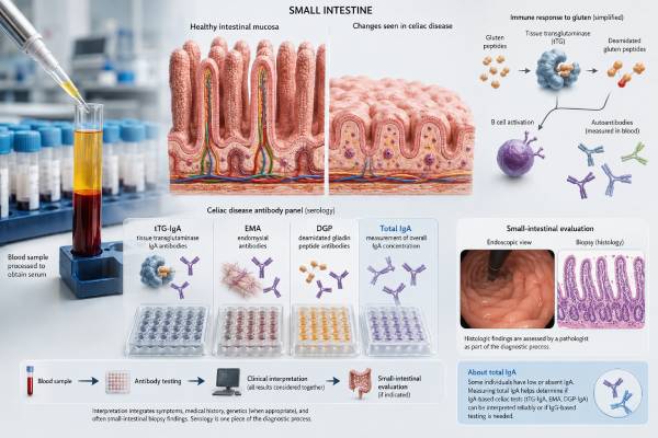
A celiac disease antibody panel helps determine whether the immune system is reacting to gluten in a way that may damage the small intestine. The most useful first-line combination is tissue transglutaminase IgA (tTG-IgA) plus total IgA. Other tests—endomysial antibody (EMA), deamidated gliadin peptide (DGP) antibodies, and IgG-based assays—answer more specific questions when initial results are strong, weak, conflicting, or affected by IgA deficiency. These blood tests can make celiac disease more or less likely, but their meaning depends on gluten intake, age, symptoms, risk factors, assay cutoffs, and whether intestinal biopsy is needed. Testing after reducing or eliminating gluten can produce a misleading negative result. A thoughtful interpretation therefore looks beyond a single “positive” or “negative” label and considers the entire diagnostic pathway, including duodenal biopsy, selected no-biopsy criteria, HLA testing, and follow-up after diagnosis.
- tTG-IgA with total IgA is the usual starting pair for people eating gluten.
- EMA-IgA is highly specific and often confirms a strongly positive or uncertain tTG-IgA result.
- DGP-IgG and tTG-IgG are mainly useful when total IgA is low or absent.
- A positive antibody panel does not automatically prove celiac disease in every patient.
- A negative panel may be unreliable after gluten restriction or when clinical suspicion remains high.
Table of Contents
- Who Should Have Celiac Antibody Testing?
- Why Gluten Intake Matters Before Testing
- tTG-IgA and Total IgA: The First-Line Pair
- EMA and DGP: When Additional Antibodies Help
- How to Interpret Positive, Negative, and Borderline Results
- Biopsy, HLA Testing, and No-Biopsy Pathways
- Follow-Up After Diagnosis and Common Testing Pitfalls
Who Should Have Celiac Antibody Testing?
Celiac disease is an immune-mediated condition triggered by gluten, a group of proteins found in wheat, barley, and rye. In a genetically susceptible person, ongoing gluten exposure can injure the lining of the small intestine. The damaged villi absorb iron, folate, calcium, vitamin D, and other nutrients less effectively. Some people develop classic digestive symptoms, but many present with problems outside the gut or have few obvious symptoms.
Testing is reasonable when symptoms or laboratory findings suggest malabsorption. Possible clues include chronic diarrhea, bulky or greasy stools, persistent bloating, unexplained abdominal discomfort, nausea, constipation, or unintended weight loss. Children may have poor growth, delayed puberty, irritability, dental enamel defects, or recurrent abdominal complaints. Adults may first come to medical attention because of iron-deficiency anemia, low bone density, recurrent mouth ulcers, abnormal liver enzymes, infertility, fatigue, peripheral neuropathy, or an itchy blistering rash called dermatitis herpetiformis.
Risk also matters. First-degree relatives of a person with celiac disease have a higher likelihood of the condition, even when they feel well. Testing may also be considered in people with type 1 diabetes, autoimmune thyroid disease, selective IgA deficiency, Down syndrome, Turner syndrome, or certain autoimmune liver disorders. These associations do not mean that every person in a risk group has celiac disease. They raise the pretest probability, which changes how a clinician interprets a borderline or negative result. Related testing for type 1 diabetes is discussed in the autoimmune diabetes antibody panel guide.
Screening should not be ordered simply because someone feels better without bread. Wheat allergy, irritable bowel syndrome, fermentable carbohydrate intolerance, and non-celiac gluten or wheat sensitivity can produce overlapping symptoms. Celiac antibodies do not diagnose those conditions. Conversely, a person can have celiac disease without dramatic symptoms, so a low symptom burden does not make testing pointless when risk is substantial.
Before ordering the panel, the clinician should clarify the question. Is the goal to evaluate active symptoms, screen a high-risk relative, explain an abnormal blood count, or reassess someone who started a gluten-free diet without testing? The answer affects which tests are useful and whether gastroenterology referral, biopsy, HLA typing, or a supervised gluten challenge may be needed.
Why Gluten Intake Matters Before Testing
Celiac antibodies are produced in response to gluten exposure. When gluten is removed, antibody levels often fall and intestinal inflammation begins to improve. That is desirable after a confirmed diagnosis, but it creates a major diagnostic problem when a person stops gluten before blood testing or biopsy. A negative tTG-IgA result after weeks or months of restriction may reflect reduced immune stimulation rather than the absence of celiac disease.
For the most reliable evaluation, serology and any planned duodenal biopsy should be performed while the patient is eating a regular gluten-containing diet. The exact amount and duration needed are not identical for everyone. A person who has already been strictly gluten-free should not restart gluten casually if previous reactions were severe, nutrition is poor, pregnancy is involved, or another medical concern exists. A gastroenterologist can decide whether a gluten challenge is appropriate and design it around the patient’s health, age, symptoms, and diagnostic goals.
The laboratory requisition should note whether the patient is eating gluten and, if possible, how consistently. “Gluten-free” is not always absolute. Some people avoid obvious bread and pasta but continue to consume sauces, seasonings, malt, shared-fryer foods, or cross-contaminated products. Others are highly adherent and may have normalized antibodies. These two situations can produce very different test reliability despite the same dietary label.
HLA-DQ2 and HLA-DQ8 testing can be useful when someone is already gluten-free. HLA results do not depend on current diet because genes do not change with gluten exposure. Absence of a celiac-permissive HLA pattern makes celiac disease very unlikely and may avoid a challenge. Presence of DQ2 or DQ8 does not prove disease because these variants are common among people who never develop celiac disease. HLA is therefore a rule-out tool in selected cases, not a stand-alone screening panel.
Do not begin a lifelong gluten-free diet based only on symptoms or one low-level antibody result. The diet is socially and financially demanding, can complicate later diagnosis, and may become nutritionally unbalanced without guidance. Confirming the diagnosis first allows appropriate monitoring, family counseling, and recognition of complications. If symptoms are severe, the clinician can often accelerate testing rather than asking the patient to endure prolonged discomfort without a plan.
tTG-IgA and Total IgA: The First-Line Pair
Tissue transglutaminase IgA is the preferred screening antibody for most children and adults. Tissue transglutaminase is an enzyme in many tissues. In celiac disease, the immune response targets modified gluten peptides and the body’s own transglutaminase 2. A high tTG-IgA concentration in a person eating gluten strongly supports celiac disease, especially when symptoms or risk factors fit.
The number must be interpreted against the laboratory’s upper limit of normal, often abbreviated ULN. A result of 80 units means little without knowing whether that laboratory’s cutoff is 10, 20, or 40 units. The ratio to the ULN is particularly important because selected diagnostic pathways use a threshold of at least 10 times the ULN. Assays from different manufacturers are not interchangeable, so a value should not be compared directly with another laboratory’s scale.
Total IgA is ordered at the same time because some people have selective IgA deficiency. If total IgA is very low or absent, the patient may be unable to produce a reliable tTG-IgA or EMA-IgA response. In that setting, a negative IgA antibody result can be falsely reassuring. The laboratory or clinician should switch to celiac-specific IgG tests, usually tTG-IgG and/or DGP-IgG, and determine whether biopsy is needed.
A low total IgA concentration is not itself evidence of celiac disease. It identifies a limitation in the IgA-based testing strategy. Similarly, a normal total IgA does not diagnose or exclude celiac disease; it shows that the tTG-IgA result can be interpreted without the specific concern of IgA deficiency.
Strong tTG-IgA positivity generally has a higher positive predictive value than a result barely above the cutoff. However, magnitude is not the only consideration. False-positive or nonspecific tTG elevations can occur, particularly at low levels, in settings such as autoimmune liver disease, other inflammatory or autoimmune conditions, and some infections. A result that conflicts with the clinical picture may need repeat testing, EMA confirmation, review of the assay, or biopsy rather than immediate labeling.
The first-line pair is efficient because it answers two questions at once: Is there a celiac-associated IgA response, and is the patient capable of mounting a dependable IgA response? Broad autoimmune testing is not a substitute. An autoantibody blood test panel may identify markers associated with other diseases, but ANA, rheumatoid factor, or unrelated autoantibodies cannot confirm celiac disease.
EMA and DGP: When Additional Antibodies Help
Endomysial antibody IgA targets tissue structures surrounding smooth muscle fibers, although the relevant autoantigen is also transglutaminase 2. EMA-IgA is usually detected by indirect immunofluorescence. It is highly specific for celiac disease when performed and interpreted correctly, but the method is labor-intensive and depends on observer expertise. For that reason, it is often used as a confirmatory test rather than the first test for everyone.
EMA is especially useful when tTG-IgA is strongly positive and a guideline-based no-biopsy pathway is being considered. It may also help when tTG-IgA is weakly or moderately positive and the clinician wants greater confidence before endoscopy. A positive EMA in a separate blood sample reduces the chance that a specimen mix-up or isolated assay issue explains a very high tTG result. A negative EMA does not automatically cancel a positive tTG, particularly if titers are low, the patient has reduced gluten intake, or technical factors are present. The discordance requires clinical review.
Deamidated gliadin peptide antibodies detect immune responses to gluten peptides altered by tissue transglutaminase. DGP-IgG is most useful in patients with IgA deficiency, often alongside tTG-IgG. DGP-IgA is generally not preferred as a routine first-line test when total IgA is adequate because it adds less diagnostic value and may reduce specificity. Current pediatric guidance also favors tTG-IgA plus total IgA as initial testing, including in IgA-competent children younger than two years.
IgG tests should not be treated as interchangeable with IgA tests in a person whose total IgA is normal. An isolated DGP-IgG positive result with negative tTG-IgA and normal total IgA has a lower positive predictive value than a concordant celiac pattern. It may represent early disease, assay variation, another inflammatory state, or a false positive. The next step depends on symptoms, age, family history, gluten exposure, antibody level, and whether endoscopy is warranted.
Some laboratories offer a reflexive cascade. They begin with total IgA and tTG-IgA, then automatically add EMA, DGP, or IgG tests according to predefined results. Reflex testing can reduce delays, but the exact algorithm varies. The report should be read line by line: which tests were performed, which were not triggered, which immunoglobulin class was measured, and what cutoff each method used. “Celiac panel negative” is not enough information if the patient is IgA deficient or had already stopped gluten.
Older “native antigliadin antibody” tests are less accurate and should not be confused with DGP assays. Likewise, stool or saliva antibody kits and many direct-to-consumer food sensitivity panels are not established replacements for validated blood serology and appropriate biopsy. The presence of multiple unrelated food IgG antibodies does not establish celiac disease.
How to Interpret Positive, Negative, and Borderline Results
A celiac panel is best interpreted as a pattern rather than a collection of independent flags. The pattern must then be combined with the patient’s gluten exposure and clinical probability.
Positive tTG-IgA with normal total IgA: This is the most familiar positive pattern. The higher the tTG-IgA is above the assay’s ULN, the stronger the association with celiac disease. A gastroenterology referral is usually appropriate. Most adults proceed to upper endoscopy with duodenal biopsies, although a carefully selected no-biopsy option may now be discussed under newer European adult guidance. Children may qualify for a pediatric no-biopsy pathway only when strict criteria are met under specialist supervision.
Weakly positive tTG-IgA: A low-level result deserves more context, not less. The clinician may verify that the patient is eating gluten, repeat the assay, obtain EMA-IgA, review liver and autoimmune conditions, and consider biopsy. The result should not be dismissed simply because symptoms are mild, but it also should not trigger a lifelong diagnosis without confirmation.
Positive tTG-IgA and positive EMA-IgA: Concordant antibodies substantially increase diagnostic confidence. Their exact levels and the ratio of tTG-IgA to the ULN matter. In children, tTG-IgA at least 10 times the ULN plus positive EMA-IgA in a second sample can support diagnosis without biopsy when the pediatric specialist and family agree. In selected adults, newer guidance permits a conditional no-biopsy approach, but eligibility is narrower and local practice may differ.
Negative tTG-IgA with normal total IgA: This makes active celiac disease less likely when the patient has been eating gluten. It does not reduce probability to zero. Early disease, limited gluten intake, immunosuppressive treatment, laboratory variation, or seronegative celiac disease may explain a negative result. When symptoms, nutritional deficiencies, family history, or biopsy findings create high suspicion, further evaluation may include endoscopy, HLA typing, repeat serology, or a supervised gluten challenge.
Negative tTG-IgA with low or absent total IgA: The IgA result may be uninterpretable. Order tTG-IgG and/or DGP-IgG. Positive IgG serology in IgA deficiency usually requires biopsy confirmation; IgG values do not meet standard antibody-only diagnostic criteria.
Isolated DGP-IgG positivity with normal IgA: This pattern is less specific. It should be interpreted cautiously, especially in a low-risk person. Repeating the core first-line tests, confirming gluten exposure, and deciding whether symptoms justify biopsy are more useful than assuming a diagnosis from one marker.
Positive antibodies with a normal biopsy: Possibilities include potential celiac disease, patchy intestinal injury missed by inadequate sampling, gluten reduction before biopsy, an incorrect serologic result, or another cause of antibody elevation. The pathology report, number and location of biopsies, antibody trend, HLA type, and diet all need review. Some patients are monitored while continuing gluten; others need repeat endoscopy or a different diagnosis.
Villous atrophy with negative antibodies: Celiac disease is one possibility, but not the only one. Medication injury, infection, immune deficiency, inflammatory disease, and other enteropathies can damage villi. A complete evaluation should avoid assuming “seronegative celiac disease” until alternatives are assessed. Broader inflammatory or autoimmune blood tests may contribute to the differential diagnosis, as described in the autoimmune disease blood test panel overview, but they do not replace intestinal investigation.
Biopsy, HLA Testing, and No-Biopsy Pathways
Upper endoscopy with duodenal biopsy remains a central diagnostic tool. During the procedure, a clinician takes several small tissue samples from the duodenal bulb and more distal duodenum. Multiple locations matter because celiac injury can be patchy. A pathologist looks for increased intraepithelial lymphocytes, crypt enlargement, and varying degrees of villous shortening or atrophy. These findings must be interpreted with the serology and clinical picture because no single microscopic change is unique to celiac disease.
Adults have traditionally required biopsy confirmation. The 2023 American College of Gastroenterology guideline allowed a “likely celiac disease” designation in symptomatic adults who could not or would not undergo endoscopy when tTG-IgA was greater than 10 times the ULN and EMA-IgA was positive in a second sample. The 2025 European Society for the Study of Coeliac Disease guideline went further by conditionally allowing diagnosis without biopsy in selected adults younger than 45 years. The adult must have IgA anti-TG2 at least 10 times the ULN, confirmation in a second blood sample, ongoing gluten intake, no concerning features that require endoscopy, and specialist-led shared decision-making. This approach may not be adopted everywhere, and it is not a reason to skip medical review.
Pediatric criteria are distinct. Under the 2020 ESPGHAN pathway, a child with tTG-IgA at least 10 times the ULN may avoid biopsy if EMA-IgA is positive in a second sample and the diagnosis is managed by an experienced pediatric team with family agreement. Symptoms and HLA-DQ2/DQ8 are not mandatory criteria in that pathway. If tTG-IgA is below 10 times the ULN, biopsies are generally required. A child should not be placed into a no-biopsy pathway based on IgG tests alone.
HLA testing asks whether the patient carries genetic variants that permit celiac disease, principally DQ2 or DQ8. A negative result has strong rule-out value. A positive result has weak rule-in value because a large share of the general population carries one of these variants while only a minority develop disease. Useful situations include an uncertain diagnosis after gluten avoidance, mismatched serology and biopsy, possible seronegative disease, or review of an old diagnosis lacking adequate documentation.
Dermatitis herpetiformis follows a related but specialized path. This intensely itchy, grouped blistering rash often affects elbows, knees, buttocks, scalp, or back. Direct immunofluorescence of normal-appearing skin next to a lesion can show granular IgA deposition and may establish the diagnosis. Celiac serology helps assess the intestinal immune response, but the skin biopsy must be taken from the correct site. Blood tests alone should not be used to label every itchy rash as dermatitis herpetiformis.
Clinical urgency depends on the person, not the antibody level alone. Prompt assessment is warranted for severe dehydration, persistent vomiting, gastrointestinal bleeding, rapid or substantial weight loss, marked weakness, severe anemia, major electrolyte abnormalities, or signs of significant malnutrition. These findings may require treatment before the diagnostic pathway is complete.
Follow-Up After Diagnosis and Common Testing Pitfalls
After celiac disease is confirmed, treatment is a strict lifelong gluten-free diet. The goal is not merely to lower an antibody number. It is to stop immune injury, restore nutrient absorption, resolve symptoms where possible, and reduce long-term complications. A dietitian experienced in celiac disease can teach label reading, cross-contact prevention, balanced substitutions, and ways to obtain adequate fiber, iron, folate, calcium, and other nutrients.
Baseline evaluation may include a complete blood count, iron studies, folate, vitamin B12, vitamin D, liver enzymes, calcium, and other tests based on symptoms. Bone-density assessment may be appropriate for adults or others with prolonged malabsorption, fractures, or additional risk factors. Associated autoimmune conditions, including thyroid disease, type 1 diabetes, and autoimmune liver disease, should be considered according to the individual’s history rather than through indiscriminate panels. The relationship between liver autoantibodies and abnormal liver tests is covered in the autoimmune liver disease antibody panel guide.
The antibody used for follow-up should usually match the reliable marker that was positive at diagnosis. For an IgA-competent patient, tTG-IgA is commonly repeated. In IgA deficiency, the relevant IgG marker may be followed instead. Falling titers support dietary response, while persistent elevation suggests ongoing gluten exposure, incomplete adherence, or less commonly another explanation. Results often take months to normalize, especially when the starting level was high.
A normal follow-up tTG does not prove complete intestinal healing. Serology can miss small, intermittent exposures and may become negative while villous injury persists. Ongoing symptoms, nutritional abnormalities, or concern for nonresponsive celiac disease may justify a detailed diet review, repeat laboratory testing, evaluation for other disorders, and sometimes repeat biopsy. Symptoms can also continue because of lactose intolerance, irritable bowel syndrome, microscopic colitis, pancreatic insufficiency, small intestinal bacterial overgrowth, or another diagnosis unrelated to active celiac disease.
Several avoidable mistakes cause confusion:
- Testing after gluten restriction: This can suppress antibodies and heal the intestine enough to produce false-negative findings.
- Skipping total IgA: An apparently negative tTG-IgA may be unreliable in selective IgA deficiency.
- Using isolated IgG tests in an IgA-competent person: This increases the risk of overinterpreting a nonspecific result.
- Ignoring the assay cutoff: “Positive” does not show whether the value is just above normal or more than 10 times the ULN.
- Starting treatment before confirmation: This makes later blood tests and biopsy harder to interpret.
- Assuming HLA positivity is diagnostic: DQ2 or DQ8 permits disease but is common without disease.
- Assuming negative serology ends the evaluation: High clinical suspicion may still require biopsy or another diagnostic pathway.
Family members should discuss testing with their clinicians rather than adopting the diet automatically. A first-degree relative may benefit from baseline serology while eating gluten and, in some cases, repeat testing later because celiac disease can develop over time. Children require age-appropriate follow-up for growth and nutrition. Adults need ongoing review for dietary adherence, deficiencies, bone health, persistent symptoms, and related conditions.
The most useful celiac antibody panel is not the largest panel. It is the right sequence of tests, performed while gluten exposure is adequate, interpreted against total IgA and the laboratory cutoff, and connected to a clear confirmation plan. That approach avoids both missed disease and an unnecessary lifelong diagnosis.
References
- European Society for the Study of Coeliac Disease 2025 Updated Guidelines on the Diagnosis and Management of Coeliac Disease in Adults. Part 1: Diagnostic Approach. United European Gastroenterology Journal. 2025.
- American College of Gastroenterology Guidelines Update: Diagnosis and Management of Celiac Disease. American Journal of Gastroenterology. 2023.
- Celiac Disease. ARUP Consult. Updated April 2025.
- Celiac Disease Reflexive Cascade, Serum. ARUP Consult. Updated April 2025.
- Diagnosis and Management of Celiac Disease. CMAJ. 2025.
- European Society Paediatric Gastroenterology, Hepatology and Nutrition Guidelines for Diagnosing Coeliac Disease 2020. Journal of Pediatric Gastroenterology and Nutrition. 2020.
Disclaimer
This article is for general educational purposes and does not replace medical diagnosis or treatment. Celiac testing should be selected and interpreted by a qualified clinician, particularly when a person has already reduced gluten, has IgA deficiency, or may need biopsy. Seek prompt medical care for severe dehydration, gastrointestinal bleeding, rapid weight loss, persistent vomiting, or signs of major malnutrition.





