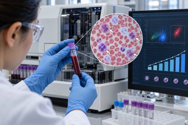
A circulating tumor cell count test measures rare intact tumor cells found in a defined volume of blood. In selected metastatic cancers, especially breast, prostate, and colorectal cancer, a higher CTC count is associated with a less favorable prognosis. The best-known CellSearch thresholds are 5 CTCs per 7.5 mL of blood for metastatic breast and prostate cancer and 3 CTCs per 7.5 mL for metastatic colorectal cancer. These values are prognostic cutoffs, not universal normal ranges and not direct measurements of how much cancer is in the body. A person can have active metastatic disease with a low or even zero detectable CTC count, while another person can have dozens or hundreds. Trends can also matter: falling counts during treatment may be associated with better outcomes, while persistently elevated counts can signal higher risk. Results should always be interpreted with imaging, symptoms, pathology, and the exact assay used.
- “High” CTC means above a validated cutoff for a specific cancer and platform, not above one universal reference range.
- CellSearch commonly uses ≥5 CTCs/7.5 mL as an unfavorable prognostic category in metastatic breast and prostate cancer.
- In metastatic colorectal cancer, ≥3 CTCs/7.5 mL is a commonly validated unfavorable CellSearch threshold.
- A high CTC count is linked to prognosis but cannot be converted directly into tumor size, stage, or number of metastases.
- A low or zero CTC count does not rule out active cancer because tumor-cell shedding and blood sampling are variable.
Table of Contents
- What a CTC Count Measures
- What Counts as a High CTC Level
- What High and Low Counts Mean for Prognosis
- CTC Count, Tumor Burden, and Changes Over Time
- Why CTC Counts Can Be Low, High, or Misleading
- How CTC Results Are Used With Treatment and Imaging
- Practical Questions Before and After Testing
What a CTC Count Measures
A CTC count estimates how many intact circulating tumor cells are present in a specific amount of blood. It is not a routine blood count and it does not measure all cancer cells in the bloodstream. The assay first enriches rare candidate cells, then applies strict criteria to decide which cells qualify as CTCs.
The most established platform, CellSearch, uses antibodies against EpCAM to capture epithelial cells. Candidate cells are then stained and classified using nuclear, cytokeratin, and white-blood-cell markers. Because the method is standardized, investigators have been able to validate particular prognostic thresholds in metastatic cancers.
The number on the report is usually written as CTCs per 7.5 mL of blood for CellSearch. Other platforms may use different blood volumes, enrichment methods, or cell definitions. A value of 4 from one technology is therefore not automatically equivalent to 4 from another.
A CTC test may provide more than a count. Research laboratories can characterize proteins, RNA, or DNA within captured cells. The count article, however, is about enumeration: how many cells were found and what that number has been shown to mean in a defined clinical setting.
What Counts as a High CTC Level
There is no universal CTC “normal range.” The most familiar thresholds come from studies of metastatic disease using CellSearch.
| Cancer setting | Common CellSearch prognostic threshold | Typical interpretation |
|---|---|---|
| Metastatic breast cancer | ≥5 CTCs per 7.5 mL | Unfavorable prognostic group in validated studies |
| Metastatic prostate cancer | ≥5 CTCs per 7.5 mL | Unfavorable prognostic group in validated studies |
| Metastatic colorectal cancer | ≥3 CTCs per 7.5 mL | Unfavorable prognostic group in validated studies |
These cutoffs should not be described as “positive for cancer.” They were developed to separate groups with different expected outcomes among patients already known to have metastatic cancer.
For metastatic breast cancer, the landmark prospective study found that patients with at least 5 CTCs per 7.5 mL before a new treatment had shorter median progression-free and overall survival than patients with fewer than 5. The threshold also remained prognostic at early follow-up.
For metastatic colorectal cancer, a prospective multicenter study used 3 CTCs per 7.5 mL. Patients at or above that level had shorter progression-free and overall survival than those below it.
The commonly used metastatic prostate cancer cutoff is also 5 CTCs per 7.5 mL. Later analyses have emphasized that CTC count can also be treated as a continuous variable rather than forcing every patient into a high-versus-low category. In other words, risk does not suddenly change biologically when a count moves from 4 to 5.
If a laboratory uses another platform, its own validated threshold should be used. Applying CellSearch cutoffs to a size-based, microfluidic, PCR-derived, or image-analysis assay can produce a misleading interpretation.
What High and Low Counts Mean for Prognosis
A high CTC count generally means the cancer is behaving in a way associated with higher risk of progression or death in validated metastatic settings. It does not tell exactly how long an individual patient will live.
Prognostic markers describe groups, not certainties. Two patients with the same CTC count can have very different outcomes because prognosis also depends on cancer subtype, metastatic sites, treatment sensitivity, performance status, organ function, other biomarkers, and available therapies.
A low count is usually associated with a more favorable prognosis compared with a high count in the same validated setting. But “favorable” does not mean the cancer is gone. A patient with 0 to 4 CTCs per 7.5 mL can still have active metastatic breast cancer that requires treatment.
Recent pooled analyses strengthen the evidence that serial CTC status carries prognostic information. In a 2025 global pooled analysis of metastatic breast cancer, repeated CellSearch measurements strongly predicted overall survival across tumor subtypes and treatment types. Patients whose CTC status improved after treatment had better outcomes than those with persistently positive results.
That is clinical validity: the biomarker reliably correlates with outcome. It is different from clinical utility, which asks whether changing therapy because of the CTC result improves the patient’s outcome. Clinical utility is more difficult to prove and remains limited for CTC-guided treatment switching.
This distinction prevents a common mistake. A high CTC result may correctly identify a higher-risk patient without providing evidence that one specific drug should be chosen solely because the count is high.
It is also important to separate prognosis from prediction. A prognostic biomarker tells something about expected outcome regardless of which therapy is chosen. A predictive biomarker helps identify whether one treatment is more likely to work than another. Standard CTC enumeration is much stronger as a prognostic tool than as a stand-alone predictive test. Molecular features found within CTCs may eventually provide more treatment-specific information, but those uses require separate validation.
The numeric cutoff itself can make the result look more absolute than it is. A count of 5 and a count of 4 fall into different CellSearch categories in metastatic breast cancer, yet the biological difference between those two samples may be small. Clinicians therefore consider the actual number, previous counts, measurement variability, and the patient’s overall course rather than treating the cutoff as a cliff.
CTC Count, Tumor Burden, and Changes Over Time
CTC count can correlate with tumor burden, but it is not a direct ruler for measuring cancer volume. Tumors differ in their ability to release cells into circulation. Blood supply, metastatic location, epithelial-marker expression, treatment, and tumor biology all affect the count.
A patient with extensive bone-predominant disease may shed a different number of detectable CTCs than someone with visceral metastases. A tumor undergoing epithelial-to-mesenchymal transition may also release cells that are less efficiently captured by EpCAM-based systems.
This means statements such as “10 CTCs equals twice as much cancer as 5 CTCs” are incorrect. A count of 100 does not mean the patient has 100 tumor deposits, and a count of zero does not mean the tumor burden is zero.
Serial trends can still be useful. Several patterns are possible:
- High to low: a drop below the prognostic cutoff after treatment may be associated with improved outcome.
- Low to high: conversion to an unfavorable category can be associated with worsening prognosis.
- High to high: persistent elevation often identifies a group with particularly poor outcomes.
- Low to low: persistent low counts generally support a more favorable prognostic group, although imaging can still show progression.
The interval between measurements matters. In metastatic breast cancer research, CTCs have been assessed within weeks after starting a new therapy, sometimes before standard imaging would normally show a clear response. That makes CTCs attractive as an early dynamic biomarker.
A dramatic change can be more informative than a small fluctuation. For example, moving from dozens of CTCs to none detected after treatment is biologically different from moving from 6 to 4, even though both cross the same categorical threshold. Conversely, a rise from 0 to 1 may reflect sampling variation rather than definite progression. The magnitude, direction, persistence, and clinical context of the change all matter.
Treatment itself can alter the count before imaging changes. Tumor-cell death, altered blood flow, or shifts in tumor phenotype can temporarily change how many cells enter the circulation. That is another reason serial values should be interpreted at validated time points rather than after arbitrary short intervals.
However, a ctDNA test may provide a different view of disease dynamics by measuring tumor-derived DNA rather than whole cells. The two biomarkers are not interchangeable and can move differently during treatment.
Why CTC Counts Can Be Low, High, or Misleading
CTCs are rare enough that sampling variation matters. A 7.5 mL tube contains only a tiny portion of the body’s total blood volume. If cells are circulating intermittently, one tube may contain fewer cells than another even without a meaningful change in disease.
Biological variation is another factor. Tumors shed different numbers of intact cells. Some CTCs travel in clusters; others undergo physical stress and die. Treatment may temporarily change shedding before it changes total tumor size.
Platform limitations also matter. CellSearch enriches EpCAM-positive epithelial cells. Tumor cells that have lost EpCAM or cytokeratin expression may escape detection. Marker-independent assays may recover different populations and therefore generate different counts.
Preanalytic problems can lower reliability. Delayed processing, incorrect collection tubes, temperature extremes, or poor sample preservation can damage cells. Because the assay depends on identifying intact cellular features, handling requirements are more demanding than for many routine chemistry tests.
A high count can also be misunderstood when used outside the population in which the threshold was validated. The classic cutoffs apply to particular metastatic cancer settings. They should not be used to screen healthy people, diagnose an unknown mass, or define recurrence after curative treatment without evidence for that use.
Early-stage disease is especially challenging. CTC counts are often very low, and a negative result has limited sensitivity. Research is exploring more sensitive rare-cell technologies and combinations with molecular residual disease testing, but standard metastatic thresholds should not be imported into the postoperative MRD setting.
How CTC Results Are Used With Treatment and Imaging
CTC count should be treated as a complementary biomarker. The oncology team interprets it alongside imaging, symptoms, examination findings, standard laboratory tests, pathology, and disease-specific tumor markers.
When imaging and CTC trends agree, interpretation is straightforward. Falling CTCs plus improving symptoms and shrinking tumors support treatment response. Rising CTCs plus radiographic growth and worsening symptoms support progression.
Discordant results require more care. A CTC count may rise before imaging changes, or imaging may show progression while the count remains low. The response should not be automatic. Clinicians may repeat the CTC assay, review scan timing, assess symptoms, check other markers, or obtain additional imaging.
Randomized studies have examined whether changing chemotherapy solely because CTC counts remain high improves outcomes. The results have not established a universal strategy in which CTC elevation by itself dictates an immediate treatment switch. That is why guidelines and expert consensus emphasize prognostic and monitoring value while remaining cautious about treatment decisions based only on enumeration.
CTC counts may be useful in clinical trials as stratification factors or intermediate biomarkers. They can help identify biologically high-risk groups and measure early changes during therapy without repeated tissue biopsy.
For individual care, the most useful question is not simply “Is my CTC high?” but “What decision would change because of this result?” If the answer is none, the test may still provide prognostic information, but the patient should understand its practical limitation.
Practical Questions Before and After Testing
Most CTC count tests require a standard venous blood draw and no fasting. The laboratory should specify the collection tube, blood volume, shipping conditions, and maximum time to processing.
Before testing, ask:
- Which CTC platform is being used?
- Is the assay validated for my cancer type and disease stage?
- What cutoff will the laboratory use, and what evidence supports it?
- Will the result affect treatment, prognosis discussion, trial eligibility, or monitoring?
- How will the result be compared with imaging and other tumor markers?
After testing, verify the exact units and sample volume. “5 CTCs” is incomplete information if the report does not say the volume and method. Ask whether the count is being interpreted categorically, as a continuous value, or as a trend from a previous measurement.
Do not compare raw numbers from different laboratories unless the methods are equivalent. If serial monitoring is planned, using the same assay and similar collection conditions makes changes easier to interpret.
A high result can be emotionally difficult because the word “unfavorable” may appear on the report. It is better understood as a statistical risk category, not a personal prediction with a fixed survival time. Modern cancer outcomes depend on many factors that a single blood biomarker cannot capture.
When discussing prognosis, ask whether the estimate comes from patients treated in an older era. Some foundational CTC studies were performed before several current targeted therapies, antibody-drug conjugates, immunotherapies, and modern supportive treatments were available. The relationship between high CTCs and higher risk remains important, but historical median survival figures should not be treated as a personal forecast today.
Likewise, a low result should not create false reassurance. If imaging, symptoms, or other evidence shows active disease, treatment and follow-up should continue according to the established cancer plan.
Timing should also be standardized when counts are used for monitoring. A baseline sample drawn before a new treatment and follow-up samples collected at clinically meaningful, protocol-consistent intervals are easier to compare than sporadic measurements obtained after different treatment cycles. Recent surgery, acute illness, transfusion, or major changes in therapy can also complicate interpretation, so the ordering clinician should know the surrounding clinical events. If a result is unexpectedly high or low and conflicts with the rest of the clinical picture, repeating the assay under comparable conditions may be more useful than making a major decision from one tube of blood. The goal is to use CTC enumeration as one reproducible data stream in longitudinal care, not as a stand-alone score that overrides stronger clinical evidence.
References
- Clinical Validity of Repeated Circulating Tumor Cell Enumeration as an Early Treatment Monitoring Tool for Metastatic Breast Cancer in the PREDICT Global Pooled Analysis 2025
- International expert consensus on the clinical integration of circulating tumor cells in solid tumors 2025 (Position Statement)
- Circulating tumor cells in breast cancer: clinical validity and utility 2024 (Review)
- Clinical applications of circulating tumor cells in patients with solid tumors 2024 (Review)
- Circulating tumor cells predict survival benefit from treatment in metastatic castration-resistant prostate cancer 2008
- Relationship of circulating tumor cells to tumor response, progression-free survival, and overall survival in patients with metastatic colorectal cancer 2008
Disclaimer
CTC thresholds are assay- and cancer-specific prognostic tools, not universal normal ranges or direct measurements of tumor burden. A high or low count should be interpreted with imaging, symptoms, pathology, treatment history, and the exact testing platform. Do not change cancer treatment based on a CTC value without guidance from the treating oncology team.