A circulating tumor DNA (ctDNA) test looks for fragments of DNA released by cancer cells into the bloodstream. It is a type of liquid biopsy that can sometimes identify tumor mutations, help select targeted treatment, track how cancer changes during therapy, or detect molecular residual disease after treatment intended to cure the cancer. The meaning of a result depends heavily on why the test was ordered. A mutation found in advanced cancer may guide a drug choice, while detectable ctDNA after surgery may indicate a higher risk of recurrence. A negative result is not proof that cancer is absent because some tumors shed very little DNA into blood. Test sensitivity also varies by cancer type, disease location, assay design, and tumor burden. For that reason, ctDNA is usually interpreted with pathology, tissue genomic testing, imaging, symptoms, and the treatment plan rather than as a stand-alone cancer test.
- ctDNA is the tumor-derived portion of cell-free DNA in plasma, not all DNA circulating in blood.
- A positive result can reveal tumor mutations or molecular evidence of disease, depending on the assay and clinical setting.
- A negative result can be false-negative, especially with low-volume, low-shedding, or certain anatomically confined cancers.
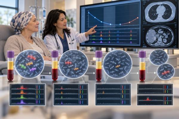
- Serial ctDNA results may help monitor response, resistance, MRD, and recurrence risk, but the appropriate action is cancer- and assay-specific.
- Tissue testing may still be needed when plasma testing is negative, inconclusive, or does not fit the clinical picture.
Table of Contents
- What ctDNA Is and How It Differs From Cell-Free DNA
- How a ctDNA Blood Test Works
- How to Understand ctDNA Results, VAF, and Tumor Fraction
- How ctDNA Is Used for Mutations, Treatment, and Monitoring
- ctDNA, MRD, and Recurrence Risk After Treatment
- False Negatives, Clonal Hematopoiesis, and Other Limitations
- What Happens After a ctDNA Result
What ctDNA Is and How It Differs From Cell-Free DNA
Most people have small fragments of DNA circulating in the cell-free portion of blood. This material is called cell-free DNA, or cfDNA. It can come from many normal tissues as cells die and release DNA. Cancer cells can also release DNA, and the tumor-derived fraction is called circulating tumor DNA, or ctDNA.
That distinction matters. A cell-free DNA cancer test analyzes a mixture that may contain both normal and tumor-derived DNA. The laboratory must identify features that make some fragments likely to come from the cancer, such as tumor-associated mutations, methylation patterns, or other molecular signals.
ctDNA usually makes up only a fraction of total cfDNA. In advanced metastatic disease, the fraction may be high enough for broad genomic profiling. In very small tumors or after curative-intent surgery, tumor DNA may be extraordinarily scarce. This is one reason assays designed for molecular residual disease often use different methods and much greater analytical sensitivity than standard broad mutation panels.
ctDNA enters blood through processes including tumor-cell death and active or passive release of DNA. Its concentration can change quickly as disease biology and treatment change. This makes ctDNA attractive as a dynamic biomarker, but it also means one blood draw is only a snapshot.
Different cancers shed different amounts of ctDNA. Tumor size is one influence, but it is not the only one. Metastatic site, vascularity, cell turnover, therapy, and tumor type all affect shedding. Brain tumors and some tumors confined to certain anatomical spaces can be especially difficult to detect in plasma.
A ctDNA test therefore does not measure cancer in the same way as a CT scan. Imaging shows anatomy. Pathology shows cells and tissue architecture. ctDNA shows molecular material in blood. The methods can complement one another, but none is automatically a substitute for the others.
How a ctDNA Blood Test Works
Testing usually begins with a venous blood draw into a tube designed to preserve the sample. The laboratory separates plasma from blood cells and extracts cfDNA. Careful processing is important because white blood cells can break down after collection and release large amounts of ordinary genomic DNA, diluting the tumor signal.
The assay then searches for predefined or broad molecular features. Common technologies include digital polymerase chain reaction and next-generation sequencing. Digital PCR can be highly sensitive when the laboratory already knows exactly which mutation to look for. NGS can examine many genes at once and is commonly used for broader cancer genomic profiling.
A ctDNA mutation panel may evaluate single-nucleotide variants and small insertions or deletions. Depending on assay design, it may also detect selected gene fusions, copy-number changes, or other genomic features. No panel detects every type of alteration equally well.
MRD assays take another approach. Some are tumor-informed, meaning the patient’s tumor tissue is sequenced first and the blood test is customized to track mutations known to be present in that tumor. Others are tumor-naive or tumor-agnostic and do not require a prior tissue profile. These approaches have different strengths, turnaround times, and sources of error.
A high-quality assay also uses error-suppression methods. Because low-frequency variants can resemble sequencing errors, laboratories may use unique molecular identifiers, replicate observations, background models, and minimum reporting thresholds to distinguish true signals from noise.
The report should identify the sample type, method, genes or targets tested, analytical limitations, and detected alterations. It may also give a variant allele fraction, an estimate of tumor fraction, or an interpretation of whether a finding is actionable. Those measurements are related but are not interchangeable.
Preanalytic details can affect results. The right tube, correct blood volume, prompt or validated delayed processing, adequate plasma yield, and avoidance of sample contamination all support reliable testing. If a sample fails quality checks, repeating the blood draw can be more appropriate than interpreting a technically inadequate result.
How to Understand ctDNA Results, VAF, and Tumor Fraction
A ctDNA report may say detected, not detected, or provide specific genetic alterations. The correct interpretation depends on the test’s purpose.
In a genomic profiling test, a positive result may list a mutation such as an activating driver alteration or a resistance mutation. The important question is whether that alteration has an established treatment implication for the patient’s cancer type and stage. A mutation can be biologically interesting without being clinically actionable.
Reports often include variant allele fraction, or VAF. VAF is the proportion of DNA molecules at a particular genomic position that carry the reported variant. A VAF of 2%, for example, means roughly 2% of the analyzable molecules at that site carried that alteration under the assay’s measurement method. It does not mean that 2% of the patient’s body is cancer or that exactly 2% of all cfDNA is tumor-derived.
Tumor fraction is a broader estimate of the proportion of cfDNA believed to come from tumor. Methods for estimating it differ across laboratories. The 2026 ASCO guideline specifically cautions against using fractional, percentage, or concentration-based ctDNA or total cfDNA measurements as surrogate measures of disease by themselves. In practical terms, a numerical rise can be clinically meaningful in a validated monitoring strategy, but it should not be treated as a universal blood-based measurement of tumor volume.
There is also no single “normal ctDNA level.” Some assays are qualitative, some report VAF, and some use patient-specific molecular thresholds. A result that is meaningful in one assay cannot automatically be compared with a number from another platform.
A not detected result means the assay did not identify a reportable tumor signal in that sample. It does not establish that no cancer exists. The tumor may not be shedding enough DNA, the relevant mutation may not be included on the panel, the alteration may be below the limit of detection, or the sample may contain too little tumor-derived DNA.
That is why a plasma-negative result in a patient who clearly has cancer is often described as non-informative rather than reassuring. When the goal is to find a treatment target in advanced cancer, tissue testing may be needed after a negative or inconclusive plasma test.
How ctDNA Is Used for Mutations, Treatment, and Monitoring
The most established use of ctDNA in many advanced solid tumors is genotyping. A blood test can sometimes identify a targetable alteration without another invasive tumor biopsy. This is useful when tissue is difficult to obtain, the biopsy would carry substantial risk, the existing sample is too small or old, or a faster result could affect an urgent treatment decision.
The 2026 ASCO guideline supports ctDNA testing for tumor genetic alterations in situations where tissue testing is challenging or not feasible, when biopsy risk is unacceptable, when tissue results would arrive too late for a clinically actionable decision, or when a drug’s regulatory indication allows or requires ctDNA testing. It also recommends seeking tissue confirmation when plasma results are negative, inconclusive, or inconsistent with the clinical scenario.
ctDNA can also capture genetic heterogeneity. A single tissue biopsy samples one location at one time. DNA in plasma may come from multiple metastatic sites, so a blood-based assay can sometimes reveal alterations present in resistant subclones that were not represented in the original biopsy.
This is especially relevant at disease progression. A cancer can evolve under treatment pressure and acquire resistance alterations. Repeat plasma testing may reveal a new molecular mechanism that supports a different targeted therapy or clinical trial. A liquid biopsy resistance mutation test is therefore often most useful when its findings can change a specific treatment decision.
Serial ctDNA can also be studied as a treatment-response marker. Falling tumor-associated DNA after effective therapy can correlate with response, while persistent or rising ctDNA may precede radiographic progression in some settings. However, the timing, assay, cancer type, and treatment all matter. There is no universal rule that a single rise means treatment has failed.
The strongest use case is one with a clear action attached to the result. If a validated mutation predicts response to an approved therapy, the result can guide care. If a monitoring signal changes but there is no evidence-based action to take, the test may provide prognostic information without improving outcomes by itself.
ctDNA, MRD, and Recurrence Risk After Treatment
After surgery, radiation, or other treatment intended to eliminate a localized cancer, imaging may show no disease while microscopic cancer cells remain. Blood-based detection of tumor DNA in this setting is called molecular or measurable residual disease, often shortened to MRD.
A ctDNA MRD test is designed to detect far smaller tumor signals than many standard genomic profiling panels. It may be performed once at a defined postoperative landmark or repeatedly during surveillance.
Across many solid-tumor studies, detectable ctDNA after curative-intent treatment is associated with a substantially higher risk of later clinical recurrence. That makes postoperative ctDNA a powerful prognostic biomarker. It can sometimes identify molecular recurrence months before a tumor becomes visible on routine imaging.
A positive MRD result, however, is not the same as a visible recurrence. It says that tumor-derived molecular material has been detected according to the assay’s criteria. The next step may include confirmatory testing, imaging, closer surveillance, systemic therapy in a setting supported by evidence, or clinical-trial enrollment.
A negative MRD result is more reassuring than a positive one but does not mean recurrence risk is zero. Sensitivity is imperfect, especially with small-volume or low-shedding disease. Repeated negative tests over time may provide more information than one negative sample, but interpretation remains disease-specific.
Clinical utility is evolving quickly. Some randomized trials have tested ctDNA-guided treatment escalation or de-escalation, and multiple cancer-specific trials are ongoing. Current guidance increasingly allows ctDNA use when a specific evidence-based action is linked to the result. It should not be assumed that every positive MRD result in every cancer automatically requires more treatment.
The timing of blood collection also matters. After surgery, normal tissue injury can temporarily increase total cfDNA and dilute the tumor fraction. Many research protocols therefore use defined postoperative windows rather than drawing immediately after an operation. The appropriate interval depends on the cancer, assay, and intended decision.
False Negatives, Clonal Hematopoiesis, and Other Limitations
The most important limitation is false-negative testing. A tumor can be present even when no ctDNA is detected. Low disease volume, low biological shedding, limited assay coverage, central nervous system–predominant disease, and technical detection limits can all contribute.
This matters most when plasma testing is used to look for a drug target. If a patient has advanced cancer and the ctDNA panel is negative, that does not prove the tumor lacks actionable alterations. When feasible, tissue testing can recover information that plasma missed.
A second limitation is clonal hematopoiesis. As people age, blood-forming stem cells can acquire mutations and expand into clones without being cancer cells from a solid tumor. DNA from those blood-cell clones enters plasma and can mimic tumor-derived mutations. Genes such as DNMT3A, TET2, and ASXL1 are classic examples, although overlap with cancer-relevant genes can occur.
Some laboratories sequence paired white blood cells or use computational filters to reduce this problem. The report and clinical context still matter, particularly when a variant appears at an unexpected frequency or does not fit the known tumor biology.
Other limitations include sequencing artifacts, differences in assay sensitivity, incomplete coverage of fusions or copy-number changes, and variation among laboratories. A technically valid result is not automatically clinically useful. The test must also have evidence that its findings accurately predict the outcome or treatment response for the intended use.
There is another source of confusion: a variant found in plasma is not necessarily inherited. Most ctDNA findings are somatic changes acquired by tumor cells. However, certain results may raise concern for a germline variant that is present in every cell. Confirmatory testing in a proper germline sample and genetic counseling may be needed rather than assuming the plasma result proves inherited risk.
Finally, ctDNA testing is not a universal screening test for healthy people. Assays designed for patients with known cancer, genomic profiling, or postoperative MRD have different performance requirements from population screening tests. Using a test outside its validated setting can change the balance of false positives and false negatives.
What Happens After a ctDNA Result
The first step is to identify why the test was ordered. The same words on a report can have very different implications in advanced cancer, postoperative surveillance, and resistance monitoring.
For a mutation-panel result, ask:
- Is the alteration considered a driver, resistance marker, or variant of uncertain significance?
- Is it actionable for this exact cancer type and stage?
- Is an approved therapy or clinical trial linked to it?
- Was tumor fraction high enough for a negative result to be informative?
- Should tissue genomic testing be performed or repeated?
For MRD or serial monitoring, ask:
- Was the blood drawn at the assay’s recommended time point?
- Is the result newly positive, persistently positive, falling, or rising?
- What evidence-based action is available if the result is positive?
- Should imaging or another ctDNA sample be obtained before changing treatment?
Consistency matters for serial testing. When possible, follow-up samples should use the same validated assay and similar timing relative to treatment. Switching platforms can make small numerical changes difficult to interpret because analytical sensitivity and reporting methods differ.
Patients should also avoid reading VAF or tumor-fraction numbers as personal survival estimates. These measures can correlate with disease biology in groups, but prognosis depends on cancer type, stage, treatment response, molecular subtype, metastatic sites, overall health, and many other factors.
A blood test can make cancer monitoring more convenient, but convenience should not replace the information that only tissue or imaging can provide. Tissue can establish histology and reveal tumor architecture. Imaging can locate disease and identify complications. ctDNA can add molecular information and can sometimes detect change earlier. Good care uses the method that best answers the clinical question.
The most useful interpretation is therefore decision-focused: What does this ctDNA result change today? A targetable mutation may change therapy. A negative plasma profile may trigger tissue testing. A validated MRD result may alter surveillance or adjuvant treatment in an evidence-supported setting. If no action is supported, the result should be discussed as prognostic or investigational rather than treated as a command to change care.
References
- Circulating Tumor DNA Testing in Solid Tumors and Lymphoma: ASCO Guideline 2026 (Guideline)
- Circulating Tumor DNA: A Pan-Cancer Biomarker in Solid Tumors with Prognostic and Predictive Value 2025 (Review)
- Circulating tumor DNA to monitor treatment response in solid tumors and advance precision oncology 2025 (Review)
- Clinical Practice Guideline for Blood-based Circulating Tumor DNA Assays 2024 (Guideline)
- ESMO recommendations on the use of circulating tumour DNA assays for patients with cancer: a report from the ESMO Precision Medicine Working Group 2022 (Guideline)
Disclaimer
ctDNA testing can provide important molecular information, but a negative result does not rule out cancer or eliminate recurrence risk. The meaning of mutations, MRD signals, and serial changes depends on the assay, cancer type, disease stage, and treatment setting. Treatment decisions should be made with the oncology team using validated evidence plus tissue, imaging, and other clinical information when appropriate.